Chinese Journal of Tissue Engineering Research ›› 2022, Vol. 26 ›› Issue (5): 724-729.doi: 10.12307/2022.118
Previous Articles Next Articles
Deep seawater promotes wound healing in diabetic mice by activating PI3K/Akt pathway
Li Weiming, Xu Qingwen, Li Yijun, Sun Yanbo, Cui Jin, Xu Pengyuan
- Second Ward of Department of Gastrointestinal Surgery, the Second Affiliated Hospital of Kunming Medical University, Kunming 650101, Yunnan Province, China
-
Received:2020-09-30Revised:2020-10-13Accepted:2021-01-19Online:2022-02-18Published:2021-10-28 -
Contact:Li Weiming, Second Ward of Department of Gastrointestinal Surgery, the Second Affiliated Hospital of Kunming Medical University, Kunming 650101, Yunnan Province, China -
About author:Li Weiming, MD, Associate chief physician, Second Ward of Department of Gastrointestinal Surgery, the Second Affiliated Hospital of Kunming Medical University, Kunming 650101, Yunnan Province, China -
Supported by:Yunnan Provincial Applied Basic Research Project, No. 2015FB057 (to LWM); the Subproject of Yunnan Provincial Surgical Clinical Nutrition Research Center, No. 2016NS272 (to LWM); High-level Talent Project of Yunnan Provincial Health and Construction Commission, No.D-2017037 (to LWM)
CLC Number:
Cite this article
Li Weiming, Xu Qingwen, Li Yijun, Sun Yanbo, Cui Jin, Xu Pengyuan . Deep seawater promotes wound healing in diabetic mice by activating PI3K/Akt pathway[J]. Chinese Journal of Tissue Engineering Research, 2022, 26(5): 724-729.
share this article
Add to citation manager EndNote|Reference Manager|ProCite|BibTeX|RefWorks
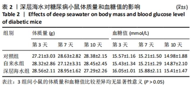
2.1 实验动物数量分析 48只小鼠在实验过程中无死亡,全部进入结果分析。 2.2 深层海水对小鼠体质量与血糖的影响 观察第3,7,10天小鼠的体质量和血糖值发现,对照组、自来水组及深层海水组小鼠的体质量和血糖值比较差异均无显著性意义(P > 0.05),见表2,说明深层海水对小鼠的体质量和血糖值没有影响。"
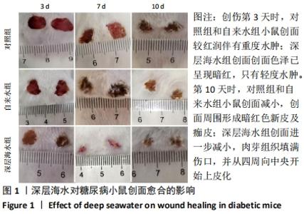
2.3 深层海水对糖尿病小鼠创面愈合的影响 创伤后不同时间点各组小鼠创面情况见图1。对照组和自来水组小鼠创伤后第3天的创面较红润伴有重度水肿;至第7天,创面色泽变为暗红、无光泽,并有肉芽的形成;到第10天,创面减小,创面周围形成暗红色新皮及痂皮。深层海水组小鼠创伤后第3天小鼠创面创面色泽已呈现暗红,只有轻度水肿;至第7天,创面明显减小,有肉芽组织增生及淡红色新皮的生成;至第10天,创面进一步减小,肉芽组织填满伤口,并从四周向中央开始上皮化,说明深层海水可促进糖尿病小鼠创面的愈合。"

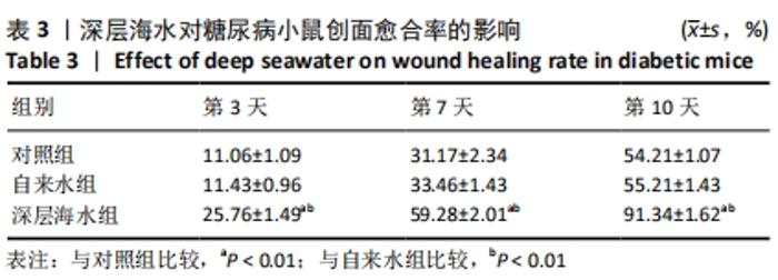
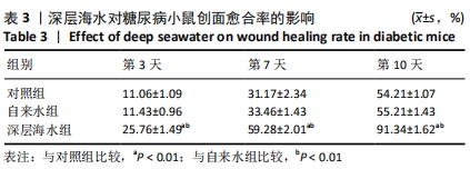
同时对小鼠创面愈合率进行统计发现,相比于对照组和自来水组,深层海水处理后可显著促进小鼠创面的愈合(P < 0.01),自来水对小鼠创面愈合的影响与对照组比较差异无显著性意义(P > 0.05),见表3。"
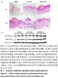
2.4 深层海水对糖尿病小鼠创面肉芽新生血管形成的影响 各组小鼠创面苏木精-伊红染色结果,见图2A。对照组和自来水组小鼠创伤后第3天创面具有大量炎性细胞浸润及少量纤维母细胞,第10天可见肉芽组织及毛细血管和纤维母细胞;深层海水组小鼠创伤后第3天就可观察到纤维母细胞、毛细血管和少量炎性细胞,第10天可见新生肉芽组织,有较多胶原纤维和少量毛细血管。 此外Western blot检测结果显示,深层海水组小鼠创伤后第3,7,10天创面组织中的血管内皮生长因子和碱性成纤维细胞生长因子蛋白表达水平明显高于对照组和自来水组(P < 0.01),自来水组小鼠创面组织中的血管内皮生长因子和碱性成纤维细胞生长因子蛋白表达水平与对照组比较差异无显著性意义(P > 0.05),见图2B。结果表明,深层海水会促进创面新生肉芽的形成,进一步说明深层海水会促进创面愈合。"
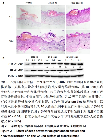
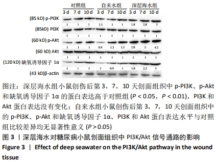
2.5 深层海水对创面组织中PI3K/Akt信号通路的影响 Western blot检测结果显示,深层海水组小鼠创伤后第3,7,10天创面组织中p-PI3K、p-Akt和缺氧诱导因子1α的蛋白表达高于对照组(P < 0.05,P < 0.01),PI3K和Akt蛋白表达没有变化;自来水组小鼠创伤后第3,7,10天创面组织中的p-PI3K、p-Akt和缺氧诱导因子1α、PI3K和Akt蛋白表达水平与对照组比较差异均无显著性意义(P > 0.05),见图3。由此可知,深层海水激活了创面组织中PI3K/Akt信号通路,通过PI3K/Akt信号通路上调缺氧诱导因子1α的表达。"

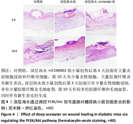
2.6 深层海水通过调控PI3K/Akt信号通路对糖尿病小鼠创面愈合的影响 基于深层海水处理糖尿病小鼠后对PI3K/Akt信号通路的影响,进一步探讨深层海水是否通过激活PI3K/Akt信号通路促进小鼠创面愈合。 2.6.1 创面苏木精-伊红染色 对照组小鼠创伤后第3天创面有大量炎症细胞浸润和纤维母细胞;第10天有少量炎性细胞、大量胶原纤维并有新生表皮。深层海水组小鼠创伤后第3天创面只有少量炎性细胞浸润,并有少量胶原纤维及毛细血管;第10天有较多的胶原纤维和毛细血管,同时伴有新生表皮的生成。深层海水+LY294002组小鼠创面苏木精-伊红染色结果与对照组相似。各组不同时间点创面组织形态观察见图4。"
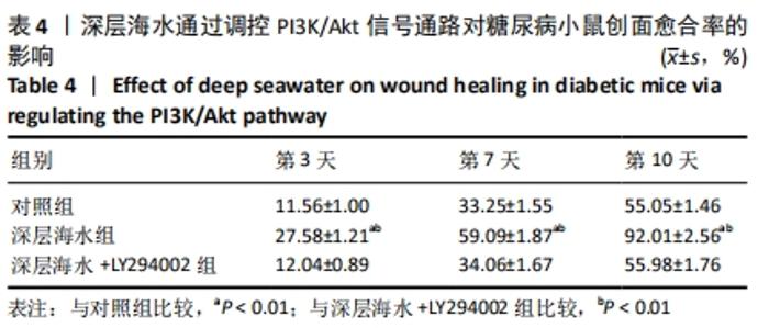
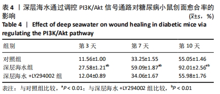
2.6.2 创面愈合率 与对照组相比,深层海水组可提高不同时间点的小鼠创面愈合率(P < 0.01),深层海水+LY294002组创面愈合率无明显变化(P > 0.05),见表4。说明PI3K/Akt信号通路对糖尿病小鼠创面愈合具有重要作用,抑制PI3K/Akt通路影响创面愈合。"
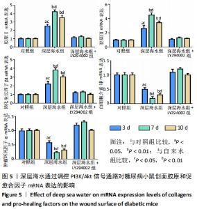
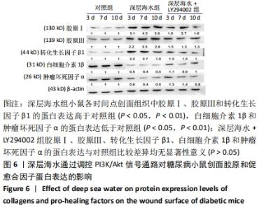
2.6.3 RT-PCR与Western blot检测结果 qRT-PCR及Western blot检测结果显示见图5,6。深层海水组小鼠各时间点创面组织中胶原Ⅰ、胶原Ⅲ和转化生长因子β1的蛋白及mRNA表达高于对照组(P < 0.05,P < 0.01),且在第7天达到最高值;白细胞介素1β和肿瘤坏死因子α的蛋白及mRNA表达低于对照组(P < 0.05,P < 0.01),且在第7天达到最低值。深层海水+LY294002组胶原Ⅰ、胶原Ⅲ、转化生长因子β1、白细胞介素1β和肿瘤坏死因子α的蛋白及mRNA表达与对照组比较差异均无显著性意义(P > 0.05),说明LY294002可逆转深层海水对胶原、促炎因子和促愈合因子表达的调控作用。由此可知,深层海水通过激活PI3K/Akt通路促进了创面组织中胶原和促愈合因子的表达,从而加速糖尿病小鼠创伤创面愈合。"

"
| [1] YU T, GAO M, YANG P, et al. Insulin promotes macrophage phenotype transition through PI3K/Akt and PPAR-gamma signaling during diabetic wound healing. J Cell Physiol. 2018;234(4):4217-4231. [2] MCBRIDE JD, JENKINS AJ, LIU X, et al. Elevated circulation levels of an antiangiogenic SERPIN in patients with diabetic microvascular complications impair wound healing through suppression of Wnt signaling. J Invest Dermatol. 2014;134(6):1725-1734. [3] MORENO-MARIN N, MERINO JM, ALVAREZ-BARRIENTOS A, et al. Aryl hydrocarbon receptor promotes liver polyploidization and inhibits PI3K, ERK, and Wnt/beta-catenin signaling. iScience. 2018;4:44-63. [4] FAN YS, LI Q, HAMDAN N, et al. Tetrahydroxystilbene glucoside regulates proliferation, differentiation, and OPG/RANKL/M-CSF expression in MC3T3-E1 cells via the PI3K/Akt pathway. Molecules. 2018;23(9):2306-2309. [5] SONG BQ, CHI Y, LI X, et al. Inhibition of notch signaling promotes the adipogenic differentiation of mesenchymal stem cells through autophagy activation and PTEN-PI3K/AKT/mTOR pathway. Cell Physiol Biochem. 2015;36(5):1991-2002. [6] YOKOTA J, CHOSA N, SAWADA S, et al. PDGF-induced PI3K-mediated signaling enhances the TGF-beta-induced osteogenic differentiation of human mesenchymal stem cells in a TGF-beta-activated MEK-dependent manner. Int J Mol Med. 2014;33(3):534-542. [7] YEH YH, HSIAO HF, YEH YC, et al. Inflammatory interferon activates HIF-1α-mediated epithelial-to-mesenchymal transition via PI3K/AKT/mTOR pathway. J Exp Clin Cancer Res. 2018;37(1):70. [8] XIAOYAN H, ZHIYING H, XINWEI J, et al. Folic Acid Represses Hypoxia-Induced Inflammation in THP-1 Cells through Inhibition of the PI3K/Akt/HIF-1α Pathway. PLoS One. 2016;11(3):e0151553. [9] CHOI YH, JIN GY, LI LC, et al. Inhibition of Protein Kinase C Delta Attenuates Allergic Airway Inflammation through Suppression of PI3K/Akt/mTOR/HIF-1 Alpha/VEGF Pathway. PLoS One. 2013;8(11):e81773. [10] DINDA M, DASGUPTA U, SINGH N, et al. PI3K-mediated proliferation of fibroblasts by Calendula officinalis tincture: implication in wound healing. Phytother Res. 2015; 29(4):607-616. [11] 农庆文,李顺堂,刘达恩,等.弱激光促进糖尿病大鼠皮肤创伤愈合及对PI3K/Akt-HIF-1α信号系统的影响[J].中国老年学杂志,2018,38(10):2459-2462. [12] JINSONG L, LEI C, JIAN X, et al. Effects of Periploca forrestii Schltr on wound healing by Src meditated Mek/Erk and PI3K/Akt signals. J Ethnopharmacol. 2019; 237:116-127. [13] 李为明,崔进,徐鹏远,等.深层海水对小鼠创面愈合的促进作用[J].重庆医学,2014,43(4):462-464. [14] 李为明,崔进,徐鹏远,等.“深层海水”对小鼠免疫功能的影响[J].天然产物研究与开发,2013,25(7):903-906. [15] 李为明,崔进,徐鹏远,等.“深层海水”对小鼠耐受力的影响[J].天然产物研究与开发,2013(6):103-106. [16] PEI Y, DEMIN Y, JIANCHENG Q, et al. High D-glucose alters PI3K and Akt signaling and leads to endothelial cell migration, proliferation and angiogenesis dysfunction. National Med J Chin. 2006;86(48):3425-3430. [17] ASSOCIATION AD. Prevention or Delay of Type 2 Diabetes: Standards of Medical Care in Diabetes—2018. Diabetes Care. 2018;41(Suppl 1):S51-S54. [18] WHITING DR, GUARIGUATA L, WEIL C, et al. IDF diabetes atlas: global estimates of the prevalence of diabetes for 2011 and 2030. Diabetes Res Clin Pract. 2011; 94(3):311-321. [19] BARROS JF, WACLAWIAK I, PECLI C, et al. Role of Chemokine Receptor CCR4 and Regulatory T Cells in Wound Healing of Diabetic Mice. J Invest Dermatol. 2018; 139(5):1161-1170. [20] BONIAKOWSKI AE, KIMBALL AS, JACOBS BN, et al. Macrophage-Mediated Inflammation in Normal and Diabetic Wound Healing. J Immunol. 2017;199(1):17. [21] BALTZIS D, ELEFTHERIADOU I, VEVES A. Pathogenesis and Treatment of Impaired Wound Healing in Diabetes Mellitus: New Insights. Adv Ther. 2014;31(8):817-836. [22] HA BG, JUNG SS, JANG YK, et al. Mineral-Enriched Deep-Seawater Modulates Lactate Metabolism via PGC-1α-Mediated Metabolic Reprogramming. Mar Drugs. 2019;17(11):611. [23] BYUNG H, EUN S, JUNG-EUN P, et al. Anti-Diabetic Effect of Balanced Deep-Seawater and Its Mode of Action in High-Fat Diet Induced Diabetic Mice. Mar Drugs. 2013;11(11):4193-4212. [24] HA BG, PARK JE, SHIN EJ, et al. Modulation of Glucose Metabolism by Balanced Deep-Seawater Ameliorates Hyperglycemia and Pancreatic Function in Streptozotocin-Induced Diabetic Mice. PLoS One. 2014;9(7):e102095. [25] SHIRAISHI H, FUJINO M, SHIRAKAWA N, et al. Effect of minerals on intestinal IgA production using deep seawater drinks. Biol Pharm Bull. 2017;40(10):1700-1705. [26] LEE CL. The advantages of deep ocean water for the development of functional fermentation food. Appl Microbiol Biotechnol. 2015;99(6):2523-2531. [27] BAK JP, KIM YM, SON J, et al. Application of concentrated deep seawater inhibits the development of atopic dermatitis-like skin lesions in NC/Nga mice. BMC Complement Altern Med. 2012;12:108. [28] YANG CC, YAO CA, LIN YR, et al. Deep-Seawater Containing Selenium Provides Intestinal Protection against Duodenal Ulcers through the Upregulation of Bcl-2 and Thioredoxin Reductase 1. PLoS One. 2014;9(7):e96006. [29] YUE Q, PAN X, LIU Y, et al. Protective effects of deep seawater against subacute alcoholic liver injury. J of hygiene Res. 2018;47(5):798-803. [30] CHUN SY, LEE KS, NAM KS. Refined deep-seawater suppresses inflammatory responses via the MAPK/AP-1 and NF-kappaB signaling pathway in LPS-treated RAW 264.7 macrophage cells. Int J Mol Sci. 2017;18(11):2282. [31] CHUN SY, KIM SNAM KS. The inhibitory effects of deep-seawater on doxorubicininduced epithelial-mesenchymal transition. Oncol Rep. 2017;38(2): 1163-1171. [32] KIMATA H, TAI H, NAKAGAWA K, et al. Improvement of skin symptoms and mineral imbalance by drinking deep seawater in patients with atopic eczema/dermatitis syndrome (AEDS). Acta Medica (Hradec Kralove). 2002;45(2):83-84. [33] YOU L, GU W, CHEN L, et al. MiR-378 overexpression attenuates high glucose-suppressed osteogenic differentiation through targeting CASP3 and activating PI3K/Akt signaling pathway. Int J Clin Exp Pathol. 2014;7(10):7249-7261. [34] MA P, GU B, XIONG W, et al. Glimepiride promotes osteogenic differentiation in rat osteoblasts via the PI3K/Akt/eNOS pathway in a high glucose microenvironment. PLoS One. 2014; 9(11):e112243. [35] ZIRU L, LEI Y, JIN Z, et al. Nobiletin protects PC12 cells from ERSinduced apoptosis in OGD/R injury via activation of the PI3K/AKT pathway. Exp Ther Med. 2018;16(2):1470-1476. [36] XIA P, XU XY. PI3K/Akt/mTOR signaling pathway in cancer stem cells: from basic research to clinical application. Am J Cancer Res. 2015;5(5):1602-1609. [37] KIM JH, BAE HC, KIM J, et al. HIF-1alpha-mediated BMP6 down-regulation leads to hyperproliferation and abnormal differentiation of keratinocytes in vitro. Exp Dermatol. 2018;27(11):1287-1293. [38] DU J, LIU L, LAY F, et al. Combination of HIF-1α gene transfection and HIF-1-activated bone marrow-derived angiogenic cell infusion improves burn wound healing in aged mice. Gene Ther. 2013;20(11):1070-1076. [39] YANG HL, TSAI YC, KORIVI M, et al. Lucidone promotes the cutaneous wound healing process via activation of the PI3K/AKT, Wnt/beta-catenin and NF-kappaB signaling pathways. Biochim Biophys Acta Mol Cell Res. 2017;1864(1):151-168. [40] ZHANG W, BAI X, ZHAO B, et al. Cell-free therapy based on adipose tissue stem cell-derived exosomes promotes wound healing via the PI3K/Akt signaling pathway. Exp Cell Res. 2018;370(2):333-342. |
| [1] | Zhang Jinglin, Leng Min, Zhu Boheng, Wang Hong. Mechanism and application of stem cell-derived exosomes in promoting diabetic wound healing [J]. Chinese Journal of Tissue Engineering Research, 2022, 26(7): 1113-1118. |
| [2] | Zhao Zixi, Xu Jun, Ding Min, Li Xiwen, Zhang Jinghang, Wang Penghua. Changes of type I and III collagen and matrix metalloproteinase 2 and 9 on the wound of diabetic foot ulcer with external application of medical collagen dressing [J]. Chinese Journal of Tissue Engineering Research, 2022, 26(10): 1544-1550. |
| [3] | Zhang Zhenkun, Li Zhe, Li Ya, Wang Yingying, Wang Yaping, Zhou Xinkui, Ma Shanshan, Guan Fangxia. Application of alginate based hydrogels/dressings in wound healing: sustained, dynamic and sequential release [J]. Chinese Journal of Tissue Engineering Research, 2021, 25(4): 638-643. |
| [4] | Wang Bo, Bai Shengchao, Li Wenbo, Wang Lin. A rabbit model of high-intensity jumping-induced tendon injury followed by post-exercise cold water immersion: inflammations and collagen remodeling in the patellar tendon [J]. Chinese Journal of Tissue Engineering Research, 2021, 25(29): 4619-4625. |
| [5] | Zhang Heng, Zhang Xianping, Dai Houjie, Huang Changzhao, Wang Ruiying. Gastrodin interferes with motor function recovery and growth associated protein-43 expression in a rabbit model of spinal cord injury [J]. Chinese Journal of Tissue Engineering Research, 2021, 25(29): 4626-4631. |
| [6] | Wen Xu, Shen Jing. Application of nano-silver antibacterial hydrogel dressing combined with recombinant human basic fibroblast growth factor on wound healing after debridement of open ankle fracture [J]. Chinese Journal of Tissue Engineering Research, 2021, 25(29): 4638-4643. |
| [7] | Liu Fang, Shan Zhengming, Tang Yulei, Wu Xiaomin, Tian Weiqun. Effects of hemostasis and promoting wound healing of ozone sustained-release hydrogel [J]. Chinese Journal of Tissue Engineering Research, 2021, 25(22): 3445-3449. |
| [8] | Chen Zhenyu, Zhang Xiaoning, Luo Yuxin, Liang Jianwei, Yan Chi. Evaluation of silk fibroin/curcumin composite film for promoting wound healing [J]. Chinese Journal of Tissue Engineering Research, 2021, 25(16): 2554-2561. |
| [9] | Zhao Xin, Shi Xin, Chen Bei, Cao Yanpeng, Chen Yaowu, Liu Xiaoren, He Yusheng, He Liyun, Li Xiying, Liu Jun. Vacuum sealing drainage enhances wound healing by up-regulating collagen type I/III ratio in rats [J]. Chinese Journal of Tissue Engineering Research, 2020, 24(32): 5122-5127. |
| [10] | Liu Yanhua, Zhu Zhou, Wan Qianbing. A drug-loading system for electrospinning wound repair: component selection and construction strategy [J]. Chinese Journal of Tissue Engineering Research, 2020, 24(28): 4465-4473. |
| [11] |
Gu Yingxuan, Huang Linfeng, Hu Xiaohui, Quan Xiaoming, Kang Liangqi, Zhou Linghan, Wang Xiaojun.
Platelet-rich plasma combined with negative pressure for chronic refractory wounds: a meta-analysis [J]. Chinese Journal of Tissue Engineering Research, 2020, 24(26): 4257-4264. |
| [12] |
Geng Kang, Ding Xiaobin, Tian Xinli, Wang Xue, Yang Yuting, Yan Hong.
Electrical stimulation promotes wound healing and angiogenesis in diabetic rats [J]. Chinese Journal of Tissue Engineering Research, 2020, 24(26): 4152-4156. |
| [13] | Liang Zhou, Wen Lichun, He Zhong, Huang Zheng, Li Kaijing, Yang Xiaoping, Liang Shihe, Pang Gexiong. Three-dimensional printing combined with virtual surgical design in the treatment of complex Pilon fractures [J]. Chinese Journal of Tissue Engineering Research, 2020, 24(24): 3786-3791. |
| [14] | Chen Sheng, Liu Jianhang, Liu Bentao, Zhang Zhaojian, Li Benche, Zhang Fan, Li Dongfei. Hot spots of animal models of cervical spondylotic myelopathy [J]. Chinese Journal of Tissue Engineering Research, 2020, 24(24): 3890-3896. |
| [15] | Zhong Shuxian, Shi Yuqing, Yang Yalan, Li Chun. Effect of soft silicone silver ion dressing applied in burn wounds [J]. Chinese Journal of Tissue Engineering Research, 2020, 24(22): 3602-3608. |
| Viewed | ||||||
|
Full text |
|
|||||
|
Abstract |
|
|||||