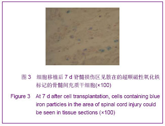
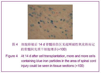
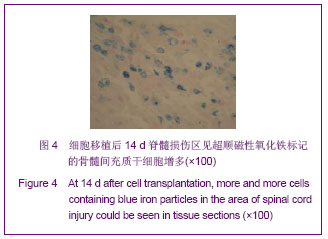
| [1] Ban DX, Ning GZ, Feng SQ, et al. Combination of activated Schwann cells with bone mesenchymal stem cells: the best cell strategy for repair after spinal cord injury in rats. Regen Med. 2011;6(6):707-720.[2] Shi CY, Ruan LQ, Feng YH, et al. Marrow mesenchymal stem cell transplantation with sodium alginate gel for repair of spinal cord injury in mice. Zhejiang Da Xue Xue Bao Yi Xue Ban. 2011;40(4):354-359. [3] Wang K, Li JD, Zhang RP, et al. Zhongguo Zuzhi Gongcheng Yanjiu yu Linchuang Kangfu. 2010;14(19):3436-3439.王凯,李健丁,张瑞平,等.超顺磁性氧化铁标记骨髓间充质干细胞诱导分化为神经细胞的体外实验[J].中国组织工程研究与临床康复,2010,14(19):3436-3439.[4] Nakamura S, Yamada Y, Baba S. Culture medium study of human mesenchymal stem cells for practical use of tissue engineering and regenerative medicine. Biomed Mater Eng. 2008;18(3):129-136.[5] Chickera S, Willert C, Mallet C, et al. Cellular MRI as a suitable, sensitive non-invasive modality for correlating in vivo migratory efficiencies of different dendritic cell populations with subsequent immunological outcomes.Int Immunol. 2012; 24(1):29-41. [6] Willenbrock S, Knippenberg S, Meier M, et al. In vivo MRI of intraspinally injected SPIO-labelled human CD34+ cells in a transgenic mouse model of ALS. In Vivo. 2012; 26(1):31-38.[7] Ren Z, Wang J, Zou C, et al. Labeling of cynomolgus monkey bone marrow-derived mesenchymal stem cells for cell tracking by multimodality imaging. Sci China Life Sci. 2011; 54(11):981-987. [8] Shuang WB, Liu Q. Zhonghua Shiyan Waike Zazhi. 2010; 27(7):1006.双卫兵,刘强.一种用于制备标准化脊髓损伤动物模型的实验装置[J].中华实验外科杂志, 2010,27(7):1006. [9] Martinez M, Brezun JM, Bonnier L,et al. A new rating scale for open-field evaluation of behavioral recovery after cervical spinal cord injury in rats.J Neurotrauma. 2009; 26(7):1043- 1053. [10] Zhang RP, Liu Q, Li J, et al. Zhongguo Yixue Yingxiangxue Zazhi. 2009;17(7):257-261.张瑞平,刘强,李晶,等.超顺磁性氧化铁标记鼠骨髓间充质干细胞的体外MR成像研究[J].中国医学影像学杂志,2009,17(7): 257-261.[11] Zhang RP, Li Q, Li JD, et al. Zhongguo Xiufu Chongjian Waike Zazhi. 2009;23(7):851-855.张瑞平,刘强,李健丁,等.超顺磁性氧化铁标记兔BMSCs生物学特性及MRI成像研究[J].中国修复重建外科杂志,2009,23(7): 851-855.[12] Paul C, Samdani AF, Betz RR, et al. Grafting of human bone marrow stromal cells into spinal cord injury: a comparison of delivery methods. Spine. 2009;34(4):328-334. |