Chinese Journal of Tissue Engineering Research ›› 2020, Vol. 24 ›› Issue (10): 1557-1562.doi: 10.3969/j.issn.2095-4344.2243

Previous Articles     Next Articles

Host response of different cross-linked hyaluronic acid composite gels and matrix metalloproteinase-9 expression

Nie Wei1, Liu Weiwei1, Liu Dawei1, Cui Xiaoxue1, Liu Shanhai1, Li Xu1, Xiao Guangli1, Wang Shiwei2, Niu Huanyun2, Li Ruizhi2   

  1. 1Tianjin Institute of Medical and Pharmaceutical Sciences, Tianjin 300020, China; 2IMEIK Technology Development Co., Ltd, Beijing 100022, China
  • Received:2019-07-08 Revised:2019-07-10 Accepted:2019-08-21 Online:2020-04-08 Published:2020-02-15
  • Contact: Nie Wei, Tianjin Institute of Medical and Pharmaceutical Sciences, Tianjin 300020, China
  • About author:Nie Wei, Associate chief technician, Tianjin Institute of Medical and Pharmaceutical Sciences, Tianjin 300020, China

Abstract:

BACKGROUND: The interaction between hydroxyl in hydroxypropyl methylcellulose and hydroxyland carboxyl in hyaluronic acid can reduce the enzymatic hydrolysis of hyaluronic acid and increase the usability when used as a skin filling material.

OBJECTIVE: To investigate the host response of different cross-linked hyaluronic acid composite gels after they were implanted in vivo and matrix metalloproteinase-9 expression.

METHODS: Twelve New Zealand white rabbits (purchased from Beijing Longan Laboratory Animal Breeding Center) were injected with cross-linked hyaluronic acid composite gels through 10 points in subcutaneous tissue at both sides of the spine. On the left side, chemically cross-linked sodium hyaluronate-hydroxypropyl methylcellulose gel was injected via 4 points (chemical cross-linking group). On the right side, physically mixed sodium hyaluronate-hydroxypropyl methylcellulose gel was injected via 4 points (physical cross-linking group). The modified sodium hyaluronate gel which was marketed was injected via 1 point on each side (control group). Subcutaneous tissues including implant material was excised at 1, 4, and 12 weeks after injection. Hematoxylin-eosin staining was performed to evaluate the degree of inflammation and fibrosis. Masson staining was performed to observe the formation of collagen fibers. Immunohistochemical staining was performed to detect the expression of matrix metalloproteinase-9. This study was approved by the Animal Ethics Committee of Tianjin Institute of Medical and Pharmaceutical Sciences (approval No. IMPS-EAEP-H-2017030).

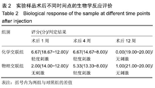
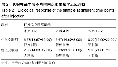
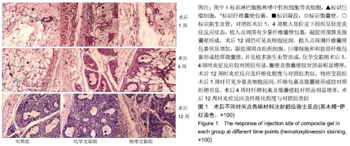
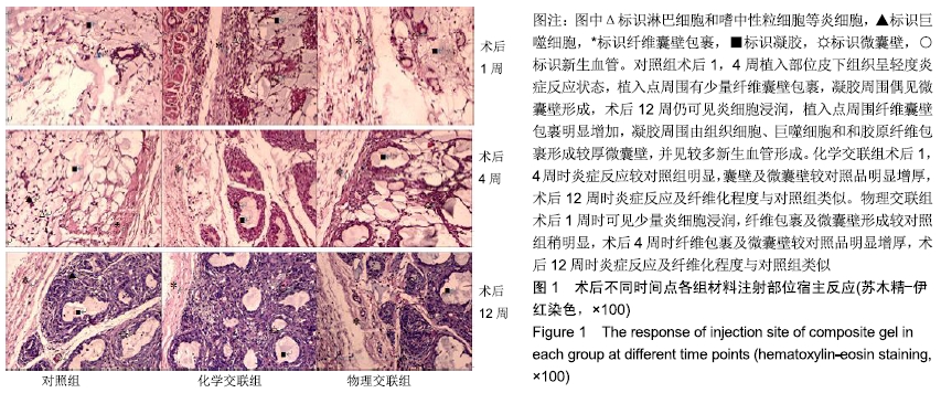
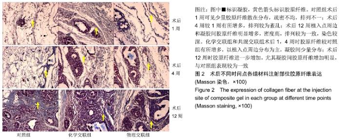
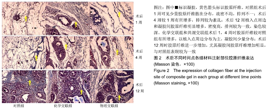
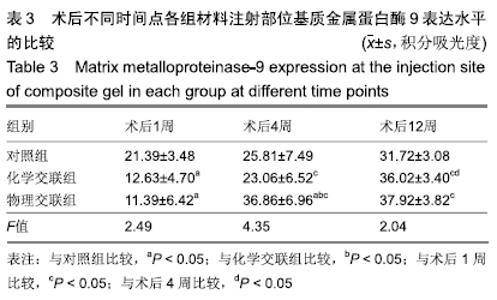
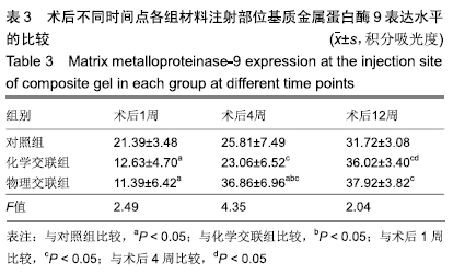
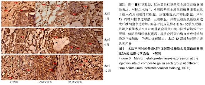
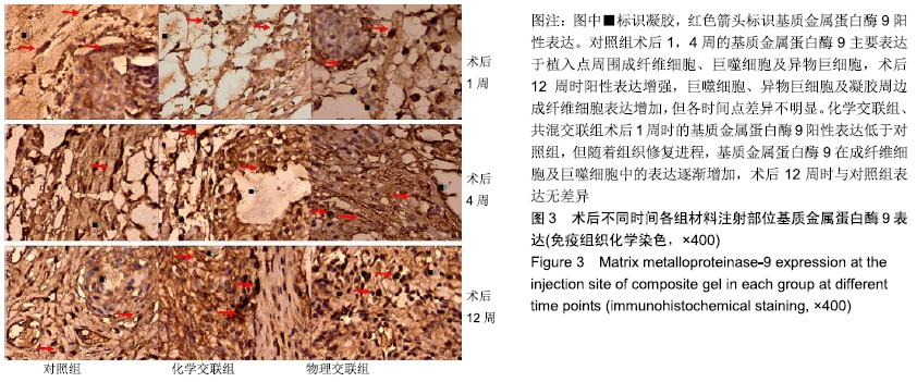
RESULTS AND CONCLUSION: Hematoxylin-eosin staining: In the chemical cross-linking group, at 1 and 4 weeks after injection, inflammatory reaction was more obvious, and the capsule wall and the microcapsule wall were thicker compared with the control group. At 12 weeks after injection, the degree of inflammation and fibrosis were similar between chemical cross-linking and control groups. At 1 week after injection, in the physical cross-linking group, a small amount of inflammatory cells were observed, and the fiber wrap and microcapsule wall formation were slightly more obvious than those in the control group. At 4 weeks after injection, the fiber wrap and microcapsule wall were thicker in the physical cross-linking group compared with the control group. At 12 weeks after injection, the degrees of inflammatory reaction and fibrosis in the physical cross-linking group were similar to those of the control group. Masson staining: In the chemical cross-linking group and physical cross-linking group, at 1 and 4 weeks after injection, the collagen fibers were increased compared with the control group and they further increased at 12 weeks, and at this time, there was no significant difference between chemical cross-linking and physical cross-linking groups. Immunohistochemical staining: in the chemical cross-linking group, matrix metalloproteinase-9 expression in the chemical cross-linking group was lower than that in the control group at 1 week after injection, and there was no significant difference at other time points. At 1 week after injection, matrix metalloproteinase-9 expression in the physical cross-linking group was lower than that in the control group. At 4 weeks after injection, matrix metalloproteinase-9 expression in the physical cross-linking group was higher than that in the control group. At 12 weeks after injection, there was no significant difference in matrix metalloproteinase-9 expression between physical cross-linking and control groups. These results showed that the biocompatibility of the two cross-linked sodium hyaluronate composite gels was good in 12 weeks after subcutaneous implantation. The expression of collagen fiber and matrix metalloproteinase-9 was gradually increased. All these are conducive to tissue remodeling. In the physical cross-linking gel, tissue reaction is smaller at the early stage, and matrix metalloproteinase-9 expression is significantly increased at the middle stage, which are more conducive to tissue repair and remodeling.

Key words: cross-linking, hyaluronic acid, gel, hydroxypropyl methyl cellulose, implantation, subcutaneous, host response, matrix metalloproteinase-9

CLC Number: