Chinese Journal of Tissue Engineering Research ›› 2019, Vol. 23 ›› Issue (34): 5538-5543.doi: 10.3969/j.issn.2095-4344.1969
Previous Articles Next Articles
Application of 3D printing technology in hip diseases
- 1School of Medicine, Nanchang University, Nanchang 330031, Jiangxi Province, China; 2Department of Orthopedics, the First Affiliated Hospital of China Medical University, Shenyang 110001, Liaoning Province, China
-
Received:2019-06-30Online:2019-12-08Published:2019-12-08 -
About author:Huang Youyi, School of Medicine, Nanchang University, Nanchang 330031, Jiangxi Province, China -
Supported by:the Liaoning Provincial Research Center for Orthopedic Translational Medicine and Network Construction-Individualized Design and Development Based on Three-Dimensional Printing, No. 2015225014 (to YW)
CLC Number:
Cite this article
Huang Youyi, Yuan Wei. Application of 3D printing technology in hip diseases[J]. Chinese Journal of Tissue Engineering Research, 2019, 23(34): 5538-5543.
share this article
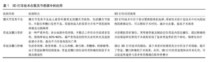
2.1 3D打印技术在复杂成人髋关节发育不良中的应用 髋关节发育不良是新生儿常见的先天性疾病之一[15],随着年龄的增长、髋关节负重的增加,患者股骨头会出现不同程度的移位。髋关节解剖异常导致髋关节应力的改变,进一步导致更严重的解剖结构改变。如果髋关节发育不良程度较轻、位置较好,异常的结构可能需要数年至数十年时间才能产生足够的关节内结构及关节外组织损伤,从而导致疼痛和功能改变[16]。对于髋关节发育不良,一般采取保守治疗、髋臼周围截骨术或人工全髋关节置换[2]。人工全髋关节置换经过几十年的临床应用,已成为最成功且经常进行的选择性手术之一[14],临床上取得了良好的短期和长期效果。严重的成人髋关节发育不良患者尤其是CroweⅢ、Ⅳ型患者,髋臼壁破坏严重,骨缺损、硬化明显,股骨头周围肌肉软组织出现不同程度的损伤,髋臼、股骨头对位不称(股骨头对位不良)[17]。严重的成人髋关节发育不良患者易继发骨性关节炎。目前,全髋关节置换是治疗非手术治疗无效的终末期髋关节关节炎临床最成功、最经济有效的骨科手术之一[18]。以往术者利用术前X射线和CT等影像结果来判断真假髋臼的位置,通过术中所见及手术经验来对比确定确定髋臼与股骨的人工假体型号。髋关节发育不良解剖变异导致的手术操作难度及风险较大,而3D打印技术的应用体现了其独特的优势[17]。 利用3D打印技术设计的个性化骨盆模型,可以在术前确定手术方案并进行模拟手术,更为直观的了解髋臼及股骨近端解剖结构及髋臼损伤程度。术前行三维CT扫描,数据导入计算机建模软件进行建模构建出所需模型后,用3D打印软件打印出髋关节模型。相关报道显示,术前模拟手术能缩短手术时间、减少出血量,患者术后髋关节Harris评分、术后负重时间也相对优于常规手术组[19-20] 。 3D打印技术也可用于指导置换假体的选择。周密的术前计划及术中对风险结构的准确定位,可以提高手术效果。Xu等[21]对10例(14髋)继发于髋关节发育不良的退行性关节疾病患者行计算机设计辅助的全髋关节置换治疗,评估其有效性。因为用计算机辅助来设计全髋关节置换的手术方案不一定都有效,所以作者对有效性进行了评估。所有患者为CroweⅡ型和HartofilakidisⅡ型髋关节脱位,通过计算机辅软件重建出1∶1骨盆模型后,指导后外侧入手术髋臼假体的选择,将术前3D计划选择的类型与实际采用的类型进行比较,发现10髋完全对应,符合率为71.4%。杨晶等[22]运用计算机辅助设计(CAD)3D打印导航模板技术在先天性髋关节发育不良的全髋关节置换术中予以初步尝试,使用CT原始数据(DICOM格式)导入3D分析软件,输出3D模型,计算真臼位置,设计真臼成型导航模板并评估该技术在矫形外科中的应用,结果显示导航组实际术中操作与在计算机图像模拟操作及成型患髋模型操作过程非常相近,导航组和非导航在手术时间、术中出血量、臼杯外展角偏移幅度方面比较差异有统计学意义。与传统治疗方法相比较,数字导航技术具有准确度高、定位时间短的优点。 近年来,国内应用3D打印技术辅助治疗髋关节发育不良已显示较好的临床短期效果[19,22-24]。结合数字骨科技术、设计和制作3D打印的骨盆模型、个性化手术导航模板指导手术能明显提升手术的精准率,患者术后运动功能的恢复相对更好。有关学者认为3D打印的多孔结构金属臼杯假体表层拥有更优越的生物学特性,更符合人体结构,对于骨细胞生长和成骨分化都有促进作用。3D打印患者专用假体用于严重的髋关节畸形有利于患者的术后恢复[25]。3D打印技术的应用,将髋关节发育不良的治疗提升到了精准治疗的个体化的水平。 2.2 3D打印技术在复杂骨盆及髋臼骨折中的应用 髋臼骨折通常为高能量损伤,多发生于创伤背景下[26-27],伴有多脏器合并伤,需要紧急处置。随着国内交通和经济的迅速发展,髋臼骨折的发病率也呈上升趋势。髋臼解剖结构复杂,术前及术中需要多次依赖X射线及CT来对判断髋臼情况,术者难以对髋臼损伤状况有全面的掌 握[27-28]。同时,复杂髋臼骨折涉及到不同骨折部位的手术入路,学习周期较长、治疗难度较大,且骨盆骨折钛板内固定术中钛板需在术中塑形以更好的匹配骨盆,术中的比对易造成患者出血量增加[29-30]。复杂髋臼骨折的治疗由于术中困难和术后不确定性,对外科医生来说是一个挑战[31]。3D打印技术在术前通过三维CT重建骨盆及髋臼影像,利用3D打印设备制造1∶1髋臼模型,有助于术者更好地了解骨折形态,规划合适的复位方法和手术入路。同时充分的术前模拟手术准备,不仅能够缩短手术时间、降低术中难度,而且通过3D打印骨盆模型将塑性钢板预弯并固定钢板,能更精确的复位骨折、提升术后恢复情况[30,32-35]。 通过3D打印技术进行复杂骨盆及髋臼骨折的术前评估及规划,在多篇报道中显示了良好效果。Hung等[30]对30例骨盆前路钢板内固定治疗骨盆骨折病例进行了回顾性分析,其中14例采用常规锁定钢板内固定,16例采用3D打印技术术前重建模型模拟手术,结果显示3D打印技术辅助治疗患者的手术时间缩短、出血量减少,两者术后影像学检查结果相似,3D打印组(1/16例)并发症发生率低于传统手术组(3/14例)。李永军等[32]对32例采用3D打印技术联合腹直肌外侧切口治疗的髋臼骨折患者进行了回顾性分析,术前应用GE LighSpeed (通用公司,美国)螺旋CT扫描,导出DICOM数据后应用Mimics软件(Materialise公司,比利时)对数据进行处理,重建3D骨盆模型,输出STL文件后用聚乳酸打印出1∶1骨盆模型,术前模拟骨折块复位、预弯钢板,加快了手术进度,减少了手术时间和术中出血量。Maini等[33]对21例髋臼移位性骨折患者进行了前瞻性随机病例对照研究,3D打印组通过快速成型技术术前对固定钢板进行预弯处理,对照组进行常规操作,结果显示3D打印组患者出血量和术后效果明显优于对照组,3D打印组所有预弯钢板与骨盆吻合良好。实时三维骨盆模型是髋臼骨折术前精确规划的一种方法。 Zhang等[34]运用Meta分析方法研究了3D打印技术对骨盆骨折治疗的影响,通过检索PubMed数据库、EMCC数据库、CBM数据库、CNKI数据库、VIP数据库及万方数据库,统计将3D打印技术用于术前规划辅助骨盆骨折手术的临床对照试验。共纳入9例临床对照试验,总共638例手术,其中3D打印手术组279例,常规组359例。结果显示,3D打印组在手术时间、术中失血和并发症率上都优于常规组,同时3D打印组盆腔骨折复位优良率和术后盆腔功能恢复均优于常规组。3D打印技术应用于盆腔骨折手术治疗具有手术时间短、术中出血少、并发症发生率低的优点,可提高盆腔骨折复位质量和术后盆腔功能恢复。 目前在骨盆骨折诊疗中,3D打印技术的主要运用及优势在于术前模拟骨折复位、预弯接骨板及辅助定位骨盆内固定螺钉。使用3D打印骨盆髋臼手术导板及定制骨盆髋臼假体治疗骨盆及髋臼骨折等技术近些年也逐渐兴起[36-37]。3D打印技术提高了手术效率,通过精确的术前规划和完善的手术流程实现了复杂骨盆骨折的个体化、精准化治疗,不仅降低了手术难度,明显减少了患者出血量,还缩短了患者康复周期。骨盆骨折修复前使用3D打印模型进行评估及规划,可作为骨盆骨折修复前准备的常规项目[27,38]。 2.3 3D打印技术在骨盆、髋臼肿瘤中的应用 恶性骨盆、髋臼周围肿瘤常呈弥漫性、浸润性生长,肿瘤组织与正常组织的边界模糊,手术方案的选择对于治疗效果至关重要[9]。若术中切除肿瘤组织过少,术后复发率较高;切除肿瘤组织过多则功能重建十分困难[10]。同时为了重建患者的骨与关节功能,会在肿瘤切除术后进行假体植入,而传统方式制造的假体有时并不能满足术中需要。假体的不匹配会给患者术后带来假体松动、肢体不等长、感染等各种并发症,甚至增加患者再手术的风险。对于复杂的骨盆及髋臼恶性肿瘤患者,进行个体化精准治疗方案十分重要。通过3D打印技术,患者病变部位的解剖结构可以清晰的显现在术者面前,避免了过度依赖临床医生手术经验的不确定性。不仅让医生在术前可直观全面地对病变情况进行充分了解,更可以促进手术团队之间及与患者的沟通交流,让患者对病情有形象的理解。通过3D打印技术的辅助,基于术前对病情的充分分析及手术预演,骨盆、髋臼肿瘤的手术结果相对较好[11]。 Wang等[5]对11例髋臼周围恶性骨肿瘤患者切除后采用个性化3D打印半骨盆假体治疗的情况进行了回顾性分析,术前根据CT和MRI等影像结果3D打印盆腔肿瘤模型和截骨引导板,在引导板的帮助下将软组织和肿瘤组织切除,并使用松质骨螺钉和锁定螺钉将3D打印的半骨盆假体与残余髋臼吻合固定,所有患者未出现严重并发症,术后出血量和手术时间均优于常规手术。通过3D打印技术手术团队可对病变区域影像进行三维重建,进而行切割、移动等手术操作预演,使得传统手术方式无法在术中准确实现的假体重建规划成为可能。 另外,3D打印假体可完美地匹配骨肿瘤切除后形成的骨缺损[8]。Liang等[39]对35例骨盆肿瘤切除和3D打印假体重建手术患者进行了回顾性分析,采用电子束熔融法制备了3种3D打印骨盆假体(钛合金材料),对骨盆肿瘤切除术后进行假体重建,在手术技术和围术期安全性方面均取得了令人满意的结果,具有良好的短期生存和功能预后。3D打印的骨盆假体具有多孔性,可以促进重建骨与患者骨的精确匹配和骨整合。3D打印辅助半骨盆假体重建关节成形术为髋臼周围恶性骨肿瘤患者提供了一种替代方案。3D打印技术应用于骨盆、髋臼周围肿瘤切除及切除后半骨盆重建取得了较好的短期效果。 近年来随着新辅助化疗的发展[40],中期恶性骨肿瘤患者保肢手术治疗后效果显著。但大面积肿瘤组织的切除及切除术后缺损骨组织的修补仍是手术所面临的难点。髋臼肿瘤不准确切除会增加死亡风险,严重影响患者预后,甚至导致高达92%的局部肿瘤复发[10]。3D打印技术的出现,推动了骨盆及髋臼恶性肿瘤治疗的发展。利用3D打印技术辅助治疗骨盆髋臼周围恶性骨肿瘤或许是未来的理想方法。3D打印技术的蓬勃发展,值得人们深入探讨骨盆及髋臼肿瘤切除术前方案的规划、恢复重建的假体设计和置换假体的选择。 综上所述,3D打印技术可显著提升髋关节疾病的治疗效果,见表1。 "

| [1]Jacobsen S,Sonne-Holm S.Hip dysplasia: a significant risk factor for the development of hip osteoarthritis.A cross-sectional survey. Rheumatology (Oxford). 2005;44(2): 211-218.[2]Gray BL,Stambough JB,Baca GR,et al.Comparison of contemporary periacetabular osteotomy for hip dysplasia with total hip arthroplasty for hip osteoarthritis.Bone Joint J.2015; 97-b(10):1322-1327.[3]Caffrey JP,Jeffords ME,Farnsworth CL,et al.Comparison of 3 Pediatric Pelvic Osteotomies for Acetabular Dysplasia Using Patient-specific 3D-printed Models.J Pediatr Orthop. 2019; 39(3):e159-e164.[4]彭昊,陈森,郑慧锋,等.生物型假体全髋关节置换术治疗成人髋关节发育不良继发骨关节炎的疗效评价[J].中国矫形外科杂志, 2013,21(15):1502-1507.[5]Wang B,Hao Y,Pu F,et al.Computer-aided designed, three dimensional-printed hemipelvic prosthesis for peri-acetabular malignant bone tumour.Int Orthop.2018;42(3):687-694.[6]Kavalerskiy GM,Murylev VY,Rukin YA,et al. Three-Dimensional Models in Planning of Revision Hip Arthroplasty with Complex Acetabular Defects. Indian J Orthop.2018;52(6):625-630.[7]Chen J,Liu H,Wang C,et al.Internal fixation of acetabular fractures in an older population using the lateral-rectus approach: short-term outcomes of a retrospective study.J Orthop Surg Res.2019;14(1):4.[8]Fang C,Cai H,Kuong E,et al. Surgical applications of three-dimensional printing in the pelvis and acetabulum: from models and tools to implants. Unfallchirurg. 2019;122(4): 278-285.[9]Punyaratabandhu T, Liacouras PC, Pairojboriboon S. Using 3D models in orthopedic oncology: presenting personalized advantages in surgical planning and intraoperative outcomes. 3D Print Med. 2018;4(1):12.[10]Abe K,Yamamoto N, Hayashi K.The usefulness of wide excision assisted by a computer navigation system and reconstruction using a frozen bone autograft for malignant acetabular bone tumors: a report of two cases.BMC Cancer. 2018;18(1):1036.[11]Zhang Y,Wen L,Zhang J,et al.Three-dimensional printing and computer navigation assisted hemipelvectomy for en bloc resection of osteochondroma: A case report.Medicine (Baltimore).2017; 96(12):e6414.[12]卢秉恒,李涤尘.增材制造(3D打印)技术发展[J].机械制造与自动化, 2013,42(4):1-4.[13]Ricles LM,Coburn JC,Di Prima M,et al.Regulating 3D-printed medical products.Sci Transl Med. 2018;10(461). pii:eaan6521. doi:10.1126/scitranslmed.aan6521.[14]Pivec R1,Johnson AJ,Mears SC,et al.Hip arthroplasty. Lancet. 2012;380(9855):1768-1777.[15]Tomlinson J, O'Dowd D, Fernandes JA. Fernandes. Managing Developmental Dysplasia of the Hip. Indian J Pediatr. 2016; 83(11):1275-1279.[16]Pun, S. Hip dysplasia in the young adult caused by residual childhood and adolescent-onset dysplasia. Curr Rev Musculoskelet Med. 2016;9(4):427-434.[17]Mao Y, Xu C, Xu J,et al.The use of customized cages in revision total hip arthroplasty for Paprosky type III acetabular bone defects.Int Orthop. 2015;39(10):2023-2030.[18]Issa K,Mont MA.Total hip replacement: mortality and risks. Lancet. 2013;382(9898):1074-1076.[19]官建中,刘亚军,吴敏,等.3D打印技术在成人DDH人工全髋关节置换术中的临床应用研究[J].中华全科医学,2016,14(7): 1080-1082.[20]Won SH, Lee YK,Ha YC,et al.Improving pre-operative planning for complex total hip replacement with a Rapid Prototype model enabling surgical simulation.Bone Joint J. 2013;95-b(11): 1458-1463.[21]Xu J,Li D,Ma RF,et al.Application of Rapid Prototyping Pelvic Model for Patients with DDH to Facilitate Arthroplasty Planning: A Pilot Study.J Arthroplasty.2015;30(11): 1963-1970.[22]杨晶,张丽娜,李飞,等.数字导航技术在成人Crowe Ⅳ型髋关节发育不良手术治疗中的应用研究[J].中国骨与关节损伤杂志,2015, 30(12):1233-1235.[23]杨清,杨毅,杨柳,等.3D打印技术用于人工全髋关节置换术治疗成人DDH的临床应用[J].实用骨科杂志, 2017,23(8):693-697.[24]涂强,曹露,丁焕文,等. 3D打印个性化手术导航模板在成人发育性髋脱位髋臼重建中的应用[J].临床外科杂志,2017,25(4):295-299.[25]Wang S,Wang L,Liu Y,et al.3D printing technology used in severe hip deformity.Exp Ther Med. 2017;14(3):2595-2599.[26]Li YT,Hung CC,Chou YC,et al.Surgical Treatment for Posterior Dislocation of Hip Combined with Acetabular Fractures Using Preoperative Virtual Simulation and Three-Dimensional Printing Model-Assisted Precontoured Plate Fixation Techniques.Biomed Res Int. 2019;2019: 3971571.[27]Chana Rodríguez F,Pérez Mañanes R,Narbona Cárceles FJ,et al.3D printing utility for surgical treatment of acetabular fractures. Rev Esp Cir Ortop Traumatol.2018. doi: 10.1016/j.recot.2018.02.007.[Epub ahead of print][28]Lin X,Xiao X,Wang Y,et al. Biocompatibility of Bespoke 3D-Printed Titanium Alloy Plates for Treating Acetabular Fractures.2018;2018:2053486.[29]Pereira GJC,Damasceno ER,Dinhane DI,et al.Epidemiology of pelvic ring fractures and injuries. Rev Bras Ortop. 2017;52(3): 260-269.[30]Hung CC,Li YT,Chou YC,et al.Conventional plate fixation method versus pre-operative virtual simulation and three-dimensional printing-assisted contoured plate fixation method in the treatment of anterior pelvic ring fracture.Int Orthop.2019;43(2):425-431.[31]Liu ZJ,Jia J,Zhang YG,et al.Internal Fixation of Complicated Acetabular Fractures Directed by Preoperative Surgery with 3D Printing Models. Orthop Surg.2017;9(2):257-260.[32]李永军,陈棉智,张志辉,等.3D打印技术联合腹直肌外侧入路手术治疗髋臼骨折[J].创伤外科杂志,2019,21(2):99-102.[33]Maini L, Sharma A,Jha S,et al.Three-dimensional printing and patient-specific pre-contoured plate: future of acetabulum fracture fixation? Eur J Trauma Emerg Surg. 2018;44(2): 215-224.[34]Zhang YD,Wu RY,Xie DD et al. Effect of 3D printing technology on pelvic fractures:a Meta-analysis. Zhongguo Gu Shang. 2018; 31(5):465-471.[35]Liu X,Zeng CJ,Lu JS,et al.Application of 3D printing and computer-assisted surgical simulation in preoperative planning for acetabular fracture. Nan Fang Yi Ke Da Xue Xue Bao.2017; 37(3):378-382.[36]Maini L,Verma T,Sharma A,et al.Evaluation of accuracy of virtual surgical planning for patient-specific pre-contoured plate in acetabular fracture fixation.Arch Orthop Trauma Surg. 2018; 138(4):495-504.[37]Liu Y,Zhou W,Xia T,et al.Application of the Guiding Template Designed by Three-dimensional Printing Data for the Insertion of Sacroiliac Screws: a New Clinical Technique.J Orthop Surg Res.2018;38(6):1090-1095.[38]Wang YC,Ma Y,Yu WZ,et al.Application of the computer-assisted virtual reduction combined with 3D printing technique in acetabular fractures. Zhongguo Gu Shang. 2017; 30(7):627-632.[39]Liang H,Ji T, Zhang Y, et al.Reconstruction with 3D-printed pelvic endoprostheses after resection of a pelvic tumour.Bone Joint J. 2017;99-b(2):267-275.[40]Chen X,Xu L,Wang Y,et al.Image-guided installation of 3D-printed patient-specific implant and its application in pelvic tumor resection and reconstruction surgery.Comput Methods Programs Biomed. 2016;125:66-78.[41]Manganaro MS,Morag Y,Weadock WJ,et al.Creating Three-dimensional Printed Models of Acetabular Fractures for Use as Educational Tools. Radiographics.2017;37(3):871-880. |
| [1] | Lu Dezhi, Mei Zhao, Li Xianglei, Wang Caiping, Sun Xin, Wang Xiaowen, Wang Jinwu. Digital design and effect evaluation of three-dimensional printing scoliosis orthosis [J]. Chinese Journal of Tissue Engineering Research, 2021, 25(9): 1329-1334. |
| [2] | Zhang Tongtong, Wang Zhonghua, Wen Jie, Song Yuxin, Liu Lin. Application of three-dimensional printing model in surgical resection and reconstruction of cervical tumor [J]. Chinese Journal of Tissue Engineering Research, 2021, 25(9): 1335-1339. |
| [3] | Xu Yulin, Shen Shi, Zhuo Naiqiang, Yang Huilin, Yang Chao, Li Yang, Zhao Heng, Zhao Lu. Biomechanical comparison of three different plate fixation methods for acetabular posterior column fractures in standing and sitting positions [J]. Chinese Journal of Tissue Engineering Research, 2021, 25(6): 826-830. |
| [4] | Liu Zhengpeng, Wang Yahui, Zhang Yilong, Ming Ying, Sun Zhijie, Sun He. Application of 3D printed interbody fusion cage for cervical spondylosis of spinal cord type: half-year follow-up of recovery of cervical curvature and intervertebral height [J]. Chinese Journal of Tissue Engineering Research, 2021, 25(6): 849-853. |
| [5] | Xu Junma, Yu Yuechao, Liu Zhi, Liu Yu, Wang Feitong. Application of 3D-printed coplanar template combined with fixed needle technique in percutaneous accurate biopsy of small pulmonary nodules [J]. Chinese Journal of Tissue Engineering Research, 2021, 25(5): 761-764. |
| [6] | Wu Zijian, Hu Zhaoduan, Xie Youqiong, Wang Feng, Li Jia, Li Bocun, Cai Guowei, Peng Rui. Three-dimensional printing technology and bone tissue engineering research: literature metrology and visual analysis of research hotspots [J]. Chinese Journal of Tissue Engineering Research, 2021, 25(4): 564-569. |
| [7] | Huang Youyi, Yuan Wei. Application of 3D printing technology in fracture and deformity of foot and ankle [J]. Chinese Journal of Tissue Engineering Research, 2021, 25(3): 438-442. |
| [8] | Lu Yiqing, He Bin, Wang Boyao, Qin Hu, Fan Lei, Wang Yunhua. Application of SLR-PLUS biological lengthened revision stem in the revision of aseptic loosening of femoral prosthesis [J]. Chinese Journal of Tissue Engineering Research, 2021, 25(27): 4318-4321. |
| [9] | Zhu Yin, Sheng Xiaolei, Sha Weiping, Zhang Xingxiang, Wang Jin, Zhu Xianwei, Wang Liming, Yan Fei. Comparison of the efficacy and biocompatibility of two anterior acetabular approaches with low-profile reconstruction plate fixation in the treatment of acetabular fractures involving quadrilateral area [J]. Chinese Journal of Tissue Engineering Research, 2021, 25(27): 4348-4353. |
| [10] | Meng Lingjie, Qian Hui, Sheng Xiaolei, Lu Jianfeng, Huang Jianping, Qi Liangang, Liu Zongbao. Application of three-dimensional printing technology combined with bone cement in minimally invasive treatment of the collapsed Sanders III type of calcaneal fractures [J]. Chinese Journal of Tissue Engineering Research, 2021, 25(24): 3784-3789. |
| [11] | Hu Jing, Xiang Yang, Ye Chuan, Han Ziji. Three-dimensional printing assisted screw placement and freehand pedicle screw fixation in the treatment of thoracolumbar fractures: 1-year follow-up [J]. Chinese Journal of Tissue Engineering Research, 2021, 25(24): 3804-3809. |
| [12] | Shu Qihang, Liao Yijia, Xue Jingbo, Yan Yiguo, Wang Cheng. Three-dimensional finite element analysis of a new three-dimensional printed porous fusion cage for cervical vertebra [J]. Chinese Journal of Tissue Engineering Research, 2021, 25(24): 3810-3815. |
| [13] | Zhang Zhenhua, Liu Zichen, Yu Baoqing. Status and problems of polycaprolactone and its composite materials in bone tissue engineering [J]. Chinese Journal of Tissue Engineering Research, 2021, 25(22): 3571-3577. |
| [14] | Chen Jie, Liao Chengcheng, Zhao Hongbo, Zhao Wei, Chen Zhiwei, Wang Yan. Application of tissue engineering urethral stent and its preparation technology in urethral reconstruction [J]. Chinese Journal of Tissue Engineering Research, 2021, 25(22): 3591-3596. |
| [15] | Lin Tianye, Yang Peng, Xiong Binglang, He Xiaoming, Yan Xinhao, Zhang Jin, He Wei, Wei Qiushi . Comparison of preoperative three-dimensional reconstruction simulation and intraoperative drawing of femoral osteotomy to measure rotation angle in vitro [J]. Chinese Journal of Tissue Engineering Research, 2021, 25(21): 3349-3353. |
| Viewed | ||||||
|
Full text |
|
|||||
|
Abstract |
|
|||||