Chinese Journal of Tissue Engineering Research ›› 2019, Vol. 23 ›› Issue (5): 749-755.doi: 10.3969/j.issn.2095-4344.1550
Previous Articles Next Articles
Underlying mechanism by which adipose-derived mesenchymal stem cell transplantation alleviates renal ischemia-reperfusion injury in rats
Lei Yu1, 2, Liu Rongan2, Zeng Fan2
- 1Southwest Medical University, Luzhou 646000, Sichuan Province, China; 2Department of ICU, Sichuan Provincial People’s Hospital, Chengdu 610072, Sichuan Province, China
-
Revised:2018-11-05Online:2019-02-18Published:2019-02-18 -
Contact:Zeng Fan, MD, Attending physician, Department of ICU, Sichuan Provincial People’s Hospital, Chengdu 610072, Sichuan Province, China -
About author:Lei Yu, Attending physician, Southwest Medical University, Luzhou 646000, Sichuan Province, China -
Supported by:the Cadre Health Care Project of Sichuan Province, No. 2015-204 (to ZF)
CLC Number:
Cite this article
Lei Yu, Liu Rongan, Zeng Fan . Underlying mechanism by which adipose-derived mesenchymal stem cell transplantation alleviates renal ischemia-reperfusion injury in rats[J]. Chinese Journal of Tissue Engineering Research, 2019, 23(5): 749-755.
share this article
Add to citation manager EndNote|Reference Manager|ProCite|BibTeX|RefWorks

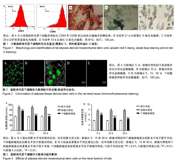
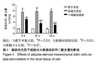
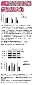
2.1 实验大鼠数量分析 实验的50只SD大鼠中体外实验5只。体内实验45只,其中15只用于假手术组,其中1只术后死亡,30只用于造模,1只移植术后死亡,3只造模不成功;最后,假手术组14只大鼠、移植对照组13只大鼠、干细胞移植组13只大鼠。最终共40只大鼠的实验数据计入结果分析。 2.2 大鼠脂肪间充质干细胞的鉴定 如图1A所示,流式细胞术检测间充质干细胞表面标志物的结果显示,所收集的大鼠脂肪间充质干细胞中黏附分子CD44和CD90的阳性率分别为97.9%和99.5%;成骨诱导21 d后,茜素红S染色后,光学显微镜下可见散在分布的深红色钙结节,见图1B,提示成骨分化阳性;成软骨诱导28 d后,阿利新蓝染色后,光镜下可见致密的块状组织被染成灰蓝色,说明组织内含有酸性黏多糖,见图1C,提示成软骨分化阳性;成脂诱导10 d后,油红O染色后,光学显微镜下可见胞质内有大小不一的黄色脂滴,见图1D,提示成脂分化阳性。 以上结果证实,分离培养的是纯度较好的脂肪间充质干细胞。 2.3 脂肪间充质干细胞在大鼠肾脏中定植的观察 如图2A,B所示,实验将用于标记干细胞的荧光染料注射至对照组大鼠体内,注射5 d后,荧光显微镜下肾脏血管球内可见绿色荧光,注射15 d后血管内绿色荧光消失;如图2C所示,注射5 d后,干细胞移植组大鼠肾组织中有沿肾小管上皮腔面分布的绿色荧光,提示大鼠肾脏内有脂肪间充质干细胞定植在肾小管;移植15和30 d,干细胞移植组大鼠的肾组织中未见绿色荧光,见图2D,E,说明随着移植时间的延长,脂肪间充质干细胞可能发生分化减弱了荧光强度。 2.4 脂肪间充质干细胞对大鼠肾功能的影响 实验取移植后5,15和30 d检测大鼠肌酐和尿素氮水平。如图3所示,与假手术组比较,移植后5,15和30 d,移植对照组和干细胞移植组大鼠肌酐和尿素氮水平升高(P < 0.01);与移植对照组比较,移植后5,15和30 d干细胞移植组大鼠肌酐和尿素氮水平降低(P < 0.01);与移植后5 d比较,干细胞移植组移植15和30 d后大鼠肌酐和尿素氮水平降低(P < 0.01)。说明移植脂肪间充质干细胞能改善肾功能。"

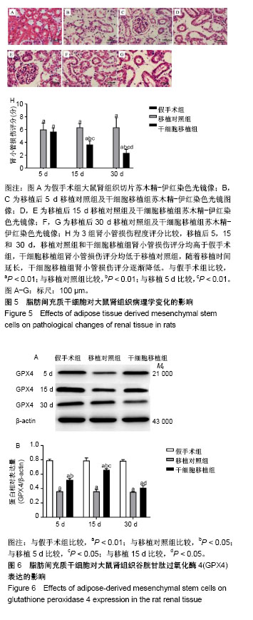
2.5 脂肪间充质干细胞对大鼠肾组织脂质过氧化水平的影响 实验检测了移植后5,15和30 d大鼠肾组织中丙二醛含量,如图4所示,与假手术组比较,移植后5,15和30 d,移植对照组和干细胞移植组大鼠肾组织丙二醛含量升高 (P < 0.01);与移植对照组比较,移植后5,15和30 d干细胞移植组大鼠肾组织丙二醛含量降低(P < 0.01);与移植后5 d比较,干细胞移植组移植15和30 d后大鼠肾组织丙二醛含量降低(P < 0.01)。说明移植脂肪间充质干细胞能降低肾组织细胞的脂质过氧化水平。"
2.6 脂肪间充质干细胞对大鼠肾组织病理学变化的影响 光学显微镜下观察肾组织苏木精-伊红染色切片,如图5A-G所示,与假手术组比较,移植对照组和干细胞移植组大鼠肾组织存在不同程度的病理变化,主要表现为肾皮质内的肾小管上皮细胞水肿、变性及坏死,肾小管管腔扩张,肾间质水肿和炎细胞浸润;与移植对照组比较,干细胞移植组随着移植时间的延长,肾组织损伤程度减轻明显。为了进一步评价肾小管的损伤程度,实验进行了肾小管损伤评分,如图5H所示,与假手术组比较,移植对照组和干细胞移植组在移植后5,15和30 d肾小管损伤评分均升高(P < 0.05);与移植对照组比较,干细胞移植组在移植后5,15和30 d肾小管评分降低(P < 0.05);与移植后5 d比较,干细胞移植组在移植后15和30 d肾小管评分降低(P < 0.05);与移植后15 d比较,干细胞移植组在移植后30 d肾小管评分降低(P < 0.05)提示移植脂肪间充质干细胞能减轻肾小管损伤。 2.7 脂肪间充质干细胞对大鼠肾组织谷胱甘肽过氧化酶4表达的影响 为了进一步探讨脂肪间充质干细胞抗脂质过氧化作用的机制,实验应用Western blot检测3组大鼠肾组织中谷胱甘肽过氧化酶4蛋白的表达情况,如图6所示,与假手术组比较,移植后5和15 d,移植对照组和干细胞移植组大鼠肾组织谷胱甘肽过氧化酶4表达降低(P < 0.05);与移植对照组比较,移植后5和15 d干细胞移植组大鼠肾组织谷胱甘肽过氧化酶4表达升高(P < 0.05);与移植后5 d比较,干细胞移植组在移植后15 d大鼠肾组织谷胱甘肽过氧化酶4表达升高(P < 0.05);与移植后15 d比较,干细胞移植组在移植后30 d大鼠肾组织谷胱甘肽过氧化酶4表达降低(P < 0.05)。提示移植脂肪间充质干细胞能促进谷胱甘肽过氧化酶4的表达,但随着时间延长作用减弱。"
| [1] Yu SQ, Ma S, Wang DH. Activation of TRPV1 prevents salt-induced kidney damage and hypertension after renal ischemia-reperfusion injury in rats. Blood Press Res. 2018;43(4):1285-1296.[2] Barakat M, Gabr MM, Zhran F, et al. Upregulation of heme oxygenase 1 (HO-1) attenuates kidney damage, oxidative stress and inflammatory reaction during renal ischemia/ reperfusion injury. Gen Physiol Biophys. 2018.37(2):193-204.[3] Wang W, Wang A, Luo G, et al. S1P1 receptor inhibits kidney epithelial mesenchymal transition triggered by ischemia/reperfusion injury via the PI3K/Akt pathway. Acta Biochim Biophys Sin (Shanghai). 2018; 50(7): 651-657.[4] Cruces P, Lillo P, Salas C, et al. Renal decapsulation prevents intrinsic renal compartment syndrome in ischemia-reperfusion-induced acute kidney injury: a physiologic approach. Crit Care Med. 2018;46(2): 216-222.[5] He Z, Tang H, You X, et al. BAPTA-AM Nanoparticle for the curing of acute kidney injury induced by ischemia/reperfusion. J Biomed Nanotechnol. 2018;14(5):868-883.[6] Ohtake T, Kobayashi S, Slavin S, et al. Human peripheral blood mononuclear cells incubated in vasculogenic conditioning medium dramatically improve ischemia/reperfusion acute kidney injury in mice. Cell Transplant. 2018;27(3):520-530.[7] Kim JS, Jang HJ, Jeong EK, et al. Crepidiastrum denticulatum extract ameliorates kidney ischemia-reperfusion injury in mice. Transplant Proc. 2018;50(4):1160-1166.[8] Choi YJ, Zhou D, Barbosa ACS, et al. Activation of constitutive androstane receptor ameliorates renal ischemia-reperfusion-induced kidney and liver injury. Mol Pharmacol. 2018;93(3):239-250.[9] Sheashaa H, Lotfy A, Elhusseini F, et al. Protective effect of adipose-derived mesenchymal stem cells against acute kidney injury induced by ischemia-reperfusion in Sprague-Dawley rats. Exp Ther Med. 2016;11(5):1573-1580.[10] Li B, Cohen A, Hudson TE, et al. Mobilized human hematopoietic stem/progenitor cells promote kidney repair after ischemia/reperfusion injury. Circulation. 2010;121(20):2211-2220.[11] Lin KC, Yip HK, Shao PL, et al. Combination of adipose-derived mesenchymal stem cells (ADMSC) and ADMSC-derived exosomes for protecting kidney from acute ischemia-reperfusion injury. Int J Cardiol. 2016;23(216):173-185.[12] Xing L, Cui R, Peng L, et al. Mesenchymal stem cells, not conditioned medium, contribute to kidney repair after ischemia-reperfusion injury. Stem Cell Res Ther. 2014;5(4):1-12.[13] Wise AF, Williams TM, Kiewiet MB, et al. Human mesenchymal stem cells alter macrophage phenotype and promote regeneration via homing to the kidney following ischemia-reperfusion injury. Am J Physiol Renal Physiol. 2014;306(10):F1222-1235.[14] Ayatollahi M, Hesami Z, Jamshidzadeh A, et al. Antioxidant effects of bone marrow mesenchymal stem cell against carbon tetrachloride- induced oxidative damage in rat livers. Int J Organ Transplant Med. 2014;5(4):166-173.[15] Kim Y, Jo SH, Kim WH, et al. Antioxidant and anti-inflammatory effects of intravenously injected adipose derived mesenchymal stem cells in dogs with acute spinal cord injury. Stem Cell Res Ther. 2015;6(1):229.[16] Zhang JB, Wang XQ, Lu GL, et al. Adipose-derived mesenchymal stem cells therapy for acute kidney injury induced by ischemia-reperfusion in a rat model. Clin Exp Pharmacol Physiol. 2017;44(12):1232-1240.[17] 侯晓琳,郁卫东,崔梅花,等.小鼠脂肪间充质干细胞的分离培养及肠道归巢[J].中国组织工程研究,2015,19(6):854-860.[18] 周丽娜,王玉新,方林,等.内皮祖细胞修复缺血再灌注大鼠肾损伤[J].中国组织工程研究,2014,18(32):5146-5151.[19] Sung PH, Chiang HJ, Wallace CG, et al. Exendin-4-assisted adipose derived mesenchymal stem cell therapy protects renal function against co-existing acute kidney ischemia-reperfusion injury and severe sepsis syndrome in rat. Am J Transl Res. 2017;9(7):3167-3183.[20] 胡红林,王共先,邹丛,等.骨髓间充质干细胞治疗肾缺血再灌注损伤的旁分泌机制[J].中国组织工程研究与临床康复,2011,15(32):5995-5998.[21] Zou X, Jiang K, Puranik AS, et al. Targeting murine mesenchymal stem cells to kidney injury molecule-1 improves their therapeutic efficacy in chronic ischemic kidney injury. Stem Cells Transl Med. 2018;7(5): 394-403.[22] Guo Q, Wang J. Effect of combination of vitamin E and umbilical cord-derived mesenchymal stem cells on inflammation in mice with acute kidney injury. Immunopharmacol Immunotoxicol. 2018;40(2):168-172.[23] Lee SJ, Ryu MO, Seo MS, et al. Mesenchymal stem cells contribute to improvement of renal function in a canine kidney injury model. In Vivo. 2017;31(6):1115-1124.[24] Tian SF, Jiang ZZ, Liu YM, et al. Human urine-derived stem cells contribute to the repair of ischemic acute kidney injury in rats. Mol Med Rep. 2017;16(4):5541-5548.[25] Jiang Y, Jahagirdar BN, Reinhardt RL, et al. Pluripotency of mesenchymal stem cells derived from adult marrow. Nature. 2002;418(6893):41-49.[26] Pavyde E, Usas A, Maciulaitis R. Regenerative pharmacology for the treatment of acute kidney injury: skeletal muscle stem/progenitor cells for renal regeneration? Pharmacol Res. 2016;113(Pt B):802-807.[27] Fateme Zhaleh, Fatemeh Amiri, Mohammad Mohammadzadeh-Vardin, et al. Nuclear factor erythroid-2 related factor 2 overexpressed mesenchymal stem cells transplantation, improves renal function, decreases injuries markers and increases repair markers in glycerol-induced Acute kidney injury rats. 2016;19(3):323-329.[28] Kuppe C, Kramann R. Role of mesenchymal stem cells in kidney injury and fibrosis. Curr Opin Nephrol Hypertens. 2016;25(4):372-377.[29] Cóndor JM, Rodrigues CE, Sousa Moreira Rd, et al. Treatment with human wharton's jelly-derived mesenchymal stem cells attenuates sepsis-induced kidney injury, liver injury, and endothelial dysfunction. Stem Cells Transl Med. 2016;5(8):1048-1057.[30] Elhusseini FM, Saad MA, Anber N, et al. Long term study of protective mechanisms of human adipose derived mesenchymal stem cells on cisplatin induced kidney injury in sprague-dawley rats. Regen Med. 2016;12(1):36-48.[31] Murata S, Sugiyama N, Maemura K, et al. Quantified kidney echogenicity in mice with renal ischemia reperfusion injury: evaluation as a noninvasive biomarker of acute kidney injury. Med Mol Morphol. 2017; 50(3):161-169.[32] Si XY, Li JJ, Yao T, et al. Transforming growth factor-β1 in the microenvironment of ischemia reperfusion-injured kidney enhances the chemotaxis of mesenchymal stem cells to stromal cell-derived factor-1 through upregulation of surface chemokine (C-X-C motif) receptor 4. Mol Med Rep. 2014;9(5):1794-1798.[33] Semedo P, Wang PM, Andreucci TH, et al. Mesenchymal stem cells ameliorate tissue damages triggered by renal ischemia and reperfusion injury. Transplant Proc. 2007;39(2):421-423.[34] Monteiro MB, Patente TA, Mohammedi K, et al. Sex-specific associations of variants in regulatory regions of NADPH oxidase-2 (CYBB) and glutathione peroxidase 4 (GPX4) genes with kidney disease in type 1 diabetes. Free Radic Res. 2013;47(10):804-810. |
| [1] | Pu Rui, Chen Ziyang, Yuan Lingyan. Characteristics and effects of exosomes from different cell sources in cardioprotection [J]. Chinese Journal of Tissue Engineering Research, 2021, 25(在线): 1-. |
| [2] | Lin Qingfan, Xie Yixin, Chen Wanqing, Ye Zhenzhong, Chen Youfang. Human placenta-derived mesenchymal stem cell conditioned medium can upregulate BeWo cell viability and zonula occludens expression under hypoxia [J]. Chinese Journal of Tissue Engineering Research, 2021, 25(在线): 4970-4975. |
| [3] | Zhang Tongtong, Wang Zhonghua, Wen Jie, Song Yuxin, Liu Lin. Application of three-dimensional printing model in surgical resection and reconstruction of cervical tumor [J]. Chinese Journal of Tissue Engineering Research, 2021, 25(9): 1335-1339. |
| [4] | Zhang Xiumei, Zhai Yunkai, Zhao Jie, Zhao Meng. Research hotspots of organoid models in recent 10 years: a search in domestic and foreign databases [J]. Chinese Journal of Tissue Engineering Research, 2021, 25(8): 1249-1255. |
| [5] | Hou Jingying, Yu Menglei, Guo Tianzhu, Long Huibao, Wu Hao. Hypoxia preconditioning promotes bone marrow mesenchymal stem cells survival and vascularization through the activation of HIF-1α/MALAT1/VEGFA pathway [J]. Chinese Journal of Tissue Engineering Research, 2021, 25(7): 985-990. |
| [6] | Shi Yangyang, Qin Yingfei, Wu Fuling, He Xiao, Zhang Xuejing. Pretreatment of placental mesenchymal stem cells to prevent bronchiolitis in mice [J]. Chinese Journal of Tissue Engineering Research, 2021, 25(7): 991-995. |
| [7] | Liang Xueqi, Guo Lijiao, Chen Hejie, Wu Jie, Sun Yaqi, Xing Zhikun, Zou Hailiang, Chen Xueling, Wu Xiangwei. Alveolar echinococcosis protoscolices inhibits the differentiation of bone marrow mesenchymal stem cells into fibroblasts [J]. Chinese Journal of Tissue Engineering Research, 2021, 25(7): 996-1001. |
| [8] | Fan Quanbao, Luo Huina, Wang Bingyun, Chen Shengfeng, Cui Lianxu, Jiang Wenkang, Zhao Mingming, Wang Jingjing, Luo Dongzhang, Chen Zhisheng, Bai Yinshan, Liu Canying, Zhang Hui. Biological characteristics of canine adipose-derived mesenchymal stem cells cultured in hypoxia [J]. Chinese Journal of Tissue Engineering Research, 2021, 25(7): 1002-1007. |
| [9] | Geng Yao, Yin Zhiliang, Li Xingping, Xiao Dongqin, Hou Weiguang. Role of hsa-miRNA-223-3p in regulating osteogenic differentiation of human bone marrow mesenchymal stem cells [J]. Chinese Journal of Tissue Engineering Research, 2021, 25(7): 1008-1013. |
| [10] | Lun Zhigang, Jin Jing, Wang Tianyan, Li Aimin. Effect of peroxiredoxin 6 on proliferation and differentiation of bone marrow mesenchymal stem cells into neural lineage in vitro [J]. Chinese Journal of Tissue Engineering Research, 2021, 25(7): 1014-1018. |
| [11] | Zhu Xuefen, Huang Cheng, Ding Jian, Dai Yongping, Liu Yuanbing, Le Lixiang, Wang Liangliang, Yang Jiandong. Mechanism of bone marrow mesenchymal stem cells differentiation into functional neurons induced by glial cell line derived neurotrophic factor [J]. Chinese Journal of Tissue Engineering Research, 2021, 25(7): 1019-1025. |
| [12] | Duan Liyun, Cao Xiaocang. Human placenta mesenchymal stem cells-derived extracellular vesicles regulate collagen deposition in intestinal mucosa of mice with colitis [J]. Chinese Journal of Tissue Engineering Research, 2021, 25(7): 1026-1031. |
| [13] | Pei Lili, Sun Guicai, Wang Di. Salvianolic acid B inhibits oxidative damage of bone marrow mesenchymal stem cells and promotes differentiation into cardiomyocytes [J]. Chinese Journal of Tissue Engineering Research, 2021, 25(7): 1032-1036. |
| [14] | Guan Qian, Luan Zuo, Ye Dou, Yang Yinxiang, Wang Zhaoyan, Wang Qian, Yao Ruiqin. Morphological changes in human oligodendrocyte progenitor cells during passage [J]. Chinese Journal of Tissue Engineering Research, 2021, 25(7): 1045-1049. |
| [15] | Wang Zhengdong, Huang Na, Chen Jingxian, Zheng Zuobing, Hu Xinyu, Li Mei, Su Xiao, Su Xuesen, Yan Nan. Inhibitory effects of sodium butyrate on microglial activation and expression of inflammatory factors induced by fluorosis [J]. Chinese Journal of Tissue Engineering Research, 2021, 25(7): 1075-1080. |
| Viewed | ||||||
|
Full text |
|
|||||
|
Abstract |
|
|||||