Chinese Journal of Tissue Engineering Research ›› 2018, Vol. 22 ›› Issue (12): 1902-1908.doi: 10.3969/j.issn.2095-4344.0810
Previous Articles Next Articles
Hypoxia induces the migration of endothelial progenitor cells
Zhao Hui, Gao Yu-zhong
- the First Affiliated Hospital of Jinzhou Medical University, Jinzhou 121000, Liaoning Province, China
-
Received:2017-11-13Online:2018-04-28Published:2018-04-28 -
Contact:Gao Yu-zhong, Chief physician, Master’s supervisor, the First Affiliated Hospital of Jinzhou Medical University, Jinzhou 121000, Liaoning Province, China -
About author:Zhao Hui, Master, Physician, the First Affiliated Hospital of Jinzhou Medical University, Jinzhou 121000, Liaoning Province, China -
Supported by:the Natural Science Foundation of Liaoning Province, No. 2014022010
CLC Number:
Cite this article
Zhao Hui, Gao Yu-zhong. Hypoxia induces the migration of endothelial progenitor cells[J]. Chinese Journal of Tissue Engineering Research, 2018, 22(12): 1902-1908.
share this article
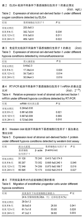
2.1 光镜下观察内皮祖细胞的形态 内皮祖细胞通常于接种后两三天开始贴壁。接种后第4天,可见细胞团聚呈“集落”样;接种后2周,可见细胞成“鹅卵石”样改变,见图1。 2.2 流式细胞检测结果与分析 结果显示,CD34阳性率为(53.21±5.14)%,CD133阳性率为(50.33±6.08)%,血管内皮生长因子受体2阳性率为(55.07±4.16)%,CD34(+)、CD133(+)、血管内皮生长因子受体2(+)的阳性率较高,可以说明实验分离的细胞符合内皮祖细胞特征,为内皮祖细胞。见图2。"
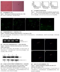
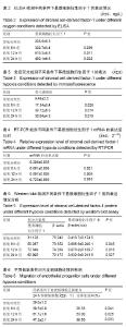
2.3 ELISA检测不同条件下基质细胞衍生因子1的表达 低氧 6,12,24 h诱导基质细胞衍生因子1的表达,6 h表达开始增强,12 h表达最强,24 h又有所减弱。与常氧对照组比较,低氧组基质细胞衍生因子1的表达显著增高(P < 0.05);低氧12 h显著高于低氧6 h和24 h(P < 0.05)。见表2。 2.4 免疫荧光检测不同条件下基质细胞衍生因子1的表达 低氧6,12,24 h诱导基质细胞衍生因子1的表达,与常氧对照组比较,低氧6 h表达开始增强,12 h表达最强,24 h又有所减弱。因基质细胞衍生因子1为胞质和胞膜表达,FITC为绿色荧光。见表3,图3。 2.5 RT-PCR检测不同条件下基质细胞衍生因子1 mRNA的表达 分析RT-PCR条带图可知,低氧6,12,24 h诱导基质细胞衍生因子1的表达,与常氧对照组比较,低氧6 h表达开始增强,12 h表达最强,24 h又有所减弱,见图4。分析表4可知,与常氧对照组相比,低氧6,12,24 h诱导基质细胞衍生因子1 mRNA的表达显著上调(P < 0.01)。 2.6 Western blot检测不同条件下基质细胞衍生因子1蛋白的表达 分析图5 Western blot条带和可知,与常氧对照组相比,低氧6,12,24 h诱导条件下基质细胞衍生因子1蛋白的表达增加,其中,低氧12 h条件下基质细胞衍生因子1蛋白的表达最强,依次为低氧24 h和低氧6 h,对基质细胞衍生因子1和β-actin蛋白积分光密度进行计算,将两者比值作为基质细胞衍生因子1的相对表达量,结果与条带的表达情况相符合,说明Western blot检测结果与ELISA检测和免疫荧光检测结果相吻合,见表5。 2.7 Transwell检测低氧对内皮祖细胞迁移的影响 分析表6可知,与常氧对照组比较,低氧组24 h穿膜细胞数显著增加(P < 0.05);低氧12 h组显著多于低氧6 h组和低氧24 h组,差异有显著性意义(P < 0.05)。见图6。"
| [1] Kütscher C,Lampert FM, Kunze M,et al.Overexpression of hypoxia-inducible factor-1 alpha improves vasculogenesis- related functions of endothelial progenitor cells. Microvascular Res.2016;105:85-92. [2] Hookham MB, Ali I H, O'Neill CL, et al. Hypoxia-induced responses by endothelial colony-forming cells are modulated by placental growth factor. Stem Cell Res Ther. 2016;7(1): 173.[3] Tsai HH,Lin CP,Lin YH,et al.High-intensity Interval training enhances mobilization/functionality of endothelial progenitor cells and depressed shedding of vascular endothelial cells undergoing hypoxia. Eur J Appl Physiol. 2016 ;116(11-12): 2375-2388. [4] Wu JR, Hsu JH, Dai ZK,et al.Activation of endothelial NO synthase by a xanthine derivative ameliorates hypoxia-induced apoptosis in endothelial progenitor cells. J Pharm Pharmacol. 2016;68(6):810-818. [5] Pham PV, Vu NB, Nguyen HT, et al. Significant improvement of direct reprogramming efficacy of fibroblasts into progenitor endothelial cells by ETV2 and hypoxia. Stem Cell Res Ther. 2016; 7(1):104. [6] Prisco AR, Hoffmann BR, Kaczorowski CC, et al. Tumor Necrosis Factor α Regulates Endothelial Progenitor Cell Migration via CADM1 and NF-kB. Stem Cells.2016;34(7): 1922. [7] 石雪峰,王立生,格日力.低氧对骨髓间充质干细胞生物学特性的影响[J]. 中国医药生物技术,2016,11(3):259-262.[8] Li L, Li C,Wang S, et al. Exosomes Derived from Hypoxic Oral Squamous Cell Carcinoma Cells Deliver miR-21 to Normoxic Cells to Elicit a Prometastatic Phenotype. Cancer Res.2016; 76(7):1770.[9] 刘婷,吕红霞,汤旭明,等.低氧通过上调Wnt5a促进人胚胎干细胞向生血内皮细胞分化[J].中国科学:生命科学,2013,43(10): 877-885. [10] Heirani-Tabasi A,Toosi SH,Mirahmadi M, et al. Hypoxia-based Stem/progenitor Cell Therapy: Focus on CXCR4/SDF-1 Axis. 2016; 1(1):32.[11] Larmore J, Black C, Seedorf G, et al. The Actin Cytoskeleton Mediates Hyperoxia Response of Patient-Derived Primary Endothelial Progenitor Cells through Thymosin BETA-4 and Hypoxia-Inducible Factor Signaling.Biophysical J.2016; 110(3):144a-144a. [12] Aday S, Besnier M, Zoldan J, et al. 198 microRNA-17 As The Target of Immobilized Vascular Endothelial Growth Factor in Endothelial Cell Survival Under Ischaemic Conditions. Heart. 2016;102(Suppl 6):A133-A134. [13] 王彦,曹洁,杨庆婵,等.间歇低氧合并肺气肿大鼠系统与内皮炎症状态及外周血内皮祖细胞水平研究[J].天津医药,2014,42(5): 427-431.[14] Yang Y, Hu S, Xu X, et al. The Vascular Endothelial Growth Factors-Expressing Character of Mesenchymal Stem Cells Plays a Positive Role in Treatment of Acute Lung Injury In Vivo. Mediators Inflamm. 2016; 2016(12):1-12.[15] 白春雨,侯玲玲,庞全海等. 内皮祖细胞的分离培养与鉴定[J]. 生物技术通报, 2010, (2):24-27.[16] Dixit P, Donnelly H, Edamatsu M, et al. Progenitor cells from atria, ventricle and peripheral blood of the same patients exhibit functional differences associated with cardiac repair. Int J Cardiol. 2017;228:412-421.[17] 张姝婷,韩潇,赵强,等.低氧对再生医学中间充质干细胞培养的研究进展[J].现代肿瘤医学, 2017,25(4):661-665.[18] Muinos-López E,López-Martínez T,González-Gil AB,et al.Hypoxia and Reactive Oxygen Species Homeostasis in Mesenchymal Progenitor Cells Define a Molecular Mechanism for Fracture Nonunion. Stem Cells.2016; 34(9):2342. [19] 谢哲凡,董航明,魏依蓝,等.低氧微环境对人肺癌细胞上皮间充质转化及迁移侵袭能力的影响[J].中国呼吸与危重监护杂志, 2016, 15(5):465-469.[20] Xu R, Sun Y, Chen Z, et al. Hypoxic Preconditioning Inhibits Hypoxia-induced Apoptosis of Cardiac Progenitor Cells via the PI3K/Akt-DNMT1-p53 Pathway. Scientific Reports.2016; 6:30922.[21] 赵文娟.氧浓度对人内皮祖细胞VEGFR-1、VEGFR-2和CXCR4受体表达调控的研究[D].山东大学, 2015.[22] Liu B, Ren KD, Peng JJ, et al. Suppression of NADPH oxidase attenuates hypoxia-induced dysfunctions of endothelial progenitor cells. Biochem Biophys Res Commun. 2017;482(4):1080-1087.[23] 龚晓明,郑澜,瞿树林. 模拟高住低练对大鼠骨髓源性内皮祖细胞增殖能力的影响[J].中国运动医学杂志,2014,33(6):530-534.[24] 买超平,阎春生,哈小琴.海拔高度对冠心病患者EPCs数量及功能的研究[J].中国循证心血管医学杂志,2015,7(6):754-757.[25] Liu C, Tsai AL, Li PC, et al. Endothelial differentiation of bone marrow mesenchyme stem cells applicable to hypoxia and increased migration through Akt and NFκB signals. Stem Cell Res Ther.2017;8(1):29.[26] 王婷,欧敏,WANGTing,等.内皮祖细胞与低氧性肺动脉高压[J]. 国际呼吸杂志, 2013, 33(11):868-871.[27] 冯勇,杨述华,肖宝钧,等.低氧诱导因子1α激动剂动员内皮祖细胞治疗兔激素性骨坏死中的疗效观察[C]//全国骨关节与风湿病暨武汉国际骨科高峰论坛. 2012.[28] Dixit P, Donnelly H, Edamatsu M, et al. Progenitor cells from atria, ventricle and peripheral blood of the same patients exhibit functional differences associated with cardiac repair. Int J Cardiol. 2017;228:412-421.[29] Isgrò A,Mezzaroma I,Aiuti A,et al. Decreased apoptosis of bone marrow progenitor cells in HIV-1-infected patients during highly active antiretroviral therapy. Aids.2004;18(9):1335-7.[30] 段滨红,向朝峰,侯庆美,等.葡萄糖对不同氧浓度下心肌细胞表达低氧诱导因子-1α相关影响研究[J].中国保健营养月刊, 2012, 22(10):3659-3660.[31] Lotfinia M,Lak S,Mohammadi GN,et al.Hypoxia Pre-Conditioned Embryonic Mesenchymal Stem Cell Secretome Reduces IL-10 Production by Peripheral Blood Mononuclear Cells. Iran Biomed J. 2017;21(1):24-31.[32] 连锋,薛松,顾萍,等.低氧环境和血清饥饿对内皮祖细胞死亡率的影响[J]. 组织工程与重建外科, 2011, 7(5):254-257.[33] 赵湜,王红祥,毛红,等. 葡萄糖对不同氧浓度下内皮祖细胞表达低氧诱导因子-1α的影响[J].中国糖尿病杂志,2011,3(1): 62-66.[34] 李梅,罗容珍,吴秋良.祖细胞归巢与低氧性肺动脉高压[J].广东医学, 2011, 32(9):1209-1211.[35] 李彩丽.肺气肿合并间歇低氧大鼠系统与内皮炎症状态及内皮祖细胞水平研究[D]. 天津医科大学, 2015.[36] Yu Y, Lin L, Zhou Y, et al. Effect of Hypoxia on Self-Renewal Capacity and Differentiation in Human Tendon-Derived Stem Cells. Medical science monitor : international medical journal of experimental and clinical research.2017;23:1334.[37] 夏爽.低氧启动子调控下VEGF_(165)基因修饰的EPCs诱导大鼠急性心肌梗死后血管新生[D].重庆医科大学, 2010.[38] Wang J, Chen Y, Yi Y, et al. Endothelial progenitor cells and neural progenitor cells synergistically protect cerebral endothelial cells from Hypoxia/reoxygenation-induced injury via activating the PI3K/Akt pathway. Molecular Brain.2016;9(1):12.[39] 郑澜,杨小渡,刘铭,等.低氧训练对大鼠心肌组织血管内皮祖细胞标记AC133表达的影响[J].中国运动医学杂志,2009,28(2): 139-141.[40] 姜振宇,宋晓燕,林海,等.人低氧诱导因子1-α转染人骨髓内皮祖细胞的体外实验[J].吉林大学学报医学版,2008, 34(6):944-948. |
| [1] | Pu Rui, Chen Ziyang, Yuan Lingyan. Characteristics and effects of exosomes from different cell sources in cardioprotection [J]. Chinese Journal of Tissue Engineering Research, 2021, 25(在线): 1-. |
| [2] | Zhang Tongtong, Wang Zhonghua, Wen Jie, Song Yuxin, Liu Lin. Application of three-dimensional printing model in surgical resection and reconstruction of cervical tumor [J]. Chinese Journal of Tissue Engineering Research, 2021, 25(9): 1335-1339. |
| [3] | Zeng Yanhua, Hao Yanlei. In vitro culture and purification of Schwann cells: a systematic review [J]. Chinese Journal of Tissue Engineering Research, 2021, 25(7): 1135-1141. |
| [4] | Jiang Xin, Qiao Liangwei, Sun Dong, Li Ming, Fang Jun, Qu Qingshan. Expression of long chain non-coding RNA PGM5-AS1 in serum of renal transplant patients and its regulation of human glomerular endothelial cells [J]. Chinese Journal of Tissue Engineering Research, 2021, 25(5): 741-745. |
| [5] | Xu Dongzi, Zhang Ting, Ouyang Zhaolian. The global competitive situation of cardiac tissue engineering based on patent analysis [J]. Chinese Journal of Tissue Engineering Research, 2021, 25(5): 807-812. |
| [6] | Wu Zijian, Hu Zhaoduan, Xie Youqiong, Wang Feng, Li Jia, Li Bocun, Cai Guowei, Peng Rui. Three-dimensional printing technology and bone tissue engineering research: literature metrology and visual analysis of research hotspots [J]. Chinese Journal of Tissue Engineering Research, 2021, 25(4): 564-569. |
| [7] | Chang Wenliao, Zhao Jie, Sun Xiaoliang, Wang Kun, Wu Guofeng, Zhou Jian, Li Shuxiang, Sun Han. Material selection, theoretical design and biomimetic function of artificial periosteum [J]. Chinese Journal of Tissue Engineering Research, 2021, 25(4): 600-606. |
| [8] | Liu Fei, Cui Yutao, Liu He. Advantages and problems of local antibiotic delivery system in the treatment of osteomyelitis [J]. Chinese Journal of Tissue Engineering Research, 2021, 25(4): 614-620. |
| [9] | Li Xiaozhuang, Duan Hao, Wang Weizhou, Tang Zhihong, Wang Yanghao, He Fei. Application of bone tissue engineering materials in the treatment of bone defect diseases in vivo [J]. Chinese Journal of Tissue Engineering Research, 2021, 25(4): 626-631. |
| [10] | Zhang Zhenkun, Li Zhe, Li Ya, Wang Yingying, Wang Yaping, Zhou Xinkui, Ma Shanshan, Guan Fangxia. Application of alginate based hydrogels/dressings in wound healing: sustained, dynamic and sequential release [J]. Chinese Journal of Tissue Engineering Research, 2021, 25(4): 638-643. |
| [11] | Chen Jiana, Qiu Yanling, Nie Minhai, Liu Xuqian. Tissue engineering scaffolds in repairing oral and maxillofacial soft tissue defects [J]. Chinese Journal of Tissue Engineering Research, 2021, 25(4): 644-650. |
| [12] | Xing Hao, Zhang Yonghong, Wang Dong. Advantages and disadvantages of repairing large-segment bone defect [J]. Chinese Journal of Tissue Engineering Research, 2021, 25(3): 426-430. |
| [13] | Jiang Tao, Ma Lei, Li Zhiqiang, Shou Xi, Duan Mingjun, Wu Shuo, Ma Chuang, Wei Qin. Platelet-derived growth factor BB induces bone marrow mesenchymal stem cells to differentiate into vascular endothelial cells [J]. Chinese Journal of Tissue Engineering Research, 2021, 25(25): 3937-3942. |
| [14] | Liu Chang, Li Datong, Liu Yuan, Kong Lingbo, Guo Rui, Yang Lixue, Hao Dingjun, He Baorong. Poor efficacy after vertebral augmentation surgery of acute symptomatic thoracolumbar osteoporotic compression fracture: relationship with bone cement, bone mineral density, and adjacent fractures [J]. Chinese Journal of Tissue Engineering Research, 2021, 25(22): 3510-3516. |
| [15] | Chen Siqi, Xian Debin, Xu Rongsheng, Qin Zhongjie, Zhang Lei, Xia Delin. Effects of bone marrow mesenchymal stem cells and human umbilical vein endothelial cells combined with hydroxyapatite-tricalcium phosphate scaffolds on early angiogenesis in skull defect repair in rats [J]. Chinese Journal of Tissue Engineering Research, 2021, 25(22): 3458-3465. |
| Viewed | ||||||
|
Full text |
|
|||||
|
Abstract |
|
|||||