Chinese Journal of Tissue Engineering Research ›› 2018, Vol. 22 ›› Issue (5): 680-685.doi: 10.3969/j.issn.2095-4344.0435
Previous Articles Next Articles
Bone marrow mesenchymal stem cell transplantation inhibits apoptosis in the rat spinal cord injured by acrylamide
Sun Jing-song1, Zhou Xue-ying2, Qu Shu-xian3, Bu Tong-jing4, Li Shuang-yue2
- 1First Affiliated Hospital of Dalian Medical University, Dalian 116011, Liaoning Province, China; 2School of Public Health, Dalian Medical University, Dalian 116044, Liaoning Province, China; 3Institute of Cancer Stem Cells, Dalian Medical University, Dalian 116044, Liaoning Province, China; 4Public Security Hospital of Dalian, Dalian 116011, Liaoning Province, China
-
Revised:2017-10-22Online:2018-02-18Published:2018-02-18 -
Contact:Li Shuang-yue, M.D., Associate professor, School of Public Health, Dalian Medical University, Dalian 116044, Liaoning Province, China -
About author:Sun Jing-song, Master, First Affiliated Hospital of Dalian Medical University, Dalian 116011, Liaoning Province, China. Zhou Xue-ying, Master, School of Public Health, Dalian Medical University, Dalian 116044, Liaoning Province, China. Sun Jing-song and Zhou Xue-ying contributed equally to this work.
CLC Number:
Cite this article
Sun Jing-song, Zhou Xue-ying, Qu Shu-xian, Bu Tong-jing, Li Shuang-yue. Bone marrow mesenchymal stem cell transplantation inhibits apoptosis in the rat spinal cord injured by acrylamide[J]. Chinese Journal of Tissue Engineering Research, 2018, 22(5): 680-685.
share this article
Add to citation manager EndNote|Reference Manager|ProCite|BibTeX|RefWorks
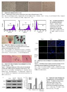
2.1 骨髓间充质干细胞的形态 原代培养细胞呈圆形,大小不一,折光性强,大多数漂浮在培养液中。培养6-8 h后细胞开始逐渐贴壁,换新的培养液除去未贴壁的细胞,在培养24 h时可见细胞形态不一,呈多角形改变,见图1A。3 d后细胞集落呈放射状排列,伸出长短不等、粗细不一的突起。胞浆丰富,核仁清晰可见,见图1B。在10-12 d细胞之间融合成片,细胞相互贴附生长,细胞排列呈螺旋状或漩涡状,见图1C。细胞生长旺盛,四至五天可传代1次,细胞形态和生长速度均未发现有明显异常,大多数呈不规则的三角形或者四边形,见图1D。 2.2 骨髓间充质干细胞表面标志物表达 通过流式细胞仪检测骨髓间充质干细胞表面标记物的表达,结果显示,第3代骨髓间充质干细胞的CD44、CD90阳性率均大于90%,而CD45的阳性率不足5%,见图2。 2.3 骨髓间充质干细胞分化能力 骨髓间充质干细胞经成骨诱导21 d后,茜素红染色显示,实验组细胞呈现广泛的橙红色,有明显的矿化结节形成,见图3A。 骨髓间充质干细胞经成脂诱导18 d后,油红O染色显示,实验组出现较多强阳性成脂细胞,其特征为细胞体积较大、呈圆形、内含鲜红色串珠样或大的融合脂滴,见图3B。 2.4 实验动物数量分析 30只SD健康大鼠,在模型制备过程中无死亡。每组3只大鼠脊髓组织用于苏木精-伊红染色,每组2只大鼠脊髓组织用于Tunel染色;每组5只大鼠脊髓组织用于Western-Blotting检测。 2.5 脊髓组织苏木精-伊红染色结果 正常对照组镜下可见大量的神经元细胞且形态正常,尼氏体清晰,细胞膜完整,细胞质染色均一,细胞核可见,见图4A。丙烯酰胺中毒组镜下可见神经元细胞数量较少,尼氏体消失,细胞膜破坏,细胞核固缩、消失,见图4B。骨髓间充质干细胞移植组镜下可见大量新生的神经元细胞,尼氏体清晰,细胞膜结构完整,细胞核清晰可见,见图4C。 2.6 Tunel法检测细胞凋亡情况 如图5所示,对照组几乎未观察到TUNEL阳性细胞,丙烯酰胺中毒组大鼠脊髓TUNEL阳性细胞显著增多,而骨髓间充质干细胞移植后脊髓TUNEL阳性细胞明显减少。 2.7 Western-Blotting检测脊髓组织Bcl-2和Bax表达水平 如图6所示,与对照组相比,丙烯酰胺中毒组大鼠脊髓组织Bcl-2的表达水平明显下降、Bax的表达水平明显升高,差异有显著性意义(P < 0.05)。骨髓间充质干细胞移植后,脊髓组织Bcl-2的表达水平升高、Bax的表达水平降低,与丙烯酰胺中毒组相比,差异有显著性意义(P < 0.05)。与对照组相比,骨髓间充质干细胞移植组脊髓组织Bcl-2表达几乎恢复至正常水平,而Bax表达略高于正常水平。"

| [1] 于素芳,谢克勤. 丙烯酰胺的神经毒性研究概况[J]. 毒理学杂志, 2005,19(3):242-244. [2] 董红运,于素芳. 丙烯酰胺的致癌性研究进展[J]. 环境与健康杂志,2012,29(9):858-860. [3] 田素民,马宇昕,刘靖,等. 丙烯酰胺亚急性染毒对断乳期雄性大鼠精子形态的影响[J]. 广东药学院学报,2014,30(1):63-66. [4] Narita Y, Inouye K. Decrease in the acrylamide content in canned coffee by heat treatment with the addition of cysteine. J Agric Food Chem. 2014;62(50):12218-12222.[5] Junqua G, Spinelli S, Gonzalez C. Occurrence and fate of acrylamide in water-recycling systems and sludge in aggregate industries. Environ Sci Pollut Res Int. 2015;22(9): 6452-6560.[6] Zuo S, Zhang T, Jiang B, et al. Reduction of acrylamide level through blanching with treatment by an extremely thermostable L-asparaginase during French fries processing.Extremophiles. 2015;19(4):841-851.[7] Mehri S, Karami HV, Hassani FV, et al. Chrysin reduced acrylamide-induced neurotoxicity in both in vitro and in vivo assessments. Iran Biomed J. 2014;18(2):101-106.[8] Song L, Wang J, Zhang W, et al. Effective suppression of acrylamide neurotoxicity by lithium in mouse. Neurochem Res. 2014;39(11):2170-2179.[9] Friedman M. Acrylamide: inhibition of formation in processed food and mitigation of toxicity in cells, animals, and humans. Food Funct. 2015;6(6):1752-1772.[10] Lee JG, Wang YS, Chou CC. Acrylamide-induced apoptosis in rat primary astrocytes and human astrocytoma cell lines. Toxicol In Vitro. 2014;28(4):562-570.[11] Liu Z, Song G, Zou C, et al. Acrylamide induces mitochondrial dysfunction and apoptosis in BV-2 microglial cells. Free Radic Biol Med. 2015;84:42-53.[12] Lai SM,Gu ZT,Zhao MM,et al. Toxic effect of acrylamide on the development of hippo¬campal neurons of weaning rats. Neural Regen Res.2017;12(10):1648-1654.[13] Nijhuis TH, Brzezicki G, Klimczak A, et al. Isogenic venous graft supported with bone marrow stromal cells as a natural conduit for bridging a 20 mm nerve gap. Microsurgery. 2010; 30(8):639-645.[14] Ruiz-López FJ, Blanquer M.Autologous bone marrow mononuclear cells as neuroprotective treatment of amyotrophic lateral sclerosis.Neural Regen Res. 2016;11(4):568-569. [15] Neirinckx V, Coste C, Rogister B, et al. Concise review: adult mesenchymal stem cells, adult neural crest stem cells, and therapy of neurological pathologies: a state of play. Stem Cells Transl Med. 2013;2(4):284-296.[16] Chiba Y, Kuroda S, Osanai T, et al. Impact of ageing on biological features of bone marrow stromal cells (BMSC) in cell transplantation therapy for CNS disorders: functional enhancement by granulocyte-colony stimulating factor (G-CSF). Neuropathology. 2012;32(2):139-148. [17] Hundepool CA, Nijhuis TH, Mohseny B, et al. The effect of stem cells in bridging peripheral nerve defects: a meta-analysis. J Neurosurg. 2014;121(1):195-209.[18] Tan H, Kang X, Lu S, et al. The therapeutic effects of bone marrow mesenchymal stem cells after optic nerve damage in the adult rat. Clin Interv Aging. 2015;10:487-490.[19] Otero L, Zurita M, Bonilla C, et al. Late transplantation of allogeneic bone marrow stromal cells improves neurologic deficits subsequent to intracerebral hemorrhage. Cytotherapy. 2011;13(5):562-571.[20] Deng P, Torrest A, Pollock K, et al. Clinical trial perspective for adult and juvenile Huntington’s disease using genetically-engineered mesenchymal stem cells.Neural Regen Res.2016;11(5): 702-705.[21] Forostyak S, Jendelova P, Kapcalova M, et al. Mesenchymal stromal cells prolong the lifespan in a rat model of amyotrophic lateral sclerosis. Cytotherapy. 2011;13(9): 1036-1046.[22] Chelluboina B, Klopfenstein JD, Pinson DM, et al. Stem cell treatment after cerebral ischemia regulates the gene expression of apoptotic molecules. Neurochem Res. 2014; 39(8):1511-1521.[23] Shi J, Ma Y, Zheng M, et al. Effect of sub-acute exposure to acrylamide on GABAergic neurons and astrocytes in weaning rat cerebellum. Toxicol Ind Health. 2012;28(1):10-20.[24] Tyl RW, Marr MC, Myers CB, et al. Relationship between acrylamide reproductive and neurotoxicity in male rats. Reprod Toxicol. 2000;14(2):147-157. [25] Charbord P. Bone marrow mesenchymal stem cells: historical overview and concepts. Hum Gene Ther. 2010;21(9): 1045-1056. ?[26] Rangappa S, Reddy VG, Bongso A, et al.Transformation of the Adult Human Mesenchymal Stem Cells into Cardiomyocyte-Like Cells in Vivo. Cardiovascular Engineering An International Journal. 2002; 2(1):7-14.[27] Jiang Y, Jahagirdar BN, Reinhardt RL, et al. Pluripotency of mesenchymal stem cells derived from adult marrow. Nature. 2002;418(6893):41-49.[28] Campagnoli C, Roberts IA, Kumar S, et al. Identification of mesenchymal stem/progenitor cells in human first-trimester fetal blood, liver, and bone marrow. Blood. 2001;98(8): 2396-2402. ?[29] Lee OK, Kuo TK, Chen WM, et al. Isolation of multipotent mesenchymal stem cells from umbilical cord blood. Blood. 2004;103(5):1669-1675.[30] Tamir A, Petrocelli T, Stetler K, et al. Stem cell factor inhibits erythroid differentiation by modulating the activity of G1-cyclin-dependent kinase complexes: a role for p27 in erythroid differentiation coupled G1 arrest. Cell Growth Differ. 2000;11(5):269-277.[31] Neuhuber B, Swanger SA, Howard L, et al. Effects of plating density and culture time on bone marrow stromal cell characteristics. Exp Hematol. 2008;36(9):1176-1185.[32] Chen ZZ, Van Bockstaele DR, Buyssens N, et al. Stromal populations and fibrosis in human long-term bone marrow cultures. Leukemia. 1991;5(9):772-781.[33] Friedenstein AJ, Chailakhyan RK, Gerasimov UV. Bone marrow osteogenic stem cells: in vitro cultivation and transplantation in diffusion chambers. Cell Tissue Kinet. 1987;20(3):263-272.[34] Deryugina EI, Müller-Sieburg CE. Stromal cells in long-term cultures: keys to the elucidation of hematopoietic development. Crit Rev Immunol. 1993;13(2):115-150.[35] Shahdadfar A, Frønsdal K, Haug T, et al. In vitro expansion of human mesenchymal stem cells: choice of serum is a determinant of cell proliferation, differentiation, gene expression, and transcriptome stability. Stem Cells. 2005; 23(9):1357-1366.[36] Ouyang L, Shi Z, Zhao S, et al. Programmed cell death pathways in cancer: a review of apoptosis, autophagy and programmed necrosis. Cell Prolif. 2012;45(6):487-498.[37] Liu H, Ding Y, Hou Y, et al. The protective effect of bone marrow mesenchymal stem cells carrying antioxidant gene superoxide dismutase on paraquat lung injury in mice. Zhonghua Lao Dong Wei Sheng Zhi Ye Bing Za Zhi. 2016; 34(1):1-7.[38] Wang Q, Sun G, Gao C, et al. Bone marrow mesenchymal stem cells attenuate 2,5-hexanedione-induced neuronal apoptosis through a NGF/AKT-dependent pathway. Sci Rep. 2016;6:34715.[39] Walensky LD. BCL-2 in the crosshairs: tipping the balance of life and death. Cell Death Differ. 2006;13(8):1339-1350.[40] Danial NN. BCL-2 family proteins: critical checkpoints of apoptotic cell death. Clin Cancer Res. 2007;13(24): 7254-7263. |
| [1] | Min Youjiang, Yao Haihua, Sun Jie, Zhou Xuan, Yu Hang, Sun Qianpu, Hong Ensi. Effect of “three-tong acupuncture” on brain function of patients with spinal cord injury based on magnetic resonance technology [J]. Chinese Journal of Tissue Engineering Research, 2021, 25(在线): 1-8. |
| [2] | Jiang Hongying, Zhu Liang, Yu Xi, Huang Jing, Xiang Xiaona, Lan Zhengyan, He Hongchen. Effect of platelet-rich plasma on pressure ulcers after spinal cord injury [J]. Chinese Journal of Tissue Engineering Research, 2021, 25(8): 1149-1153. |
| [3] | Chai Le, Lü Jianlan, Hu Jintao, Hu Huahui, Xu Qingjun, Yu Jinwei, Quan Renfu. Signal pathway variation after induction of inflammatory response in rats with acute spinal cord injury [J]. Chinese Journal of Tissue Engineering Research, 2021, 25(8): 1218-1223. |
| [4] | Geng Qiudong, Ge Haiya, Wang Heming, Li Nan. Role and mechanism of Guilu Erxianjiao in treatment of osteoarthritis based on network pharmacology [J]. Chinese Journal of Tissue Engineering Research, 2021, 25(8): 1229-1236. |
| [5] | Hou Jingying, Yu Menglei, Guo Tianzhu, Long Huibao, Wu Hao. Hypoxia preconditioning promotes bone marrow mesenchymal stem cells survival and vascularization through the activation of HIF-1α/MALAT1/VEGFA pathway [J]. Chinese Journal of Tissue Engineering Research, 2021, 25(7): 985-990. |
| [6] | Liang Xueqi, Guo Lijiao, Chen Hejie, Wu Jie, Sun Yaqi, Xing Zhikun, Zou Hailiang, Chen Xueling, Wu Xiangwei. Alveolar echinococcosis protoscolices inhibits the differentiation of bone marrow mesenchymal stem cells into fibroblasts [J]. Chinese Journal of Tissue Engineering Research, 2021, 25(7): 996-1001. |
| [7] | Geng Yao, Yin Zhiliang, Li Xingping, Xiao Dongqin, Hou Weiguang. Role of hsa-miRNA-223-3p in regulating osteogenic differentiation of human bone marrow mesenchymal stem cells [J]. Chinese Journal of Tissue Engineering Research, 2021, 25(7): 1008-1013. |
| [8] | Lun Zhigang, Jin Jing, Wang Tianyan, Li Aimin. Effect of peroxiredoxin 6 on proliferation and differentiation of bone marrow mesenchymal stem cells into neural lineage in vitro [J]. Chinese Journal of Tissue Engineering Research, 2021, 25(7): 1014-1018. |
| [9] | Zhu Xuefen, Huang Cheng, Ding Jian, Dai Yongping, Liu Yuanbing, Le Lixiang, Wang Liangliang, Yang Jiandong. Mechanism of bone marrow mesenchymal stem cells differentiation into functional neurons induced by glial cell line derived neurotrophic factor [J]. Chinese Journal of Tissue Engineering Research, 2021, 25(7): 1019-1025. |
| [10] | Pei Lili, Sun Guicai, Wang Di. Salvianolic acid B inhibits oxidative damage of bone marrow mesenchymal stem cells and promotes differentiation into cardiomyocytes [J]. Chinese Journal of Tissue Engineering Research, 2021, 25(7): 1032-1036. |
| [11] | Wan Ran, Shi Xu, Liu Jingsong, Wang Yansong. Research progress in the treatment of spinal cord injury with mesenchymal stem cell secretome [J]. Chinese Journal of Tissue Engineering Research, 2021, 25(7): 1088-1095. |
| [12] | Wang Shiqi, Zhang Jinsheng. Effects of Chinese medicine on proliferation, differentiation and aging of bone marrow mesenchymal stem cells regulating ischemia-hypoxia microenvironment [J]. Chinese Journal of Tissue Engineering Research, 2021, 25(7): 1129-1134. |
| [13] | Kong Desheng, He Jingjing, Feng Baofeng, Guo Ruiyun, Asiamah Ernest Amponsah, Lü Fei, Zhang Shuhan, Zhang Xiaolin, Ma Jun, Cui Huixian. Efficacy of mesenchymal stem cells in the spinal cord injury of large animal models: a meta-analysis [J]. Chinese Journal of Tissue Engineering Research, 2021, 25(7): 1142-1148. |
| [14] | Li Shibin, Lai Yu, Zhou Yi, Liao Jianzhao, Zhang Xiaoyun, Zhang Xuan. Pathogenesis of hormonal osteonecrosis of the femoral head and the target effect of related signaling pathways [J]. Chinese Journal of Tissue Engineering Research, 2021, 25(6): 935-941. |
| [15] | Xie Yang, Zhang Shujiang, Liu Menglan, Luo Ying, Yang Yang, Li Zuoxiao. Mechanism by which rapamycin protects spinal cord neurons in experimental autoimmune encephalomyelitis mice [J]. Chinese Journal of Tissue Engineering Research, 2021, 25(5): 695-700. |
| Viewed | ||||||
|
Full text |
|
|||||
|
Abstract |
|
|||||