Chinese Journal of Tissue Engineering Research ›› 2013, Vol. 17 ›› Issue (24): 4451-4456.doi: 10.3969/j.issn.2095-4344.2013.24.012
Previous Articles Next Articles
Three-dimensional finite element analysis of the maxillary first premolar loaded with different biological forces
Sun Hong-li, Yang Jian-jun, Xu Guo-hao, Gu Fang
- Medical College of Qingdao University, Qingdao 266021, Shandong Province, China
-
Received:2012-09-16Revised:2012-10-13Online:2013-06-11Published:2013-06-11 -
Contact:Gu Fang, Professor, Master’s supervisor, Medical College of Qingdao University, Qingdao 266021, Shandong Province, China gufang61@163.com -
About author:Sun Hong-li★, Studying for master’s degree, Medical College of Qingdao University, Qingdao 266021, Shandong Province, China bugoubb@163.com -
Supported by:General Project of National Natural Science Foundation of China, No. 81171408*; Science and Technology Planning Project of Universities of Shandong Province, No. JI0LF25*
CLC Number:
Cite this article
Sun Hong-li, Yang Jian-jun, Xu Guo-hao, Gu Fang. Three-dimensional finite element analysis of the maxillary first premolar loaded with different biological forces[J]. Chinese Journal of Tissue Engineering Research, 2013, 17(24): 4451-4456.
share this article

上颌第一前磨牙在不同角度力下各轴向最大位移见表1,总位移分布见图4-6。"
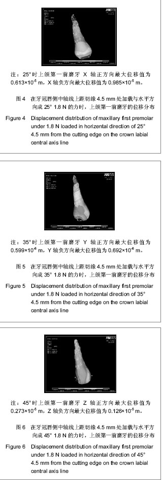

由上述结果可以看出,在力值相同时,不同角度下X轴负方向位移量大于X轴正方向位移量,且随着角度的减小正负方向位移量差值减小。这说明在45°,35°,25°时,牙齿X轴位移为X轴负方向,且随着角度减小位移量减小。即牙齿产生龈向压低移动趋势,且随角度减小压低位移量减小。 在不同角度下Y轴方向上牙尖移动方向(负方向移动)与牙根移动方向(正方向移动)相反,负方向的位移量大于正方向位移量,且随着角度的减小正负方向位移量差值增大。这说明在45°,35°,25°时,牙齿Y轴位移为Y轴负方向,且随着角度减小位移量增大。即牙齿产生远中移动趋势,且随角度减小牙齿倾斜移动的角度增大。 在不同角度下Z轴方向上牙冠近中处移动方向(正方向移动)与牙冠远中处移动方向(负方向移动)相反,正方向的位移量大于负方向的位移量,且随着角度的减小正负方向位移量差值增大。即牙齿产生远中舌向旋转趋势,且随角度减小牙齿舌向旋转角度增大。"

| [1]Hu W,Xun CL,Qiu LX,et al. Kouqiang Zhengjixue. 2008;15(1):42-46.胡炜,寻春雷,邱立新,等. 利用种植体支抗压低修复前过长的后牙[J].口腔正畸学,2008,15(1):42-46.http://www.cnki.net/KCMS/detail/detail.aspx?QueryID=0&CurRec=1&recid=&filename=KQQZ200801015&dbname=CJFD2008&DbCode=CJFQ&urlid=&yx=[2]Ni ZY,Lin XP,Hu RD,et al. Kouqiang Yixue Yanjiu. 2005;21(4):435-437.倪振宇,林新平,胡荣党,等.应用微型种植体支抗压低磨牙[J].口腔医学研究,2005,21(4):435-437.http://www.cnki.net/KCMS/detail/detail.aspx?QueryID=4&CurRec=1&recid=&filename=KQYZ200504031&dbname=CJFD2005&DbCode=CJFQ&urlid=&yx=[3]Chen XM,Zhao YF. Beijing:Science Press,1996.陈新民,赵云凤.口腔生物力学[M].北京:科学出版社,1996.http://www.sciencep.com/[4]Lu XH,Ling JQ,Cai B,et al. Zhonghua Laonian Kouqiang Yixue Zazhi. 2003;1(4):200-201.卢新华, 凌均棨,蔡斌,等.下尖牙三维有限元模型的建立与应力分析[J].中华老年口腔医学杂志,2003,1(4):200-201.http://www.cnki.net/KCMS/detail/detail.aspx?QueryID=8&CurRec=1&recid=&filename=ZHKQ200304002&dbname=CJFD2003&DbCode=CJFQ&urlid=&yx=[5]Zhang T,Liu HC,Wang YR,et al. Zhonghua Kouqiang Yixue Zazhi. 2000;35(5):374-376.张彤,刘洪臣,王延荣,等.上颌骨复合体三维有限元模型的建立[J].中华口腔医学杂志,2000,35(5):374-376.http://www.cnki.net/KCMS/detail/detail.aspx?QueryID=12&CurRec=1&recid=&filename=ZHKY200005023&dbname=cjfd2000&DbCode=CJFQ&urlid=&yx=[6] Motoyoshi M, Hirabayashi M, Shimazaki T,et al. An experimental study on mandibular expansion: increases in arch width and perimeter. Eur J Orthod. 2002;24(2):125-130.http://www.ncbi.nlm.nih.gov/pubmed?term=An%20%20experimenta%20l%20%20study%20%20on%20%20mandibular%20%20%20expansion%EF%BC%9Aincreases%20in%20arch%20width%20and%20perimeter[7]Yan LJ,Liu HC,Bai L,et al. Kouqiang Hemian Xiufuxue Zazhi. 2002;3(3):149-151.阎黎津,刘洪臣,白露,等.下颌固定义齿的三维有限元模型的建立[J].口腔颌面修复学杂志,2002,3(3):149-151.http://www.cnki.net/KCMS/detail/detail.aspx?QueryID=16&CurRec=1&recid=&filename=KHXF200203005&dbname=cjfd2002&DbCode=CJFQ&urlid=&yx=[8]Li J,Yang DR,Dong FS,et al. Xiandai Kouqiang Yixue Zazhi. 2008;22(5):550,523.李健,杨冬茹,董福生,等. CT扫描结合 Mimics三维成像软件对上颌第二磨牙的三维重建[J]. 现代口腔医学杂志,2008,22(5):550,523.http://www.cnki.net/KCMS/detail/detail.aspx?QueryID=20&CurRec=1&recid=&filename=XDKY200805040&dbname=CJFD2008&DbCode=CJFQ&urlid=&yx=[9] McGuinness NJ, Wilson AN, Jones ML,et al. A stress analysis of the periodontal ligament under various orthodontic loadings. Eur J Orthod. 1991;13(3):231-242.http://www.ncbi.nlm.nih.gov/pubmed?term=A%20stress%20analysis%20of%20the%20periodontal%20ligament%20under%20various%20orthodontic%20loadings[10] Farah JW, Craig RG, Meroueh KA. Finite element analysis of three- and four-unit bridges. J Oral Rehabil. 1989;16(6):603-611.http://www.ncbi.nlm.nih.gov/pubmed/2689617[11] Langer B, Langer L, Herrmann I,et al. The wide fixture: a solution for special bone situations and a rescue for the compromised implant. Part 1. Int J Oral Maxillofac Implants. 1993;8(4):400-408.http://www.ncbi.nlm.nih.gov/pubmed?term=The%20wide%20fixture%3AA%20solution%20for%20special%20bone%20situations%20and%20rescue%20for%20the%20compromised%20implant.%20Part%201[12]He X,Tian LG,Mao YL,et al. Shanghai Kouqiang Yixue. 2011;20(3):321-323.何翔,田立国,毛永灵,等. 局部正畸联合微螺钉支抗在对颌牙伸长邻牙倾斜种植修复中的应用[J]. 上海口腔医学, 2011,20(3):321-323.http://www.cnki.net/KCMS/detail/detail.aspx?QueryID=24&CurRec=1&recid=&filename=SHKY201103033&dbname=CJFD2011&DbCode=CJFQ&urlid=&yx=[13] Zhang JW. Zhongguo Shequ Yishi. 2012;5(14):69.张金望.微螺钉种植体支抗在 Angle氏Ⅱ类牙颌畸形治疗中的临床应用[J].中国社区医师:医学专业, 2012,14(5):69.http://www.cnki.net/KCMS/detail/detail.aspx?QueryID=28&CurRec=1&recid=&filename=ZGSQ201205066&dbname=CJFD2012&DbCode=CJFQ&urlid=&yx=[14] Ohnishi H, Yagi T, Yasuda Y,et al. A mini-implant for orthodontic anchorage in a deep overbite case. Angle Orthod. 2005;75(3):444-452.http://www.ncbi.nlm.nih.gov/pubmed?term=.A%20%20mini-implant%20for%20%20orthodontic%20anchorage%20in%20a%20deep%20overbite%20%20case[15] Kim TW, Kim H, Lee SJ. Correction of deep overbite and gummy smile by using a mini-implant with a segmented wire in a growing Class II Division 2 patient. Am J Orthod Dentofacial Orthop. 2006;130(5):676-685.http://www.ncbi.nlm.nih.gov/pubmed?term=Correction%20%20of%20%20deep%20%20overbite%20%20%20and%20gummy%20%20smile%20%20by%20%20using%20%20a%20mini%20-implant%20%20with%20a%20segmented%20wire%20in%20a%20growing%20Class%20II%20Division%202%20%20patient[16] Upadhyay M, Nagaraj K, Yadav S,et al. Mini-implants for en masse intrusion of maxillary anterior teeth in a severe Class II division 2 malocclusion. J Orthod. 2008;35(2):79-89.http://www.ncbi.nlm.nih.gov/pubmed?term=for%20%20enmasse%20intrusion%20of%20maxillary%20%20anterior%20%20teeth%20in%20a%20severe%20%20%20Class%20II%20division%202%20%20malocclusio[17]Wang J,Ni LX,Ai L,et al. Shiyong Fangshexue Zazhi. 2006;22(5):535-536.王疆,倪龙兴,艾林,等.结合Micro-CT技术的上颌第一前磨牙三维模型的建立[J].实用放射学杂志,2006,22(5):535-536.http://www.cnki.net/KCMS/detail/detail.aspx?QueryID=32&CurRec=1&recid=&filename=SYFS200605008&dbname=cjfd2006&DbCode=CJFQ&urlid=&yx=[18] Jang SH, Kim WY. Defining a new annotation object for DICOM image: a practical approach. Comput Med Imaging Graph. 2004;28(7):371-375.http://www.ncbi.nlm.nih.gov/pubmed?term=Defining%20a%20new%20annotation%20object%20for%20DICOM%20image%EF%BC%9Aa%20practical%20approach[DOI]: http://www.medicalimagingandgraphics.com/article/S0895-6111(04)00081-3/abstract[19] Escott EJ, Rubinstein D. Free DICOM image viewing and processing software for your desktop computer: what's available and what it can do for you. Radiographics. 2003;23(5):1341-1357.http://www.ncbi.nlm.nih.gov/pubmed/12975521[20]Tong MJ,Hu DK. Guowai Yixue:Shengwu Yixue Gongcheng Fence. 1999;22(5):303-307.童明杰,胡大可. 认知医学数字图像通讯标准——DICOM [J].国外医学:生物医学工程分册,1999,22(5):303-307.http://www.cnki.net/KCMS/detail/detail.aspx?QueryID=36&CurRec=1&recid=&filename=GWSW199905009&dbname=cjfd1999&DbCode=CJFQ&urlid=&yx=[21]Wei HT,Zhang TF,Zeng CG,et al. Baiqiuen Yike Daxue Xuebao. 2000;26(2):150-151.魏洪涛,张天夫,曾晨光,等.牙颌三维有限元模型生成方法的探讨[J].白求恩医科大学学版,2000,26(2):150-151.http://www.cnki.net/KCMS/detail/detail.aspx?QueryID=40&CurRec=1&recid=&filename=BQEB200002023&dbname=cjfd2000&DbCode=CJFQ&urlid=&yx=[22] Mao XY,Qin YK,Wang ZY,et al. Huaxi Kouqiang Yixue Zazhi. 1996;14(4):299.毛祥彦,芩远坤,王政严,等. 全下颌牙种植义齿及支持组织应力的三维有限元分析──Ⅰ.三维有限元模型的建立[J].华西口腔医学杂志,1996,14(4):299.http://www.cnki.net/KCMS/detail/detail.aspx?QueryID=44&CurRec=1&recid=&filename=HXKQ604.018&dbname=CJFD1996&DbCode=CJFQ&urlid=&yx=[23]Yi L,Zhang N,Zhao SL,et al. Zhongguo Meirong Yixue. 2007;16(4):494-496.怡力,张娜,赵守亮,等. 下颌第一磨牙三维有限元模型的建立[J]. 中国美容医学, 2007,16(4):494-496.http://so.med.wanfangdata.com.cn/ViewHTML/PeriodicalPaper_zgmryxzz200704024.aspx[24]Shi JN. Beijing:Higher Education Press. 2000:166-168.史俊南.现代口腔内科学[M].北京:高等教育出版社,2000:166-168. http://www.hep.com.cn/portal/product/index?bk=15878-00[25]Cheng GD. Beijing:Science Press. 1989:123-129.程耿东.有限元法的概念和应用[M].北京:科学出版社,1989:123-129. http://www.sciencep.com/[26] Li N,Zhang Y. Zhongguo Shequ Yishi, 2010;12(14):121.李娜,张扬.上颌第一磨牙及其支持组织三维有限元模型的建立[J].中国社区医师:医学专业,2010,12(14):121.http://www.cnki.net/KCMS/detail/detail.aspx?QueryID=56&CurRec=2&recid=&filename=ZGSQ201014133&dbname=CJFD2010&DbCode=CJFQ&urlid=&yx=[27] Zhang H,Wang DW,Zhu SL,et al. Zhongshan Daxue Xuebao:Yixue Kexue Ban. 2006;27(z2):11-13.张弘,王大为,朱双林,等. 不同方向加载力移动上颌尖牙的三维有限元分析[J]. 中山大学学报:医学科学版,2006,27(z2):11-13.http://www.cnki.net/KCMS/detail/detail.aspx?QueryID=60&CurRec=1&recid=&filename=ZSYK2006S2006&dbname=CJFD2006&DbCode=CJFQ&urlid=&yx=[28] Fritz U, Ehmer A, Diedrich P.Clinical suitability of titanium microscrews for orthodontic anchorage-preliminary experiences. J Orofac Orthop. 2004;65(5):410-418.http://www.ncbi.nlm.nih.gov/pubmed?term=Clinical%20suitability%20of%20titanium%20microscrews%20for%20orthodontic%20anchorage-preliminary%20experiences[29] Motoyoshi M, Yano S, Tsuruoka T,et al. Biomechanical effect of abutment on stability of orthodontic mini-implant. A finite element analysis. Clin Oral Implants Res. 2005;16(4):480-485. http://www.ncbi.nlm.nih.gov/pubmed?term=Biomechanical%20effect%20of%20abutment%20on%20stability%20of%20orthodontic%20mini-implant%EF%BC%8EA%20finite%20element%20analysis[30]Jin SM,Wang XX,Zhang LN,et al. Shandong Daxue Xuebao:Yixueban. 2010;48(12):67-74.靳淑梅,王旭霞,张丽娜,等.不同工况下埋伏牙的位移趋势及牙周应力的三维有限元分析[J]. 山东大学学报:医学版,2010,48(12):67-74.http://www.cnki.net/KCMS/detail/detail.aspx?QueryID=64&CurRec=1&recid=&filename=SDYB201012016&dbname=CJFD2010&DbCode=CJFQ&urlid=&yx=[31]Zhu SJ,Zhou YH,Fu MK,et al. Kouqiang Zhengjixue. 2007;11(4):169-173.朱胜吉,周彦恒,傅民魁,等. 应用种植体支抗正畸治疗的初步临床研究[J].口腔正畸学,2007,11(4):169-173.http://so.med.wanfangdata.com.cn/ViewHTML/PeriodicalPaper_kqzjx200404007.aspx |
| [1] | Xu Feng, Kang Hui, Wei Tanjun, Xi Jintao. Biomechanical analysis of different fixation methods of pedicle screws for thoracolumbar fracture [J]. Chinese Journal of Tissue Engineering Research, 2021, 25(9): 1313-1317. |
| [2] | Jiang Yong, Luo Yi, Ding Yongli, Zhou Yong, Min Li, Tang Fan, Zhang Wenli, Duan Hong, Tu Chongqi. Von Mises stress on the influence of pelvic stability by precise sacral resection and clinical validation [J]. Chinese Journal of Tissue Engineering Research, 2021, 25(9): 1318-1323. |
| [3] | Lu Dezhi, Mei Zhao, Li Xianglei, Wang Caiping, Sun Xin, Wang Xiaowen, Wang Jinwu. Digital design and effect evaluation of three-dimensional printing scoliosis orthosis [J]. Chinese Journal of Tissue Engineering Research, 2021, 25(9): 1329-1334. |
| [4] | Chen Xinmin, Li Wenbiao, Xiong Kaikai, Xiong Xiaoyan, Zheng Liqin, Li Musheng, Zheng Yongze, Lin Ziling. Type A3.3 femoral intertrochanteric fracture with augmented proximal femoral nail anti-rotation in the elderly: finite element analysis of the optimal amount of bone cement [J]. Chinese Journal of Tissue Engineering Research, 2021, 25(9): 1404-1409. |
| [5] | Zhou Jihui, Li Xinzhi, Zhou You, Huang Wei, Chen Wenyao. Multiple problems in the selection of implants for patellar fracture [J]. Chinese Journal of Tissue Engineering Research, 2021, 25(9): 1440-1445. |
| [6] | Pei Lili, Sun Guicai, Wang Di. Salvianolic acid B inhibits oxidative damage of bone marrow mesenchymal stem cells and promotes differentiation into cardiomyocytes [J]. Chinese Journal of Tissue Engineering Research, 2021, 25(7): 1032-1036. |
| [7] | Yang Weiqiang, Ding Tong, Yang Weike, Jiang Zhengang. Combined variable stress plate internal fixation affects changes of bone histiocyte function and bone mineral density at the fractured end of goat femur [J]. Chinese Journal of Tissue Engineering Research, 2021, 25(6): 890-894. |
| [8] | Song Chengjie, Chang Hengrui, Shi Mingxin, Meng Xianzhong. Research progress in biomechanical stability of lateral lumbar interbody fusion [J]. Chinese Journal of Tissue Engineering Research, 2021, 25(6): 923-928. |
| [9] | Xu Yulin, Shen Shi, Zhuo Naiqiang, Yang Huilin, Yang Chao, Li Yang, Zhao Heng, Zhao Lu. Biomechanical comparison of three different plate fixation methods for acetabular posterior column fractures in standing and sitting positions [J]. Chinese Journal of Tissue Engineering Research, 2021, 25(6): 826-830. |
| [10] | Cai Qunbin, Zou Xia, Hu Jiantao, Chen Xinmin, Zheng Liqin, Huang Peizhen, Lin Ziling, Jiang Ziwei. Relationship between tip-apex distance and stability of intertrochanteric femoral fractures with proximal femoral anti-rotation nail: a finite element analysis [J]. Chinese Journal of Tissue Engineering Research, 2021, 25(6): 831-836. |
| [11] | Xie Chongxin, Zhang Lei. Comparison of knee degeneration after anterior cruciate ligament reconstruction with or without remnant preservation [J]. Chinese Journal of Tissue Engineering Research, 2021, 25(5): 735-740. |
| [12] | Liu Bo, Chen Xianghe, Yang Kang, Yu Huilin, Lu Pengcheng. Mechanism of DNA methylation in exercise intervention for osteoporosis [J]. Chinese Journal of Tissue Engineering Research, 2021, 25(5): 791-797. |
| [13] | Zhang Guomei, Zhu Jun, Hu Yang, Jiao Hongwei. Stress of three-dimensional finite element models of E-MAX porcelain inlay [J]. Chinese Journal of Tissue Engineering Research, 2021, 25(4): 537-541. |
| [14] | Nie Shaobo, Li Jiantao, Sun Jien, Zhao Zhe, Zhao Yanpeng, Zhang Licheng, Tang Peifu. Mechanical stability of medial support nail in treatment of severe osteoporotic intertrochanteric fracture [J]. Chinese Journal of Tissue Engineering Research, 2021, 25(3): 329-333. |
| [15] | Tan Jiachang, Yuan Zhenchao, Wu Zhenjie, Liu Bin, Zhao Jinmin. Biomechanical analysis of elastic nail combined with end caps and wire fixation for long oblique femoral shaft fractures [J]. Chinese Journal of Tissue Engineering Research, 2021, 25(3): 334-338. |
| Viewed | ||||||
|
Full text |
|
|||||
|
Abstract |
|
|||||