Chinese Journal of Tissue Engineering Research ›› 2017, Vol. 21 ›› Issue (20): 3123-3128.doi: 10.3969/j.issn.2095-4344.2017.20.002
Previous Articles Next Articles
Radiofrequency combined with platelet-rich plasma for repair of white meniscal tears
Zhou Wei-feng1, Zhu Lin2, Jiang Xue-feng1, Wang Ya-bin1
- 1Department of Orthopaedics, 2Central Laboratory, Jiangyin People’s Hospital, Jiangyin 214400, Jiangsu Province, China
-
Revised:2017-02-22Online:2017-07-18Published:2017-07-28 -
Contact:Wang Ya-bin, Master, Associate chief physician, Department of Orthopaedics, Jiangyin People’s Hospital, Jiangyin 214400, Jiangsu Province, China -
About author:Zhou Wei-feng, Studying for master’s degree, Department of Orthopaedics, Jiangyin People’s Hospital, Jiangyin 214400, Jiangsu Province, China; Zhu Lin, Master, Master, Research assistant, Central Laboratory, Jiangyin People’s Hospital, Jiangyin 214400, Jiangsu Province, China. Zhou Wei-feng and Zhu Lin contributed equally to this work. -
Supported by:the Project of Health Scientific Research of Jiangsu Province, No. Z201217; the Science and Technology Project of Jiangsu Province, No. (2011)85-22
CLC Number:
Cite this article
Zhou Wei-feng, Zhu Lin, Jiang Xue-feng, Wang Ya-bin. Radiofrequency combined with platelet-rich plasma for repair of white meniscal tears[J]. Chinese Journal of Tissue Engineering Research, 2017, 21(20): 3123-3128.
share this article
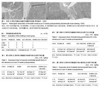
2.1 实验动物数量分析 实验选用新西兰大白兔48只,分为4组,实验过程无脱失,全部进入结果分析。 2.2 标本大体观察 术后观察4组兔膝关节滑膜不同程度增生,均无切口感染;单纯缝合组12周后半月板裂口均未愈合,创口内充满滑液,未见组织填充,半月板表面变粗糙,探针探查无任何连接;射频组12周后半月板伤口部分被组织填充,裂口较术前变窄;富血小板血浆组12周后半月板伤口均已基本被组织填充,探针可挑开创口,愈合组织的颜色比正常半月板组织深,裂口较术前变窄,其表面稍向内凹陷;射频联合富血小板血浆组12周后伤口基本愈合。 2.3 组织学观察 术后12周,如图1所示:单纯缝合组的半月板裂口仍然未见愈合,裂口边缘的软骨细胞处于不活跃、低代谢状态,细胞体积小,细胞死亡,软骨囊消失,胶原纤维进一步减少,半月板变薄,颜色变苍白,韧性和弹性下降;射频组半月板创口被部分填塞,镜下见半月板组织愈合呈瘢痕样愈合;富血小板血浆组半月板创口被填塞,创口粘合,镜下见半月板组织愈合呈瘢痕样愈合;射频联合富血小板血浆组半月板创口闭合,愈合组织内可见软骨细胞,有软骨囊形成,胶原纤维排列较前规则。 2.4 各组愈合效果的评级比较 术后12周将各组半月板愈合情况,按Henning CE的评价标准对愈合效果进行评级,具体结果见表1。对4组愈合效果采用秩和检验进行比较(P=0.002),差异有显著性意义,因此可以认为各组间愈合效果不同。再进行组间两两比较,发现仅在单纯缝合组与射频联合富血小板血浆组之间差异有显著性意义(P=0.001),其余各组之间愈合效果没有差别。因此,可以认为射频联合富血小板血浆治疗半月板白区撕裂与单纯缝合比较,更有利于半月板的愈合。 2.5 单位质量半月板组织中转化生长因子、血管内皮生长因子、血小板衍生生长因子的含量 见表2-4。在术后第3周和第12周,如表2所示:各实验组转化生长因子β1含量较单纯缝合组均增加(P < 0.05)。在相同组别,与3周时相比单位质量半月板组织中转化生长因子β1含量在12周时均明显下降(P < 0.01)。 从表3可以看出在3周时射频组、富血小板血浆组及射频联合富血小板血浆组血管内皮生长因子的表达较单纯缝合组明显增高(P < 0.01);在12周时,仅富血小板血浆组和射频联合富血小板血浆组血管内皮生长因子含量较单纯缝合组明显增加(P < 0.01)。12周时各组血管内皮生长因子含量已明显下降(P < 0.01)。 表4说明在3周时各实验组,尤其是富血小板血浆组和射频联合富血小板血浆组,血小板衍生生长因子的表达较单纯缝合组增高。12周时各组血小板衍生生长因子表达均明显下降(P < 0.01)。"

| [1] Papalia R, Del Buono A, Osti L, et al. Meniscectomy as a risk factor for knee osteoarthritis: a systematic review. Br Med Bull. 2011;99:89-106.[2] Nepple JJ, Dunn WR, Wright RW. Meniscal repair outcomes at greater than five years: a systematic literature review and meta-analysis. J Bone Joint Surg Am.2012;94(24): 2222-2227.[3] Petty CA, Lubowitz JH. Does arthroscopic partial meniscectomy result in knee osteoarthritis? A systematic review with a minimum of 8 years' follow-up. Arthroscopy. 2011;27(3):419-424.[4] Lee CS, Tasto JP, Healey RM, et al. Radiofrequency stimulation for potential healing of meniscal injuries in the avascular zone. Am J Orthop (Belle Mead NJ).2014;43(12): 292-298.[5] Oda S, Otsuki S, Kurokawa Y, et al. A new method for meniscus repair using type I collagen scaffold and infrapatellar fat pad. J Biomater Appl. 2015;29(10): 1439-1448.[6] Scordino LE, Deberardino TM. Biologic enhancement of meniscus repair. Clin Sports Med.2012;31(1):91-100.[7] Jang SH, Ha JK, Lee DW, et al. Fibrin clot delivery system for meniscal repair. Knee Surg Relat Res. 2011;23(3): 180-183.[8] Kamimura T, Kimura M. Meniscal Repair of Degenerative Horizontal Cleavage Tears Using Fibrin Clots: Clinical and Arthroscopic Outcomes in 10 Cases. Orthop J Sports Med 2014;2(11):2325967114555678.[9] McCrum CL, Vangsness CT. Postmeniscectomy Meniscus Growth With Stem Cells: Where Are We Now? Sports Med Arthrosc.2015;23(3):139-142.[10] Yu H, Adesida AB, Jomha NM. Meniscus repair using mesenchymal stem cells - a comprehensive review. Stem Cell Res Ther.2015;6:86.[11] Ding Z, Huang H. Mesenchymal stem cells in rabbit meniscus and bone marrow exhibit a similar feature but a heterogeneous multi-differentiation potential: superiority of meniscus as a cell source for meniscus repair. BMC Musculoskelet Disord.2015;16: 65.[12] Nakagawa Y, Muneta T, Kondo S, et al. Synovial mesenchymal stem cells promote healing after meniscal repair in microminipigs. Osteoarthritis Cartilage.2015;23(6): 1007-1017.[13] Schwartz JA, Wang W, Goldstein T, et al.Tissue Engineered Meniscus Repair: Influence of Cell Passage Number, Tissue Origin, and Biomaterial Carrier. Cartilage.2014;5(3): 165-171.[14] Hutchinson ID, Rodeo SA, Perrone GS, et al. Can platelet-rich plasma enhance anterior cruciate ligament and meniscal repair? J Knee Surg.2015;28(1):19-28.[15] Metcalf KB, Mandelbaum BR, McIlwraith CW. Application of Platelet-Rich Plasma to Disorders of the Knee Joint. Cartilage. 2013;4(4):295-312.[16] Lee CS, Tasto JP, Healey RM, et al. Radiofrequency stimulation for potential healing of meniscal injuries in the avascular zone. Am J Orthop (Belle Mead NJ).2014;43(12): E292-298.[17] Ochi M, Uchio Y, Okuda K, et al. Expression of cytokines after meniscal rasping to promote meniscal healing. Arthroscopy. 2001;17(7):724-731.[18] Pavlovich RI. Meniscal tissue repair using radiofrequency. Orthopedics. 2010;33(6):405-406.[19] Sabarish R, Lavu V, Rao SR. A Comparison of Platelet Count and Enrichment Percentages in the Platelet Rich Plasma (PRP) Obtained Following Preparation by Three Different Methods. J Clin Diagn Res.2015;9(2):ZC10-12.[20] Petersen W, Pufe T, Stärke C, et al. Locally applied angiogenic factors--a new therapeutic tool for meniscal repair. Ann Anat.2005;187(5-6):509-519.[21] Henning CE, Lynch MA, Clark JR. Vascularity for healing of meniscus repairs. Arthroscopy.1987;3(1):13-18.[22] Slavkin HC and Bartold PM. Challenges and potential in tissue engineering. Periodontol. 2000 2006;41:9-15.[23] Anitua E, Sánchez M, Nurden AT, et al. New insights into and novel applications for platelet-rich fibrin therapies. Trends Biotechnol.2006;24(5):227-234.[24] Ishida K, Kuroda R, Miwa M, et al. The regenerative effects of platelet-rich plasma on meniscal cells in vitro and its in vivo application with biodegradable gelatin hydrogel. Tissue Eng. 2007;13(5):1103-1112.[25] Lee HR, Park KM, Joung YK, et al. Platelet-rich plasma loaded hydrogel scaffold enhances chondrogenic differentiation and maturation with up-regulation of CB1 and CB2. J Control Release 2012;159(3):332-337.[26] Chen WH, Lo WC, Hsu WC, et al. Synergistic anabolic actions of hyaluronic acid and platelet-rich plasma on cartilage regeneration in osteoarthritis therapy. Biomaterials. 2014;35(36):9599-9607.[27] Narita A, Takahara M, Sato D, et al. Biodegradable gelatin hydrogels incorporating fibroblast growth factor 2 promote healing of horizontal tears in rabbit meniscus. Arthroscopy. 2012;28(2):255-263.[28] Lee HR, Shon OJ, Park SI, et al. Platelet-Rich Plasma Increases the Levels of Catabolic Molecules and Cellular Dedifferentiation in the Meniscus of a Rabbit Model. Int J Mol Sci.2016;17(1):pii: E120.[29] Zellner J, Mueller M, Berner A, et al. Role of mesenchymal stem cells in tissue engineering of meniscus. J Biomed Mater Res A.2010;94(4):1150-1161.[30] Zellner J, Taeger CD, Schaffer M, et al. Are applied growth factors able to mimic the positive effects of mesenchymal stem cells on the regeneration of meniscus in the avascular zone? Biomed Res Int.2014;2014:537686.[31] Shin KH, Lee H, Kang S, et al. Effect of Leukocyte-Rich and Platelet-Rich Plasma on Healing of a Horizontal Medial Meniscus Tear in a Rabbit Model. Biomed Res Int. 2015;2015: 179756.[32] Ruiz Ibán MÁ, Comellas Melero N, Martinez-Botas J, et al. Growth factor expression after lesion creation in the avascular zone of the meniscus: a quantitative PCR study in rabbits. Arthroscopy.2014;30(9):1131-1138.[33] Becker R, Pufe T, Kulow S, et al. Expression of vascular endothelial growth factor during healing of the meniscus in a rabbit model.J Bone Joint Surg Br. 2004;86(7):1082-1087.[34] Kawamura S, Ying L, Kim HJ, et al. Macrophages accumulate in the early phase of tendon-bone healing. J Orthop Res. 2005;23(6):1425-1432.[35] Swenson TM. The use of exogenous fibrin clot to supplement meniscal surgery techniques. Orthopedics. 2007;30(9): 718-723.[36] Grana WA, Szivek JA, Schnepp AB, et al. A comparison of the effects of radiofrequency treatment and mechanical shaving for meniscectomy. Arthroscopy. 2006;22(8):884-888.[37] Pavlovich RI. Radiofrequency: future applications for current knowledge. Int Orthop. 2005;29(1):65-66.[38] Hatayama K,Higuchi H,Kimura M,et al.Histologic changes after meniscal repair using radiofrequency energy in rabbits. Arthroscopy. 2007;23(3):299-304. |
| [1] | Yao Xiaoling, Peng Jiancheng, Xu Yuerong, Yang Zhidong, Zhang Shuncong. Variable-angle zero-notch anterior interbody fusion system in the treatment of cervical spondylotic myelopathy: 30-month follow-up [J]. Chinese Journal of Tissue Engineering Research, 2022, 26(9): 1377-1382. |
| [2] | Zhang Jinglin, Leng Min, Zhu Boheng, Wang Hong. Mechanism and application of stem cell-derived exosomes in promoting diabetic wound healing [J]. Chinese Journal of Tissue Engineering Research, 2022, 26(7): 1113-1118. |
| [3] | An Weizheng, He Xiao, Ren Shuai, Liu Jianyu. Potential of muscle-derived stem cells in peripheral nerve regeneration [J]. Chinese Journal of Tissue Engineering Research, 2022, 26(7): 1130-1136. |
| [4] | He Yunying, Li Lingjie, Zhang Shuqi, Li Yuzhou, Yang Sheng, Ji Ping. Method of constructing cell spheroids based on agarose and polyacrylic molds [J]. Chinese Journal of Tissue Engineering Research, 2022, 26(4): 553-559. |
| [5] | He Guanyu, Xu Baoshan, Du Lilong, Zhang Tongxing, Huo Zhenxin, Shen Li. Biomimetic orientated microchannel annulus fibrosus scaffold constructed by silk fibroin [J]. Chinese Journal of Tissue Engineering Research, 2022, 26(4): 560-566. |
| [6] | Chen Xiaoxu, Luo Yaxin, Bi Haoran, Yang Kun. Preparation and application of acellular scaffold in tissue engineering and regenerative medicine [J]. Chinese Journal of Tissue Engineering Research, 2022, 26(4): 591-596. |
| [7] | Kang Kunlong, Wang Xintao. Research hotspot of biological scaffold materials promoting osteogenic differentiation of bone marrow mesenchymal stem cells [J]. Chinese Journal of Tissue Engineering Research, 2022, 26(4): 597-603. |
| [8] | Shen Jiahua, Fu Yong. Application of graphene-based nanomaterials in stem cells [J]. Chinese Journal of Tissue Engineering Research, 2022, 26(4): 604-609. |
| [9] | Zhang Tong, Cai Jinchi, Yuan Zhifa, Zhao Haiyan, Han Xingwen, Wang Wenji. Hyaluronic acid-based composite hydrogel in cartilage injury caused by osteoarthritis: application and mechanism [J]. Chinese Journal of Tissue Engineering Research, 2022, 26(4): 617-625. |
| [10] | Li Hui, Chen Lianglong. Application and characteristics of bone graft materials in the treatment of spinal tuberculosis [J]. Chinese Journal of Tissue Engineering Research, 2022, 26(4): 626-630. |
| [11] | Gao Cangjian, Yang Zhen, Liu Shuyun, Li Hao, Fu Liwei, Zhao Tianyuan, Chen Wei, Liao Zhiyao, Li Pinxue, Sui Xiang, Guo Quanyi. Electrospinning for rotator cuff repair [J]. Chinese Journal of Tissue Engineering Research, 2022, 26(4): 637-642. |
| [12] | Guan Jian, Jia Yanfei, Zhang Baoxin , Zhao Guozhong. Application of 4D bioprinting in tissue engineering [J]. Chinese Journal of Tissue Engineering Research, 2022, 26(3): 446-455. |
| [13] | Liu Jiali, Suo Hairui, Yang Han, Wang Ling, Xu Mingen. Influence of lay-down angles on mechanical properties of three-dimensional printed polycaprolactone scaffolds [J]. Chinese Journal of Tissue Engineering Research, 2022, 10(16): 2612-2617. |
| [14] | Huang Bo, Chen Mingxue, Peng Liqing, Luo Xujiang, Li Huo, Wang Hao, Tian Qinyu, Lu Xiaobo, Liu Shuyun, Guo Quanyi . Fabrication and biocompatibility of injectable gelatin-methacryloyl/cartilage-derived matrix particles composite hydrogel scaffold [J]. Chinese Journal of Tissue Engineering Research, 2022, 10(16): 2600-2606. |
| [15] | Li Xuan, Sun Yimin, Li Longbiao, Wang Zhenming, Yang Jing, Wang Chenglin, Ye Ling. Manufacturing of nano-modified polycaprolactone microspheres and its biological effects in dental pulp cells [J]. Chinese Journal of Tissue Engineering Research, 2022, 26(10): 1530-1536. |
| Viewed | ||||||
|
Full text |
|
|||||
|
Abstract |
|
|||||