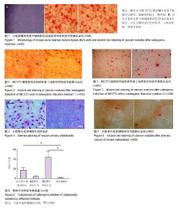
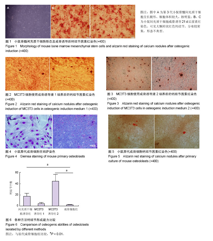
| [1] Corrado A, Neve A, Macchiarola A, et al. RANKL/OPG ratio and DKK-1 expression in primary osteoblastic cultures from osteoarthritic and osteoporotic subjects.J Rheumatol. 2013; 40(5):684-694.[2] Golden D, Saria EA, Hansen MF. Regulation of Osteoblast Migration Involving Receptor Activator of Nuclear Factor-kappa B (RANK) Signaling. J Cell Physiol. 2015; 230(12):2951-2960.[3] Drager J, Ramirez-GarciaLuna JL, Kumar A, et al. Hypoxia Biomimicry to Enhance Monetite Bone Defect Repair. Tissue Eng Part A. 2017 May 31. [Epub ahead of print][4] Sun C, Liu F, Cen S, et al. Tensile strength suppresses the osteogenesis of periodontal ligament cells in inflammatory microenvironments. Mol Med Rep. 2017 May 29. [Epub ahead of print][5] Zhang J, Yu X, Yu Y, et al. MicroRNA expression analysis during FK506-induced osteogenic differentiation in rat bone marrow stromal cells. Mol Med Rep. 2017 May 31. [Epub ahead of print][6] Luo G, Xu B, Huang Y. Icariside II promotes the osteogenic differentiation of canine bone marrow mesenchymal stem cells via the PI3K/AKT/mTOR/S6K1 signaling pathways. Am J Transl Res. 2017;9(5):2077-2087.[7] Morsczeck C, Reichert TE. The dexamethasone induced osteogenic differentiation of dental follicle cells. Histol Histopathol. 2017:11907.[8] Wei K, Xie Y, Chen T, et al. ERK1/2 signaling mediated naringin-induced osteogenic differentiation of immortalized human periodontal ligament stem cells. Biochem Biophys Res Commun. 2017 May 26. [Epub ahead of print][9] Genchi GG, Sinibaldi E, Ceseracciu L, et al. Ultrasound- activated piezoelectric P (VDF-TrFE)/boron nitride nanotube composite films promote differentiation of human SaOS-2 osteoblast-like cells. Nanomedicine. 2017 May 26. [Epub ahead of print][10] Jayakumar P, Di Silvio L. Osteoblasts in bone tissue engineering. Proc Inst Mech Eng H. 2010;224(12):1415-1440.[11] Yin X, Chen Z, Liu Z, et al. Tissue transglutaminase (TG2) activity regulates osteoblast differentiation and mineralization in the SAOS-2 cell line. Braz J Med Biol Res. 2012;45(8): 693-700.[12] Makowski AJ, Uppuganti S, Wadeer SA, et al. The loss of activating transcription factor 4 (ATF4) reduces bone toughness and fracture toughness. Bone. 2014;62:1-9.[13] Guañabens N, Gifre L, Peris P. The role of Wnt signaling and sclerostin in the pathogenesis of glucocorticoid-induced osteoporosis. Curr Osteoporos Rep. 2014;12(1):90-97.[14] Li K, Yan J, Wang C, et al. Graphene modified titanium alloy promote the adhesion, proliferation and osteogenic differentiation of bone marrow stromal cells. Biochem Biophys Res Commun. 2017 May 23. [Epub ahead of print][15] Cai X, Ten Hoopen S, Zhang W, et al. Influence of highly porous electrospun PLGA/PCL/nHA fibrous scaffolds on the differentiation of tooth bud cells in vitro. J Biomed Mater Res A. 2017 May 24. [Epub ahead of print][16] Deng Z, Han H, Yang J, et al. Fabrication and Characterization of Carbon Fiber-Reinforced Nano-Hydroxyapatite/Polyamide46 Biocomposite for Bone Substitute. Med Sci Monit. 2017;23:2479-2487.[17] Wang X, Li G, Liu Y, et al. Biocompatibility of biological material polylactic acid with stem cells from human exfoliated deciduous teeth. Biomed Rep. 2017;6(5):519-524.[18] Zambuzzi WF, Fernandes GV, Iano FG, et al. Exploring anorganic bovine bone granules as osteoblast carriers for bone bioengineering: a study in rat critical-size calvarial defects. Braz Dent J. 2012;23(4):315-321.[19] Premnath P, Tan B, Venkatakrishnan K. Direct patterning of free standing three dimensional silicon nanofibrous network to facilitate multi-dimensional growth of fibroblasts and osteoblasts. J Biomed Nanotechnol. 2013;9(11):1875-1881.[20] Gao A, Hang R, Huang X, et al. The effects of titania nanotubes with embedded silver oxide nanoparticles on bacteria and osteoblasts. Biomaterials. 2014;35(13): 4223-4235.[21] Katsuyama M, Demura M, Katsuyama H, et al. Genistein and menaquinone-4 treatment-induced alterations in the expression of mRNAs and their products are beneficial to osteoblastic MC3T3-E1 cell functions. Mol Med Rep. 2017 May 25. [Epub ahead of print][22] Li JY, Liu SG, Xiao GN, et al. Fibroblast growth factor receptor 1 propagates estrogen and fluid shear stress driven proliferation and differentiation response in MC3T3-E1 cells. Mol Biol (Mosk). 2017;51(2):342-355.[23] Yu C, Zhuang J, Dong L, et al. Effect of hierarchical pore structure on ALP expression of MC3T3-E1 cells on bioglass films. Colloids Surf B Biointerfaces. 2017;156:213-220.[24] Gapski R, Martinez EF. Behavior of MC3T3-E1 Osteoblastic Cells Cultured on Titanium and Zirconia Surfaces: An In Vitro Study. Implant Dent. 2017;26(3):373-377.[25] Lamoureux F, Baud'huin M, Rodriguez Calleja L, et al. Selective inhibition of BET bromodomain epigenetic signalling interferes with the bone-associated tumour vicious cycle. Nat Commun. 2014;5:3511.[26] Wang PP, Zhu XF, Yang L, et al. Puerarin stimulates osteoblasts differentiation and bone formation through estrogen receptor, p38 MAPK, and Wnt/β-catenin pathways. J Asian Nat Prod Res. 2012;14(9):897-905.[27] 孔祥鹤,牛银波,武祥龙,等.黄芪总黄酮对大鼠原代成骨细胞的影响及其机制研究[J].化学与生物工程, 2012,29(6): 26-30,45.[28] Zheng D, Peng S, Yang SH, et al. The beneficial effect of Icariin on bone is diminished in osteoprotegerin-deficient mice. Bone. 2012;51(1):85-92.[29] Wittrant Y, Theoleyre S, Couillaud S, et al. Relevance of an in vitro osteoclastogenesis system to study receptor activator of NF-kB ligand and osteoprotegerin biological activities. Exp Cell Res. 2004;293(2):292-301.[30] 李玲慧,丁道芳,杜国庆,等.不同胶原酶消化对原代成骨细胞获得率及活性的比较[J].中国骨伤, 2013, 26(4): 328-331.[31] Xu R, Zhang C, Shin DY, et al. c-Jun N-terminal kinases (JNKs) are critical mediators of osteoblast activity in vivo. J Bone Miner Res. 2017 May 31.[Epub ahead of print] |