Chinese Journal of Tissue Engineering Research ›› 2017, Vol. 21 ›› Issue (2): 209-214.doi: 10.3969/j.issn.2095-4344.2017.02.009
Previous Articles Next Articles
Directional cartilage scaffold for the repair of articular cartilage injury caused by exercise
- 1School of Sport of Jinzhong University, Jinzhong 030619, Shanxi Province, China; 2Department of Physical Education, Shanxi Medical University, Taiyuan 030001, Shanxi Province, China
-
Received:2016-12-13Online:2017-01-18Published:2017-02-27 -
About author:Zhao Xia, M.D., Lecturer, School of Sport of Jinzhong University, Jinzhong 030619, Shanxi Province, China -
Supported by:the Natural Science Foundation of Shanxi Province, No. 2014010023
CLC Number:
Cite this article
Zhao Xia1, Guo Qiu-lin2. Directional cartilage scaffold for the repair of articular cartilage injury caused by exercise[J]. Chinese Journal of Tissue Engineering Research, 2017, 21(2): 209-214.
share this article

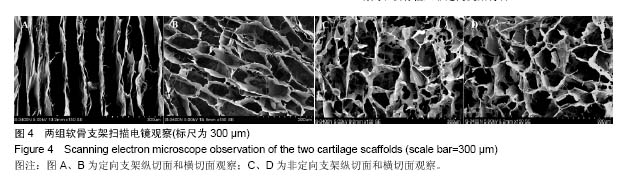
2.1 实验动物数量分析 48只兔进行动物分组实验,随机分为2组,每组24只。全部进入结果分析,中途无脱失。 2.2 软骨支架扫描电镜观察 2组不同的支架扫描电镜下存在明显的差异。定向支架组纵切面上可见排列相对蒸气的微管样结构,且方向一致,不同的结构之间存在微孔相互贯通;而非定向支架组纵切面与横切面表现为多孔蜂窝状结构,见图4。"
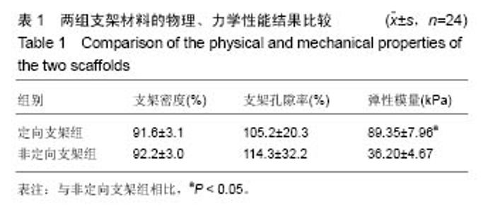
2.3 两组支架材料的物理、力学性能比较 2组支架密度、支架孔隙率差异无显著性意义(P > 0.05);定向支架组支架材料弹性模量显著高于非定向支架组(P < 0.05),见表1。"
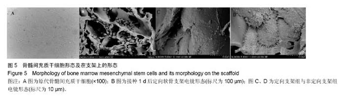
2.4 骨髓间充质干细胞形态及在支架上的形态 倒置显微镜下观察原代骨髓间充质干细胞24 h后开始部分贴壁, 3 d后细胞呈圆形,7 d后原代细胞占据培养瓶底大部,细胞为多角形(图5A)。扫描电子显微镜下,骨髓间充质干细胞在定向及非定向软骨支架黏附较好,接种1 d后细胞未铺开,为球状(图5B)。定向支架组可见大量球星细胞黏附在微观侧壁,细胞规则具有方向性和规律性;非定向软骨支架中可见大量球星细胞黏附在微管侧壁,细胞比较随机(图5C、D)。"


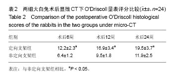
2.5 两组大白兔一般观察 所有大白兔术后均存在跛行现象,切口部位均未出现红肿、化脓等。定向支架组大白兔术后3 d恢复较好,活跃程度适中,未发生截瘫、狂躁等不良反应;非定向支架组2只大白兔术后出现腹泻。 2.6 两组大白兔术后显微CT下O'Driscoll量表评分比较 定向支架组大白兔术后6,12,24周显微CT下O'Driscoll量表评分均显著高于非定向支架组(P < 0.05),见表2。"

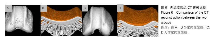
术后24周定向支架组大白兔CT下三维重建可见缺损部位平坦、光滑,新生组织与周围正常软骨界限消失;冠状面二维扫面可见新生类软骨厚度与周围正常骨十分接近。非定向组大白兔3D下显示软骨表面比较平整,难以确定原位存在缺损;冠状面2D显示修复软骨与正常软骨基本相同,见图6。"
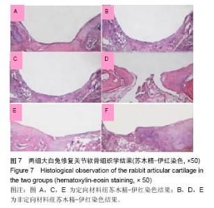
2.7 两组大白兔修复关节软骨组织学观察 定向支架组术后6周苏木精-伊红染色可见软骨缺损部位填充大量类软骨样组织;术后12周缺损部位存在大量类软骨样组织,可见经典软骨陷窝,细胞排列呈垂直方法;术后24周缺损部位消失,软骨细胞呈垂直方向排列,与正常组织几乎相同。 非定向支架组术后6周苏木精-伊红染色可见缺损局部存在大量类软骨样组织;术后12周软骨缺损部位存在大量类软骨样组织充填;术后24周缺损部位存在典型软骨陷窝,与周围软骨组织存在差异,见图7。"
| [1]Vo N,Niedernhofer LJ,Nasto LA,et al.An overview of underlying causes and animal models for the study of age-related degenerative disorders of the spine and synovial joints.J Orthop Res.2013;31(6):831-837. [2]Capeci CM,Turchiano M,Strauss EJ,et al.Osteochondral allografts: applications in treating articular cartilage defects in the knee. Bull Hosp Jt Dis.2013;71(1):60-67. [3]张永涛,金丹.组织工程骨软骨复合体构建的研究进展[J].中国修复重建外科杂志,2011,25(9):1120-1124. [4]Lim HC,Bae JH,Song SH,et al.Current treatments of isolated articular cartilage lesions of the knee achieve similar outcomes. Clin Orthop Relat Res. 2012;470(8): 2261-2267. [5]Kelly TA,Roach BL,Weidner ZD,et al.Tissue-engineered articular cartilage exhibits tension-compression nonlinearity reminiscent of the native cartilage.J Biomech. 2013;46(11): 1784-1791. [6]Gardner OF,Archer CW,Alini M,et al.Chondrogenesis of mesenchymal stem cells for cartilage tissue engineering. Histol Histopathol. 2013;28(1):23-42. [7]Schwarz S,Koerber L,Elsaesser AF,et al.Decellularized cartilage matrix as a novel biomatrix for cartilage tissue- engineering applications. Tissue Eng Part A. 2012; 18(21-22):2195-2209. [8]Cheng H,Byrska-Bishop M,Zhang CT,et al.Stem cell membrane engineering for cell rolling using peptide conjugation and tuning of cell-selectin interaction kinetics. Biomaterials. 2012;33(20):5004-5012. [9]刘清宇,王富友,杨柳.关节软骨组织工程支架的研究进展[J].中国修复重建外科杂志, 2012, 26(10):1247-1250. [10]李元城,张卫国,秦建华,等.微流控芯片上胰岛素样生长因子1和碱性成纤维细胞生长因子对兔关节软骨细胞增殖的影响[J].解放军医学杂志, 2013, 38(6):476-480. [11]Fortier LA, Barker JU, Strauss EJ, et al. The role of growth factors in cartilage repair. Clin Orthop Relat Res. 2011; 469 (10):2706-2710.[12]Kwon DR, Park GY, Lee SU. The effects of intra-articular platelet-rich plasma injection according to the severity of collagenase-induced knee osteoarthritis in a rabbit model. Ann Rehabil Med. 2012;36(4):458-465.[13]Lee HR, Park KM, Joung YK, et al. Platelet-rich plasma loaded hydrogel scaffold enhances chondrogenic differentiation and maturation with up-regulation of CB1 and CB2. J Control Release. 2012;159(3):332-337.[14]Kon E, Buda R, Filardo G, et ai. Platelet-rich plasma: intra-articular knee injections produced favorable results on degenerative cartilage lesions. Knee Surg Sports Traumatol Arthrosc. 2010;518(4):472-479.[15]Gobbi A, Karnatzikos G, Mahajan V, et al. Platelet-rich plasma treatment in symptomatic patients with knee osteoarthritis: preliminary results in a group of active patients. Sports Health. 2012;4(2): 162-172.[16]Filardo G, Kon E, Buda R, et al. Platelet-rich plasma intra-articular knee injections for the treatment of degenerative cartilage lesions and osteoarthritis. Knee Surg Sports Traumatol Arthrosc. 2011;19(4):528-535.[17]Kon E, Mandelbaum B, Buda R, et al. Platelet-rich plasma intra-articular injection versus hyaluronic acid viscosupplementation as treatments for cartilage pathology: from early degeneration to osteoarthritis. Arthroscopy. 2011;27(11): 1490-1501.[18]熊小龙, 伍亮, 相大勇, 等.富血小板血浆对大鼠跟腱断裂早期愈合的影响[J].中国修复重建外科杂志, 2012,26(4):466-471.[19]Milano G, Deriu L, Sanna PE,et al. Repeated platelet concentrate injections enhance reparative response of microfractures in the treatment of chondral defects of the knee: an experimental study in an animal model. Arthroscopys. 2012;28(5):688-701.[20]Seixa CI,Soler C,Carillo JM,et al. Effect of autologous platelet-rich plasma on the repair of full-thickness articular defects in rabbits. Knee Surg Sports Traumatol Arflirosc. 2013;21(8):1730-1736.[21]Zhu Y,Yuan M,Meng HY,et al. Basic science and clinical application of platelet-rich plasma for cartilage defects and osteoarthritis: a review. Osteoarthritis Cartilage. 2013;21(11):1627-1637.[22]Andia I,Maffulli N. Platelet-rich plasma for managing pain and inflammation in osteoarthritis.Nat Rev Rheumatol. 2013;9(12): 721-730.[23]Filardo G,Kon E,Pereira RM,et al. Platelet-rich plasma intra-articular injections for cartilage degeneration and osteoarthritis: single- versus double-spinning approach. Knee Surg Sports Traumatol Arthrosc. 2012;20(10):2082-2091.[24]Abrams GD,Frank RM,Fortier LA,et al. Platelet-rich plasma for articular cartilage repair. Sports Med Arthroscs. 2013; 21(4):213-219.[25]Chang KV,Hung CY,Aliwarga F,et al. Comparative Effectiveness of Platelet-Rich Plasma Injections for Treating Knee Joint Cartilage Degenerative Pathology: A Systematic Review and Meta-Analysis. Arch Phys Med Rehabil. 2014; 95(3):562-575.[26]Dold AP,Zywiel MG,Taylor DW,et al. Platelet-rich plasma in the management of articular cartilage pathology: a systematic review. Clin J Sport Med. 2014;24(1):31-43. |
| [1] | Wang Jianping, Zhang Xiaohui, Yu Jinwei, Wei Shaoliang, Zhang Xinmin, Xu Xingxin, Qu Haijun. Application of knee joint motion analysis in machanism based on three-dimensional image registration and coordinate transformation [J]. Chinese Journal of Tissue Engineering Research, 2022, 26(在线): 1-5. |
| [2] | Tan Xinfang, Guo Yanxing, Qin Xiaofei, Zhang Binqing, Zhao Dongliang, Pan Kunkun, Li Yuzhuo, Chen Haoyu. Effect of uniaxial fatigue exercise on patellofemoral cartilage injury in a rabbit [J]. Chinese Journal of Tissue Engineering Research, 2022, 26(在线): 1-6. |
| [3] | Zhang Yufang, Lü Meng, Mei Zhao. Construction and verification of a full spine biomechanical model of adolescent scoliosis [J]. Chinese Journal of Tissue Engineering Research, 2022, 26(9): 1351-1356. |
| [4] | Bai Zixing, Cao Xuhan, Sun Chengyi, Yang Yanjun, Chen Si, Wen Jianmin, Lin Xinxiao, Sun Weidong. Construction and biomechanical analysis of ankle joint finite element model in gait cycle [J]. Chinese Journal of Tissue Engineering Research, 2022, 26(9): 1362-1366. |
| [5] | Liu Feng, Feng Yi. Finite element analysis of different Kirschner wire tension bands on transverse patella fractures during gait cycle [J]. Chinese Journal of Tissue Engineering Research, 2022, 26(9): 1367-1371. |
| [6] | Yao Xiaoling, Peng Jiancheng, Xu Yuerong, Yang Zhidong, Zhang Shuncong. Variable-angle zero-notch anterior interbody fusion system in the treatment of cervical spondylotic myelopathy: 30-month follow-up [J]. Chinese Journal of Tissue Engineering Research, 2022, 26(9): 1377-1382. |
| [7] | Wu Cong, Jia Quanzhong, Liu Lun. Relationship between transforming growth factor beta1 expression and chondrocyte migration in adult articular cartilage after fragmentation [J]. Chinese Journal of Tissue Engineering Research, 2022, 26(8): 1167-1172. |
| [8] | An Weizheng, He Xiao, Ren Shuai, Liu Jianyu. Potential of muscle-derived stem cells in peripheral nerve regeneration [J]. Chinese Journal of Tissue Engineering Research, 2022, 26(7): 1130-1136. |
| [9] | Zhang Jinglin, Leng Min, Zhu Boheng, Wang Hong. Mechanism and application of stem cell-derived exosomes in promoting diabetic wound healing [J]. Chinese Journal of Tissue Engineering Research, 2022, 26(7): 1113-1118. |
| [10] | Duan Chao, Shang Xiaoqiang, Duan Xianglin, Yang Ping, Tao Shengxiang. Stability of patellar claw versus loop plate combined with patellar claw for the treatment of comminuted fractures of the lower pole of the patella [J]. Chinese Journal of Tissue Engineering Research, 2022, 26(6): 934-937. |
| [11] | Wen Mingtao, Liang Xuezhen, Li Jiacheng, Xu Bo, Li Gang. Mechanical stability of Sanders II type calcaneal fractures fixed by two internal fixation methods [J]. Chinese Journal of Tissue Engineering Research, 2022, 26(6): 838-842. |
| [12] | Huang Hao, Hong Song, Wa Qingde. Finite element analysis of the effect of femoral component rotation on patellofemoral joint contact pressure in total knee arthroplasty [J]. Chinese Journal of Tissue Engineering Research, 2022, 26(6): 848-852. |
| [13] | Zheng Yongze, Zheng Liqin, He Xingpeng, Chen Xinmin, Li Musheng, Li Pengfei, Lin Ziling. Extended finite element modeling analysis of femoral neck fracture based on ABAQUS software [J]. Chinese Journal of Tissue Engineering Research, 2022, 26(6): 853-857. |
| [14] | Liu Yuhang, Zhou Jianqiang, Xu Xuebin, Qu Xingyue, Li Ziyu, Li Kun, Wang Xing, Li Zhijun, Li Xiaohe, Zhang Shaojie. Establishment and validation of finite element model of lower cervical spine in 6-year-old children [J]. Chinese Journal of Tissue Engineering Research, 2022, 26(6): 870-874. |
| [15] | Lin Xuchen, Zhu Hainian, Wang Zengshun, Qi Tengmin, Liu Limin, Suonan Angxiu. Effect of xanthohumol on inflammatory factors and articular cartilage in a mouse mode of osteoarthritis [J]. Chinese Journal of Tissue Engineering Research, 2022, 26(5): 676-681. |
| Viewed | ||||||
|
Full text |
|
|||||
|
Abstract |
|
|||||