| [1] Guo SZ,Ren XJ,Wu B,et al.Preparation of the acellular scaffold of the spinal cord and the study of biocompatibility. Spinal Cord.2010;48(7):576-581.
[2] Weber B,Emmert MY,Schoenauer R,et al.Tissue engineering on ma-trix: future of autologous tissue replacement. Semin Immunopathol.2011;33(3):307-315.
[3] Nishibe T,Kondo Y,Muto A,et a1.Optimal prost hetic graft design for sni&ll diameter vascular grafts.Vascular.2007;(6): 356-360.
[4] Vavken P,Joshi S,Murray MM.TRITON‐X is most effective among three decellularization agents for ACL tissue engineering. J Orthop Res.2009;27(12):1612-1618.
[5] Yu BT, Li WT,Song BQ,et al. Comparative study of the Triton X-100-sodium deoxycholate method and detergent-enzymatic digestion method for decellularization ofporcine aortic valves.Eur Rev Med Pharmacol Sci.2013;17(16):2179-2184.
[6] Cebotari S,Tudorache I,Jaekel T,et al.Detergent decellularization of heart valves for tissue engineering: toxicological effects of residual detergents on human endothelial cells.Artif Organs. 2010;34(3):206-210.
[7] Deeken CR,White AK,Bachman SL,et al.Method of preparing a decellularized porcine tendonusing tributyl phosphate.J Biomed Mater Res B Appl Biomater.2011;96(2):199-206.
[8] 张国安,宁方刚,钟京鸣,等.反复冻融配合超声振荡洗涤制备猪脱细胞真皮基质[J].中国组织工程研究与临床康复,2007,11(41): 8280-8284.
[9] Azhim A,Yamagami K,Muramatsu K,et al.The use of sonication treatment to comletely decellularize aorta tissue.Conf Proc IEEE Eng Med Biol Soc.2011;2011: 2468-2471.
[10] Hopkinson A,Shanmuganathan VA,Gray T,et al. Optimization ofamniotic membran e (AM) denuding for tissue engineering. Tissue Eng Part C Methods.2008;14(4):371-381.
[11] Liu TX,Wang Z.Collagen crosslinking of porcine sclera using genipin.Acta Ophthalmol.2013;91(4):e253-257.
[12] 蒋涛,任先军,阴洪,等.京尼平交联对大鼠脱细胞脊髓生物学特性的影响[J].第三军医大学学报,2013,35(20):2164-2167.
[13] Bi L,Cao Z,Hu Y,et al.Effects of different cross-linking conditions on the properties of genipin-cross-linked chitosan/collagen scaffolds for cartilage tissue engineering. J Mater Sci Mater Med.2011;22(1):51-62.
[14] Yahyouche A,Zhidao X,Czernuszka JT,et al.Macrophage- mediated degradation of crosslinked collagen scaffolds.Acta biomaterialia.2011;7(1):278-286.
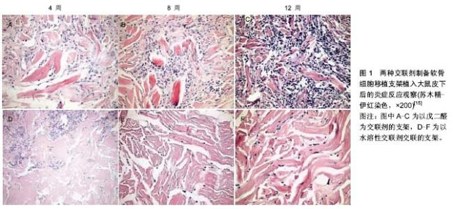
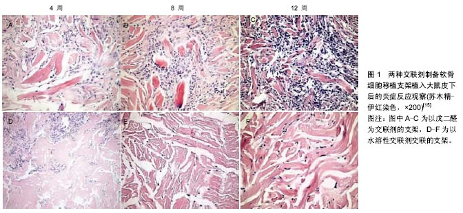
[15] 綦惠,杰永生,陈磊,等.两种交联剂制备软骨细胞移植支架的比较[J].中国医药生物技术,2013,8(6):408-413.
[16] 郭树章,任先军,蒋涛,等.脱细胞脊髓天然支架的制备及形态学观察[J].中国矫形外科杂志,2007,15(3):226-228.
[17] Singh P,Schwarzbauer JE.Fibronectin matrix assembly is essential for cell condensation during chondrogenesis.J Cell Sci.2014;127(Pt 20):4420-4428.
[18] Turney SG,Bridgman PC.Laminin stimulates and guides axonal outgrowth via growth cone myosin II activity. Nat Neurosci. 2005;8:717-719.
[19] King VR,Alovskaya A,Wei DY,et al.The use of injectable forms of fibrin and fibronectin to support axonal ingrowth after spinal cord injury. Biomaterials. 2010;31(15):4447-4456.
[20] King VR,Hewazy D,Alovskaya A,et al.The neuroprotective effects of fibronectin mats and fibronectin peptides following spinal cord injury in the rat. Neuroscience. 2010;168(2): 523-530.
[21] Lin CY,Lee YS,Lin VW,et al.Fibronectin inhibits chronic pain development after spinal cord injury.J Neurotrauma.2012; 29(3):589-599.
[22] Xia M, Zhu Y. Fibronectin enhances spinal cord astrocyte proliferation by elevating P2Y1 receptor expression.J Neurosci Res.2014;92(8):1078-1090.
[23] Deng L,Walker C,Wen X,et al. Laminin-coated filaments co-grafted with Schwann cells and GDNF direct axonal growth following rat thoracic spinal cord injury (729.2). FASEB J.2014;28(1 Supplement):729.
[24] Menezes K,Nascimento MA,Gonçalves JP,et al.Human Mesenchymal Cells from Adipose Tissue Deposit Laminin and Promote Regeneration of Injured Spinal Cord in Rats. PloS One.2014;9(5):e96020.
[25] Menezes K,de Menezes JR,Nascimento MA,et al.Polylaminin, a polymeric form of laminin, promotes regeneration after spinal cord injury. FASEB J.2010;24(11): 4513-4522.
[26] Yuan N,Tian W,Sun L,et al.Neural stem cell transplantation in a double-layer collagen membrane with unequal pore sizes for spinal cord injury repair. Neural Regen Res.2014;9(10): 1014.
[27] Sekeljic V,Andjus PR.Tenascin-C and its functions in neuronal plasticity.Int J Biochem Cell Biol.2012;44:825-829.
[28] Tsai HL,Chiu WT,Fang CL,et al.Different forms of tenascin-C with tenascin-R regulate neural differentiation in bone marrow-derived human mesenchymal stemcells.Tissue Eng Part A.2014;20(13-14):1908-1921.
[29] Crapo PM,Medberry CJ,Reing JE,et al.Biologic scaffolds composed of central nervous system extracellular matrix. Biomaterials.2012;33(13):3539-3547.
[30] Liu J,Chen J,Liu B,et al.Acellular spinal cord scaffold seeded with mesenchymal stem cells promotes long-distance axon regeneration and functional recovery in spinal cord injured rats.J Neurol Sci.2013;325(1):127-136.
[31] Chen J,Joon Lee H,Jakovcevski I,et al.The extracellular matrix glycoprotein tenascin-C is beneficial for spinal cord regeneration.Mol Ther.2010;18(10):1769-1777.
[32] Crapo PM,Medberry CJ,Reing JE,et al.Biologic scaffolds composed of central nervous system extracellular matrix. Biomaterials.2012;33(13):3539-3547. |