Chinese Journal of Tissue Engineering Research ›› 2026, Vol. 30 ›› Issue (36): 9393-9401.doi: 10.12307/2026.381
Previous Articles Next Articles
Mechanism of high glucose-induced osteoblast cuproptosis
Lian Yong1, Xie Zhixing1, Lu Jiaxin1, Pan Zhaofeng1, Chen Qingzhen2, Shao Min2
- 1The Third Clinical Medical College of Guangzhou University of Chinese Medicine, Guangzhou 510405, Guangdong Province, China; 2The Third Affiliated Hospital of Guangzhou University of Chinese Medicine, Guangzhou 510405, Guangdong Province, China
-
Received:2025-07-06Revised:2025-09-19Online:2026-12-28Published:2026-05-20 -
Contact:Shao Min, MD, Chief physician, Master’s supervisor, Doctoral supervisor, the Third Affiliated Hospital of Guangzhou University of Chinese Medicine, Guangzhou 510405, Guangdong Province, China -
About author:Lian Yong, MD candidate, the Third Clinical Medical College of Guangzhou University of Chinese Medicine, Guangzhou 510405, Guangdong Province, China -
Supported by:Guangdong Provincial Special Innovation Project for Ordinary Higher Education Institutions, No. 2024KTSCX115 (to CQZ)

CLC Number:
Cite this article
Lian Yong, Xie Zhixing, Lu Jiaxin, Pan Zhaofeng, Chen Qingzhen, Shao Min. Mechanism of high glucose-induced osteoblast cuproptosis[J]. Chinese Journal of Tissue Engineering Research, 2026, 30(36): 9393-9401.
share this article
Add to citation manager EndNote|Reference Manager|ProCite|BibTeX|RefWorks
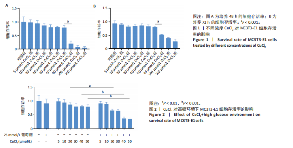
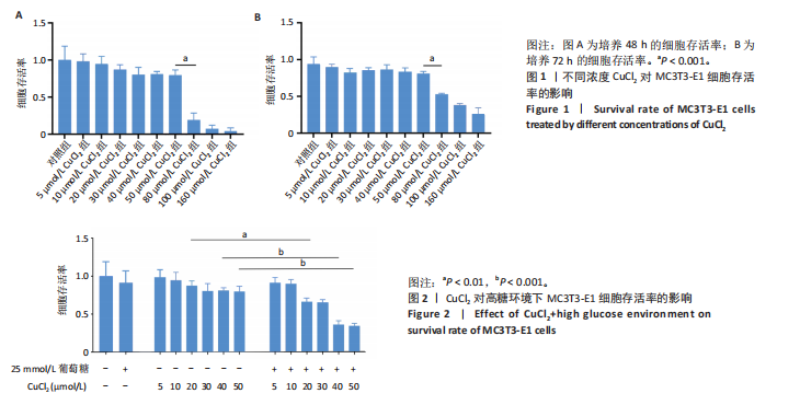
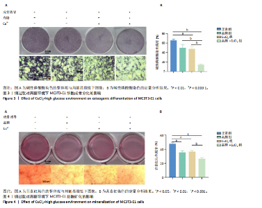
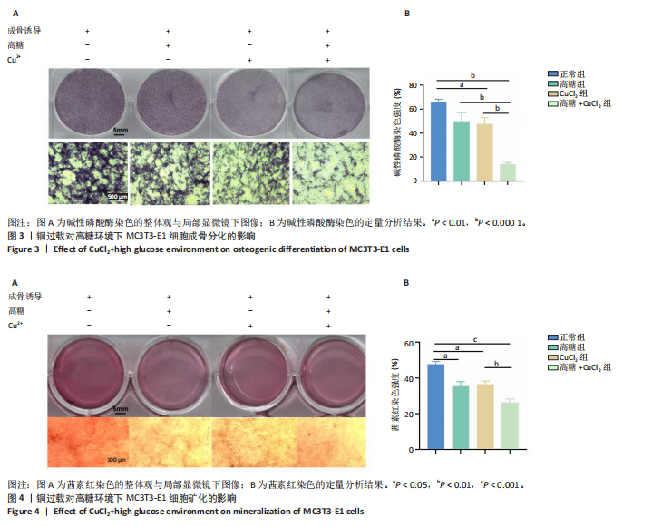
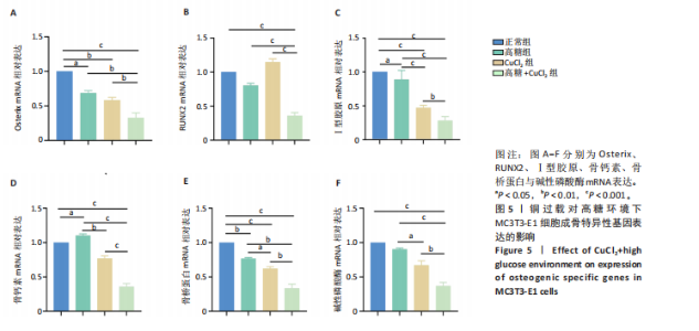
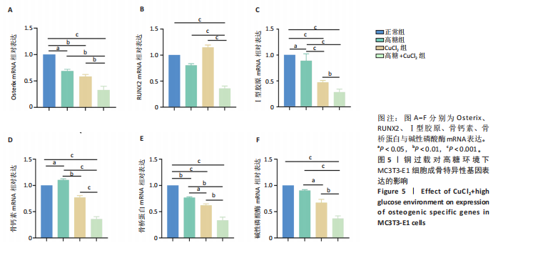
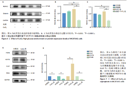
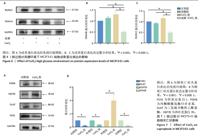
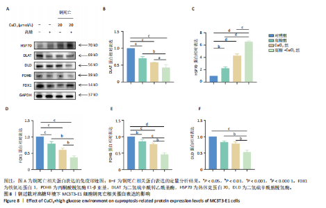
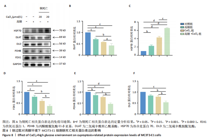
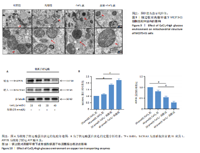
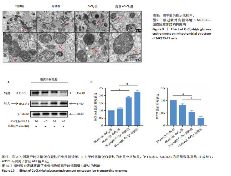
2.1 CuCl2对MC3T3-E1细胞的毒性作用 CCK-8检测结果显示,培养48,72 h,80 μmol/L CuCl2对细胞有绝对的毒性作用,与对照组比较,80,100,160 μmol/L CuCl2组细胞存活率显著降低(P < 0.001),见图1。 2.2 CuCl2对高糖环境下MC3T3-E1细胞存活率的影响 CCK-8检测结果显示,与对照组比较,单纯的高糖环境与不同浓度CuCl2对MC3T3-E1细胞存活率无明显影响(P > 0.05);20 μmol/L CuCl2+高糖组细胞存活率低于20 μmol/L CuCl2组(P < 0.01),40 μmol/L CuCl2+高糖组细胞存活率低于40 μmol/L CuCl2组(P < 0.001),50 μmol/L CuCl2+高糖组细胞存活率低于50 μmol/L CuCl2组(P < 0.001),见图2。结果表明高糖环境加重了CuCl2的细胞毒性作用。 2.3 铜过载对高糖环境下MC3T3-E1细胞成骨分化的影响 碱性磷酸染色结果显示,CuCl2组、高糖+CuCl2组碱性磷酸酶表达低于对照组(P < 0.01或P < 0.000 1),高糖+CuCl2组碱性磷酸酶表达低于CuCl2组、高糖组(P < 0.000 1),见图3所示,说明铜过载使MC3T3-E1细胞的成骨分化明显受到抑制,联合高糖环境后抑制程度更显著。 2.4 铜过载对高糖环境下MC3T3-E1细胞矿化的影响 茜素红染色结果显示,高糖组、CuCl2组、高糖+CuCl2组细胞矿化水平均低于对照组(P < 0.05或P < 0.001),高糖+CuCl2组细胞矿化水平低于高糖组、CuCl2组(P < 0.01),见图4所示,说明铜过载使MC3T3-E1细胞的成骨矿化明显受到抑制,联合高糖环境后抑制程度更显著。 2.5 铜过载对高糖环境下MC3T3-E1细胞成骨特异性基因表达的影响 qRT-PCR检测结果显示,与对照组比较,高糖组Osterix、Ⅰ型胶原、骨桥蛋白mRNA表达降低(P < 0.05),骨钙素mRNA表达升高(P < 0.05);CuCl2组Osterix、Ⅰ型胶原、骨桥蛋白mRNA表达降低(P < 0.01或P < 0.001),见图5所示。高糖+CuCl2组骨钙素、RUNX2、Ⅰ型胶原、Osterix、骨桥蛋白、碱性磷酸酶mRNA表达均低于高糖组、CuCl2组(P < 0.01或P < 0.001),见图5所示。 2.6 铜过载对高糖环境下MC3T3-E1细胞成骨蛋白表达的影响 Western Blot检测结果显示,高糖+CuCl2组RUNX2、Osterix蛋白表达均低于其他3组(P < 0.001或P < 0.000 1),见图6所示。20 μmol/L CuCl2单独干预有促进MC3T3-E1细胞成骨蛋白表达的趋势,但无统计学差异,而 CuCl2联合高糖可显著抑制MC3T3-E1细胞成骨蛋白表达,抑制成骨分化。 2.7 铜过载对MC3T3-E1细胞铜死亡相关蛋白表达的影响 Western Blot检测结果显示,与对照组比较,CuCl2组热休克蛋白70的蛋白表达升高(P < 0.000 1),FDX1、二氢硫辛酰转乙酰基酶、丙酮酸脱氢酶E1-β亚基蛋白表达降低(P < 0.001),如图7所示,说明20 μmol/L CuCl2可诱导MC3T3-E1细胞铜死亡。 2.8 铜过载对高糖环境下MC3T3-E1细胞铜死亡相关蛋白表达的影响 Western Blot检测结果显示,与对照组比较,高糖组与CuCl2组热休克蛋白70的蛋白表达升高(P < 0.01,P < 0.0001),高糖组与CuCl2组二氢硫辛酰转乙酰基酶、FDX1、丙酮酸脱氢酶E1-β亚基的蛋白表达降低(P < 0.01或P < 0.001);与高糖组、CuCl2组比较,高糖+CuCl2组热休克蛋白70的蛋白表达升高(P < 0.001,P < 0.000 1),二氢硫辛酰转乙酰基酶、FDX1、丙酮酸脱氢酶E1-β亚基、二氢硫辛酰胺脱氢酶的蛋白表达均降低(P < 0.05,P < 0.01,P < 0.001,P < 0.000 1),见图8所示。 2.9 铜过载对高糖环境下MC3T3-E1细胞线粒体结构的影响 透射电镜下可见CuCl2组细胞内线粒体数量明显减少,线粒体结构也发生明显变化,主要表现为线粒体嵴消失、线粒体皱缩、体积变小、线粒体膜结构破坏,联合高糖后加重了这些结构的恶变,见图9。 2.10 铜过载对高糖环境下成骨细胞铜离子转运酶蛋白表达的影响 Western Blot检测结果显示,20 μmol/L CuCl2+高糖组SLC31A1蛋白表达高于20 μmol/L CuCl2组(P < 0.001),ATP7B蛋白表达低于20 μmol/L CuCl2组(P < 0.001);40 μmol/L CuCl2+高糖组SLC31A1蛋白表达高于40 μmol/L CuCl2组(P < 0.001),ATP7B蛋白表达低于40 μmol/L CuCl2组(P < 0.001),见图10所示,说明高糖环境促进了铜离子转入酶SLC31A1的表达,抑制了铜离子转出酶ATP7B的表达。"
"
"
"
"
"
| [1] TSVETKOV P, COY S, PETROVA B, et al. Copper induces cell death by targeting lipoylated TCA cycle proteins. Science. 2022;375(6586): 1254-1261. [2] LUTSENKO S, ROY S, TSVETKOV P. Mammalian copper homeostasis: physiological roles and molecular mechanisms. Physiol Rev. 2025; 105(1):441-491. [3] JOMOVA K, MAKOVA M, ALOMAR SY, et al. Essential metals in health and disease. Chem Biol Interact. 2022;367:110173. [4] EVANS GW. Copper homeostasis in the mammalian system. Physiol Rev. 1973;53(3):535-570. [5] EVANS GW, BUBOIS RS, HAMBIDGE KM. Wilson’s disease: identification of an abnormal copper-binding protein. Science. 1973;181(4105): 1175-1176. [6] TIAN Z, JIANG S, ZHOU J, et al. Copper homeostasis and cuproptosis in mitochondria. Life Sci. 2023;334:122223. [7] JIA D, LIU L, LIU W, et al. Copper metabolism and its role in diabetic complications: A review. Pharmacol Res. 2024;206:107264. [8] 高红丽,秦玉凤,张玥晗,等.铜代谢与口腔疾病的诊断及治疗[J].中国组织工程研究,2025,29(20):4316-4324. [9] 朱赛雅,刘晶,余晨.线粒体铜稳态失衡与纤维化疾病的研究进展[J].生理学报,2024,76(4):597-604. [10] CHANG W, LI P. Copper and Diabetes: Current Research and Prospect. Mol Nutr Food Res. 2023;67(23):e2300468. [11] POURESMAEIL V, AL ABUDI A H, MAHIMID AH, et al. Evaluation of Serum Selenium and Copper Levels with Inflammatory Cytokines and Indices of Oxidative Stress in Type 2 Diabetes. Biol Trace Elem Res. 2023;201(2):617-626. [12] WU K, CHEN L, KONG Y, et al. The association between serum copper concentration and prevalence of diabetes among US adults with hypertension (NHANES 2011-2016). J Cell Mol Med. 2024;28(8): e18270. [13] CUI Z, ZHOU H, LIU K, et al. Dietary Copper and Selenium Intakes and the Risk of Type 2 Diabetes Mellitus: Findings from the China Health and Nutrition Survey. Nutrients. 2022;14(10):2055. [14] ZHANG Z, JI C, WANG YN, et al. Maresin1 Suppresses High-Glucose-Induced Ferroptosis in Osteoblasts via NRF2 Activation in Type 2 Diabetic Osteoporosis. Cells. 2022;11(16):2560. [15] LIN Y, SHEN X, KE Y, et al. Activation of osteoblast ferroptosis via the METTL3/ASK1-p38 signaling pathway in high glucose and high fat (HGHF)-induced diabetic bone loss. FASEB J. 2022;36(3):e22147. [16] LI Y, SHRESTHA A, ZHANG H, et al. Impact of diabetes mellitus simulations on bone cell behavior through in vitro models. J Bone Miner Metab. 2020;38(5):607-619. [17] HALL DO, CAMMACK R, RAO KK. Role for ferredoxins in the origin of life and biological evolution. Nature. 1971;233(5315):136-138. [18] 李朝霞,张晓峰,徐西林,等.铜死亡与骨质疏松[J].中国骨质疏松杂志,2024,30(1):98-103. [19] SCHULZ V, BASU S, FREIBERT SA, et al. Functional spectrum and specificity of mitochondrial ferredoxins FDX1 and FDX2. Nat Chem Biol. 2023;19(2):206-217. [20] STEINHILPER R, BOSS L, FREIBERT SA, et al. Two-stage binding of mitochondrial ferredoxin-2 to the core iron-sulfur cluster assembly complex. Nat Commun. 2024;15(1):10559. [21] JOSHI PR, SADRE S, GUO XA, et al. Lipoylation is dependent on the ferredoxin FDX1 and dispensable under hypoxia in human cells. J Biol Chem. 2023;299(9):105075. [22] JIA M, SEN S, WACHNOWSKY C, et al. Characterization of [2Fe-2S]-Cluster-Bridged Protein Complexes and Reaction Intermediates by use of Native Mass Spectrometric Methods. Angew Chem Int Ed Engl. 2020;59(17):6724-6728. [23] WANG T, LIU Y, LI Q, et al. Cuproptosis-related gene FDX1 expression correlates with the prognosis and tumor immune microenvironment in clear cell renal cell carcinoma. Front Immunol. 2022;13:999823. [24] GUOWEI L, XIUFANG L, QIANQIAN X, et al. The FDX1 methylation regulatory mechanism in the malignant phenotype of glioma. Genomics. 2023;115(2):110601. [25] PARSANATHAN R. Copper’s dual role: unravelling the link between copper homeostasis, cuproptosis, and cardiovascular diseases. Hypertens Res. 2024;47(5):1440-1442. [26] CHEN J, SUN Q, WANG Y, et al. Revealing the key role of cuproptosis in osteoporosis via the bioinformatic analysis and experimental validation of cuproptosis-related genes. Mamm Genome. 2024;35(3):414-431. [27] TU B, SONG K, ZHOU ZY, et al. SLC31A1 loss depletes mitochondrial copper and promotes cardiac fibrosis. Eur Heart J. 2025. doi: 10.1093/eurheartj/ehaf130. [28] DAS A, ASH D, FOUDA AY, et al. Cysteine oxidation of copper transporter CTR1 drives VEGFR2 signalling and angiogenesis. Nat Cell Biol. 2022; 24(1):35-50. [29] HUO S, WANG Q, SHI W, et al. ATF3/SPI1/SLC31A1 Signaling Promotes Cuproptosis Induced by Advanced Glycosylation End Products in Diabetic Myocardial Injury. Int J Mol Sci. 2023;24(2):1667. [30] QIU Z, LIU Q, WANG L, et al. The copper transporter, SLC31A1, transcriptionally activated by ELF3, imbalances copper homeostasis to exacerbate cisplatin-induced acute kidney injury through mitochondrial dysfunction. Chem Biol Interact. 2024;393:110943. [31] CHEN X, LI K, XIAO Y, et al. SP1/CTR1-mediated oxidative stress-induced cuproptosis in intervertebral disc degeneration. Biofactors. 2024; 50(5):1009-1023. [32] SHI X, NI H, TANG L, et al. Comprehensive Gene Analysis Reveals Cuproptosis-Related Gene Signature Associated with M2 Macrophage in Staphylococcus aureus-Infected Osteomyelitis. J Inflamm Res. 2024; 17:3057-3077. [33] CHEN T, GAO Z, WANG Y, et al. Identification and immunological role of cuproptosis in osteoporosis. Heliyon. 2024;10(5):e26759. [34] WANG LJ, NI XH, ZHANG F, et al. Osteoblast Response to Copper-Doped Microporous Coatings on Titanium for Improved Bone Integration. Nanoscale Res Lett. 2021;16(1):146. [35] QI J, ZHENG Z, HU L, et al. Development and characterization of cannabidiol-loaded alginate copper hydrogel for repairing open bone defects in vitro. Colloids Surf B Biointerfaces. 2022;212:112339. [36] KHALKHALI P, OMIDI M, MASSON-MEYERS DS, et al. Promoting Angiogenesis/Osteogenesis by a New Copper/Magnesium Hydroxide Hybrid Nanoparticle: In Vitro and In Vivo Investigation. J Biomed Mater Res A. 2025;113(1):e37855. [37] SAFAROVA YANTSEN Y, NESSIPBEKOVA A, SYZDYKOVA A, et al. Strontium- and Copper-Doped Ceramic Granules in Bone Regeneration-Associated Cellular Processes. J Funct Biomater. 2024;15(11):352. [38] XIE C, SUN Q, CHEN J, et al. Cu-Tremella fuciformis polysaccharide-based tumor microenvironment-responsive injectable gels for cuproptosis-based synergistic osteosarcoma therapy. Int J Biol Macromol. 2024;270(Pt 2):132029. [39] MEDEIROS C, WALLACE JM. High glucose-induced inhibition of osteoblast like MC3T3-E1 differentiation promotes mitochondrial perturbations. PLoS One. 2022;17(6):e0270001. [40] ZHANG P, LIAO J, WANG X, et al. High glucose promotes apoptosis and autophagy of MC3T3-E1 osteoblasts. Arch Med Sci. 2023;19(1):138-150. |
| [1] | Sun Lei, Zhang Qi, Zhang Yu. Pro-osteoblastic effect of chlorogenic acid protein microsphere/polycaprolactone electrospinning membrane [J]. Chinese Journal of Tissue Engineering Research, 2026, 30(8): 1877-1884. |
| [2] | Dong Shiming, Xie Zhenzi, Tao Rongrong, Mardan·Mamat, Ma Hairong. Type VI collagen: a multifunctional regulator in bone homeostasis and tissue engineering [J]. Chinese Journal of Tissue Engineering Research, 2026, 30(35): 9239-9247. |
| [3] | Chen Di, Xu Mengling, Rao Binchan, Zhu Liying, Li Xing, Xu Yongjie, Pan Wei. Effects and mechanisms of glycemic variability on apoptosis in mouse hippocampal neuronal HT-22 cells [J]. Chinese Journal of Tissue Engineering Research, 2026, 30(25): 6566-6574. |
| [4] | Li Huaying, Li Hao, Peng Wuxun, Dong Wentao. Mechanism of cuproptosis in the diagnosis and treatment of orthopedic-related diseases [J]. Chinese Journal of Tissue Engineering Research, 2026, 30(24): 6275-6281. |
| [5] | Wu Lingjie, Zheng Kaiyuan, Wang Guangrong, Yin Chong . Strategies for the application of miRNA-targeted therapy in the treatment of osteoporosis [J]. Chinese Journal of Tissue Engineering Research, 2026, 30(22): 5792-5803. |
| [6] | Li Congcong, Wufanbieke·Baheti, Zhao Li, Chen Xiaotao, Kong Chuifan, Yu Min. Physicochemical properties and biocompatibility of hydroxyapatite/graphene oxide/interleukin-4 composite coating materials [J]. Chinese Journal of Tissue Engineering Research, 2026, 30(2): 404-413. |
| [7] | Li Ruiying, Xia Hong. Visual analysis of cuproptosis research: global landscape of hotspots and frontiers [J]. Chinese Journal of Tissue Engineering Research, 2026, 30(17): 4529-4541. |
| [8] | Zhao Yu, Xue Yun, Huang Jiajun, Wu Diyou, Yang Bin, Huang Junqing. Total flavonoids from Semen Cuscutae inhibits osteoblast apoptosis in hormone-induced femoral head avascular necrosis [J]. Chinese Journal of Tissue Engineering Research, 2026, 30(17): 4289-4298. |
| [9] | Sun Long, Wu Haiyang, Tong Linjian, Liu Rui, Yang Weiguang, Xiao Jian, Liu Lice, Sun Zhiming. Regulatory mechanism of leptin in bone metabolism [J]. Chinese Journal of Tissue Engineering Research, 2026, 30(12): 3100-3108. |
| [10] | Shi Tengbo, Tang Yanfeng, Zhang Mengyu, Wang Xingfei, Li Chenyang, Shi Jinyu, Guo Chaowei, Li Yanzhou, He Zike, Wang Shangzeng. Effect of high-dose low molecular weight heparin on the healing of femoral shaft fractures [J]. Chinese Journal of Tissue Engineering Research, 2026, 30(12): 2957-2964. |
| [11] | Li Mingzhe, Ye Xiangling, Wang Bing, Yu Xiang. Preparation and osteogenic properties of liquid crystal display light-cured polylactic acid scaffold loaded with nano-tantalum [J]. Chinese Journal of Tissue Engineering Research, 2025, 29(4): 670-677. |
| [12] |
Li Yunzhe, Niu Zefan, Wang Zirou, Ai Chongyi, Chen Gang, Wang Xinxing.
Asperosaponin VI promotes osteogenic differentiation of MC3T3-E1 cells under hypoxia environment #br#
#br#
[J]. Chinese Journal of Tissue Engineering Research, 2025, 29(35): 7481-7489.
|
| [13] | Yin Hang, Song Kui. Effect of crocin hydrogel on chondrocytes and MC3T3-E1 cells [J]. Chinese Journal of Tissue Engineering Research, 2025, 29(34): 7293-7300. |
| [14] | Tang Haoxu, Liang Yingjie, Li Ce, Ding Penglin, Qian Minlong, Yuan Lingli. Deferoxamine alleviates the inhibitory effect of glucocorticoids on osteogenic differentiation [J]. Chinese Journal of Tissue Engineering Research, 2025, 29(32): 6821-6827. |
| [15] | Lu Xiuli, Xu Huazhen, Chen Yuxing, Yao Nan, Hu Zixuan, Huang Dane. Mechanism of Jiangu Formula in treating osteoporosis based on osteoclast-osteoblast coupling [J]. Chinese Journal of Tissue Engineering Research, 2025, 29(32): 6828-6835. |
| Viewed | ||||||
|
Full text |
|
|||||
|
Abstract |
|
|||||