Chinese Journal of Tissue Engineering Research ›› 2026, Vol. 30 ›› Issue (36): 9381-9392.doi: 10.12307/2026.908
Aucubin promotes osteogenic differentiation of mouse cranial pre-osteoblasts MC3T3-E1
Wang Houyuan1, 2, Xiao Jiacong1, 2, Shao Shoujia1, 3, He Paian1, 3, Yang Nan1, 3, Jiang Ziwei1, 2, 3
- ¹Guangzhou University of Chinese Medicine, Guangzhou 510405, Guangdong Province, China; 2The First Clinical Medical College of Guangzhou University of Chinese Medicine, Guangzhou 510405, Guangdong Province, China; 3The First Affiliated Hospital of Guangzhou University of Chinese Medicine, Guangzhou 510405, Guangdong Province, China
-
Received:2025-10-29Revised:2026-03-14Online:2026-12-28Published:2026-05-20 -
Contact:Jiang Ziwei, PhD, Chief physician, Doctoral supervisor, Guangzhou University of Chinese Medicine, Guangzhou 510405, Guangdong Province, China; The First Clinical Medical College of Guangzhou University of Chinese Medicine, Guangzhou 510405, Guangdong Province, China; The First Affiliated Hospital of Guangzhou University of Chinese Medicine, Guangzhou 510405, Guangdong Province, China -
About author:Wang Houyuan, PhD candidate, Guangzhou University of Chinese Medicine, Guangzhou 510405, Guangdong Province, China; The First Clinical Medical College of Guangzhou University of Chinese Medicine, Guangzhou 510405, Guangdong Province, China -
Supported by:National Natural Science Foundation of China, No. 81974575 (to JZW)

CLC Number:
Cite this article
Wang Houyuan, Xiao Jiacong, Shao Shoujia, He Paian, Yang Nan, Jiang Ziwei. Aucubin promotes osteogenic differentiation of mouse cranial pre-osteoblasts MC3T3-E1[J]. Chinese Journal of Tissue Engineering Research, 2026, 30(36): 9381-9392.
share this article
Add to citation manager EndNote|Reference Manager|ProCite|BibTeX|RefWorks
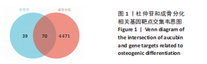
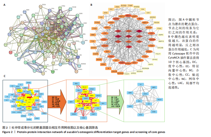
2.1 网络药理学分析结果 2.1.1 筛选靶点相关基因 合并SwissTargetPrediction与Pharmmapper数据库中的重复项后,发现109个杜仲苷作用靶点。同时,通过GeneCards等数据库共检索到成骨分化相关靶点4 541个。使用Venny 2.1.0分析工具共发现70个交集靶点,见图1。 2.1.2 蛋白-蛋白互作网络 将杜仲苷与成骨分化相关基因的交集靶点导入STRING数据库,建立蛋白-蛋白互作网络,选择中等置信度,得到70个节点和366条边,见图2A,随后将STRING分析的互作网络导入Cytoscape软件中进行可视化(图2B)。为进一步筛选关键基因,将靶点基因输入CytoNCA插件,并选取介数中心性、贴近中心性、度中心性、特征向量中心性、网络中心性和局部平均连通性6类中心性算法进行系统评价,共获得10个核心基因,包括Janus激酶1(JAK1)、Janus激酶2(JAK2)、信号转导与转录激活因子3 (STAT3)、半胱天冬酶3(CASP3)、基质金属蛋白酶2(MMP2)、基质金属蛋白酶9(MMP9)、BCL-2样蛋白1(BCL2L1)、白细胞介素2(IL2)、细胞间黏附分子1 (ICAM1)和H-Ras蛋白(HRAS),见图2C。"
"
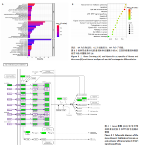
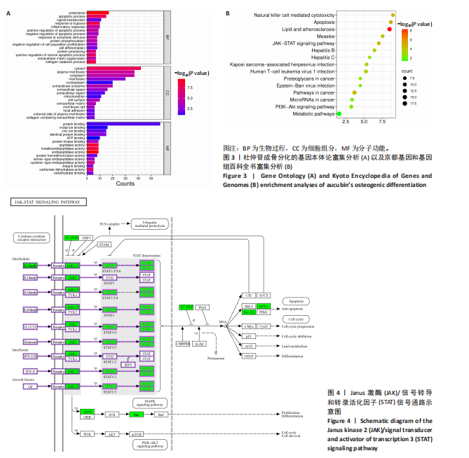
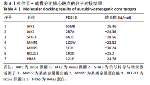
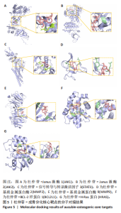
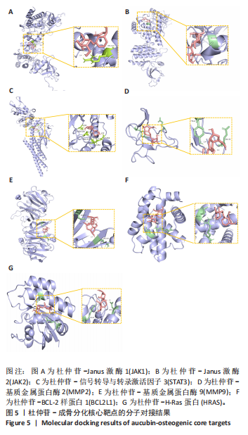
2.1.3 GO及KEGG富集分析 将相关基因导入DAVID数据库进行GO、KEGG富集分析[23]。GO功能注释共获得226个显著条目,其中生物过程148个、细胞组分32个、分子功能46个。结合条目显著性排名,进一步筛选前15个条目,并借助微生信平台进行可视化[24],生成GO功能条形图,见图3A,该结果提示杜仲苷可能通过调控缺氧应答通路、细胞凋亡进程、炎症反应及蛋白质磷酸化等生物学途径参与成骨分化的调控。KEGG共富集到63条显著通路,选取富集程度前15的通路进行可视化分析,见图3B。杜仲苷调控成骨分化主要涉及凋亡通路、Janus激酶-信号转导与转录激活因子信号通路、磷脂酰肌醇3激酶-蛋白激酶B信号通路以及代谢通路等。根据显著性、基因比例、核心靶点数以及相关文献研究,选择Janus激酶2/信号转导与转录激活因子3信号通路以及氧化应激进行实验验证,根据图4可见发挥作用的关键靶点为Janus激酶2、信号转导与转录激活因子3等。 2.1.4 分子对接 将杜仲苷与10个核心靶点进行分子对接分析,除白细胞介素2(IL2)、半胱天冬酶3(CASP3)及细胞间黏附分子1(ICAM1)结合能为0外,其余靶点的结合能均小于–21 kJ/mol,提示结合力较强。具体结合能结果见表4。将对接后的化合物导入Pymol中并进行可视化,见图5。 2.2 体外细胞实验结果 2.2.1 过氧化氢对MC3T3-E1细胞增殖的影响 不同浓度过氧化氢处理导致MC3T3-E1细胞增殖率发生时间依赖性变化:在12,24,48 h时,过氧化氢组细胞增殖率均显著低于对照组,"
"
"
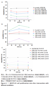
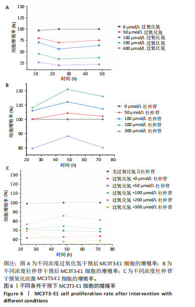
其中24 h时增殖率最低,48 h时出现回升趋势(图6A)。过氧化氢对细胞增殖的抑制作用表现出浓度依赖性(0-400 μmol/L梯度),当浓度≥100 μmol/L时,抑制作用达到了统计学显著性。基于此,100 μmol/L过氧化氢处理24 h能有效诱导氧化应激,细胞增殖显著受到抑制,同时可避免高浓度过氧化氢干预后细胞增殖率过低影响后续实验,适合用于构建氧化应激模型。 2.2.2 杜仲苷对MC3T3-E1细胞增殖的影响 干预24 h后,300 μmol/L杜仲苷组细胞增殖率低于对照组,其余各组(杜仲苷浓度分别为50,100,200 μmol/L)细胞增殖率均高于对照组;干预48 h后,300 μmol/L杜仲苷组细胞增殖率仍低于对照组,其余各组细胞增殖率均较24 h显著升高,且均高于对照组;干预72 h后,300 μmol/L杜仲苷组细胞增殖率仍低于对照组,其余各组细胞增殖率虽较48 h有所下降,但仍高于对照组(图6B)。综上,杜仲苷浓度在300 μmol/L以下对细胞无明显毒性,且当杜仲苷浓度为200 μmol/L时能够显著促进细胞增殖。 2.2.3 杜仲苷缓解氧化应激对MC3T3-E1细胞增殖的抑制作用 在过氧化氢干预的基础上加入不同浓度的杜仲苷,干预24 h后,杜仲苷组细胞增殖率均高于模型组;干预48 h后,300 μmol/L杜仲苷组细胞增殖被抑制,其余组别(杜仲苷浓度为50,100,200 μmol/L)细胞增殖率较模型组均有所提高;干预72 h后, 50,100,200 μmol/L杜仲苷组细胞增殖率仍显著高于模型组,且增殖率较48 h降低,300 μmol/L杜仲苷组细胞增殖率仍低于模型组(图6C)。结果发现在使用200 μmol/L杜仲苷干预氧化应激条件下的MC3T3-E1细胞48 h后,细胞增殖率最高。"
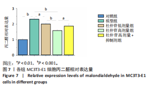
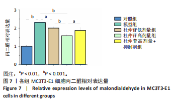
综合2.2.2和2.2.3的实验结果,在保证MC3T3-E1细胞生长活性的条件下,选择200 μmol/L杜仲苷干预48 h作为后续实验的最佳条件。 2.2.4 杜仲苷对氧化应激条件下MC3T3-E1细胞成骨能力的增强作用 (1)丙二醛水平:模型组MC3T3-E1细胞丙二醛水平较对照组显著升高(P < 0.001),提示氧化应激模型构建成功。杜仲苷低、高剂量组丙二醛水平较模型组显著降低(P < 0.01,P < 0.001),且杜仲苷高剂量组丙二醛水平显著低于杜仲苷低剂量组(P < 0.001)。杜仲苷高剂量+抑制剂组丙二醛水平较杜仲苷高剂量组显著升高(P < 0.01),结果表明杜仲苷可剂量依赖性降低氧化应激水平,且该作用可被Janus激酶2/信号转导与转录激活因子3通路抑制剂AG490阻断,见图7。"
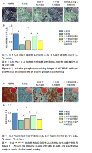
(2)成骨分化结果:碱性磷酸酶是成骨分化早期标志物,染色结果(图8A)显示:对照组染色较深,模型组染色明显变浅;杜仲苷低、高剂量组染色深度随剂量递增而加深,且杜仲苷高剂量组染色效果优于杜仲苷低剂量组;杜仲苷高剂量+抑制剂组染色较杜仲苷高剂量组浅。定量分析结果(图8B)表明:模型组碱性磷酸酶相对表达显著低于对照组(P < 0.001);杜仲苷低、高剂量组碱性磷酸酶相对表达均显著高于模型组(P < 0.001),且杜仲苷高剂量组显著高于杜仲苷低剂量组(P < 0.001);杜仲苷高剂量+抑制剂组碱性磷酸酶相对表达较杜仲苷高剂量组显著降低(P < 0.001),与杜仲苷低剂量组无统计学差异(P > 0.05)。上述结果提示,在氧化应激条件下,MC3T3-E1细胞的成骨分化受到抑制;杜仲苷能够以剂量依赖性的方式促进细胞成骨分化,而这一作用可能被Janus激酶2/信号转导与转录激活因子3通路抑制剂所阻断。 (3)成骨矿化结果:茜素红染色可反映成骨矿化能力。茜素红染色结果(图9A)显示:对照组矿化结节丰富,模型组显著减少;杜仲苷低、高剂量组矿化结节较模型组增多,且杜仲苷高剂量组矿化结节多于杜仲苷低剂量组;杜仲苷高剂量组+抑制剂组矿化结节少于杜仲苷高剂量组。定量结果(图9B)显示:模型组矿化结节形成显著少于对照组(P < 0.001);杜仲苷低、高剂量组矿化结节均多于模型组(P < 0.05,P < 0.001),且杜仲苷高剂量组矿化结节形成多于杜仲苷低剂量组(P < 0.01);杜仲苷高剂量+抑制剂组矿化结节形成较杜仲苷高剂量组显著减少(P < "
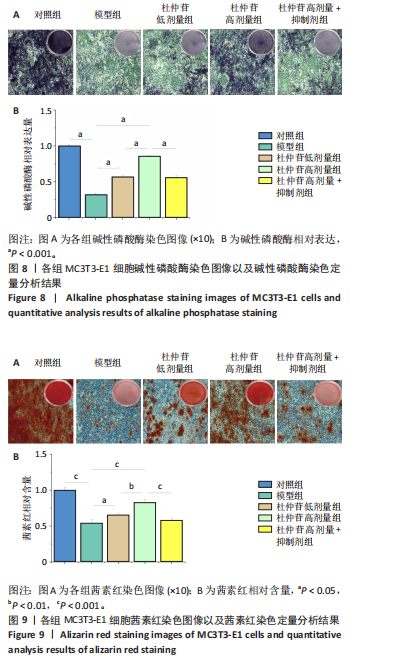
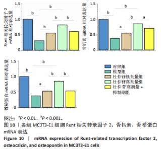
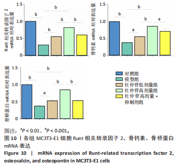
0.001),与杜仲苷低剂量组无差异(P > 0.05),表明杜仲苷可能呈剂量依赖性促进成骨矿化,且该作用可被抑制剂阻断。 (4)RT-qPCR检测结果:见图10。与对照组相比,模型组Runt相关转录因子2、骨钙素、骨桥蛋白 mRNA相对表达量均显著降低(P < 0.001),提示过氧化氢造模后抑制成骨分化相关基因表达。杜仲苷低、高剂量组上述基因表达水平均较模型组显著升高(P < 0.01, P < 0.001),且杜仲苷高剂量组显著高于杜仲苷低剂量组(P < 0.001),呈现剂量依赖性促基因表达趋势。当联合应用通路抑制剂AG490时,杜仲苷高剂量+抑制剂组的3种基因表达量较杜仲苷高剂量组显著下调( P < 0.01,P < 0.001)。综上,杜仲苷通过剂量依赖性上调 Runt相关转录因子2、骨钙素、骨桥蛋白基因表达促进成骨分化,该作用依赖特定信号通路,并可被Janus激酶2/信号转导与转录激活因子3通路抑制剂AG490阻断。"
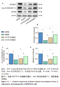
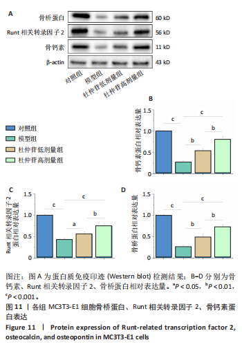
(5)Western blot 检测结果:成骨相关蛋白表达如图11所示。模型组骨钙素、Runt相关转录因子2、骨桥蛋白表达量均较对照组显著降低(P < 0.001);杜仲苷低、高剂量组上述蛋白表达较模型组显著升高(P < 0.05,P < 0.01,P < 0.001),且呈剂量依赖性,杜仲苷高剂量组上述蛋白表达高于杜仲苷低剂量组(P < 0.01)。"
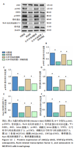
加入Janus激酶2/信号转导与转录激活因子3通路抑制剂AG490干预后,如图12所示:杜仲苷高剂量组骨钙素、Runt相关转录因子2、骨桥蛋白、磷酸化Janus激酶2、磷酸化信号转导与转录激活因子3蛋白表达明显高于模型组(P < 0.001),杜仲苷高剂量+抑制剂组骨钙素、Runt相关转录因子2、骨桥蛋白、磷酸化Janus激酶2、磷酸化信号转导与转录激活因子3蛋白表达较杜仲苷高剂量组明显降低(P < 0.001)。"
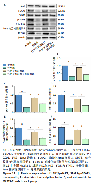

上述结果提示,杜仲苷可通过上调成骨相关蛋白表达及激活 Janus激酶2/信号转导与转录激活因子3 通路(促进磷酸化Janus激酶2、磷酸化信号转导与转录激活因子3表达)改善氧化应激诱导的成骨功能抑制,且该作用呈剂量依赖性;抑制剂可阻断杜仲苷的上述效应,提示杜仲苷可能通过激活Janus激酶2/信号转导与转录激活因子3通路参与调控氧化应激条件下MC3T3-E1细胞的成骨分化。"

| [1] ADEJUYIGBE B, KALLINI J, CHIOU D, et al. Osteoporosis: Molecular Pathology, Diagnostics, and Therapeutics. Int J Mol Sci. 2023;24(19):14583. [2] WU X, LIANG S, ZHU X, et al. CoQ10 suppression of oxidative stress and cell senescence increases bone mass in orchiectomized mice. Am J Transl Res. 2020;12(8):4314-4325. [3] KIMBALL JS, JOHNSON JP, CARLSON DA. Oxidative Stress and Osteoporosis. J Bone Joint Surg Am. 2021;103(15):1451-1461. [4] CHENNA S, KOOPMAN WJH, PREHN JHM, et al. Mechanisms and mathematical modeling of ROS production by the mitochondrial electron transport chain. Am J Physiol Cell Physiol. 2022;323(1):C69-C83. [5] IANTOMASI T, ROMAGNOLI C, PALMINI G, et al. Oxidative Stress and Inflammation in Osteoporosis: Molecular Mechanisms Involved and the Relationship with microRNAs. Int J Mol Sci. 2023;24(4):3772. [6] REICHMANN D, VOTH W, JAKOB U. Maintaining a Healthy Proteome during Oxidative Stress. Mol Cell. 2018;69(2):203-213. [7] 董子杰,浦飞飞,张慧浩,等.氧化应激反应在骨质疏松高危人群中的作用及机制研究进展[J].骨科,2025,16(1):86-90. [8] CHANDRA A, RAJAWAT J. Skeletal Aging and Osteoporosis: Mechanisms and Therapeutics. Int J Mol Sci. 2021;22(7):3553. [9] DA W, TAO L, ZHU Y. The Role of Osteoclast Energy Metabolism in the Occurrence and Development of Osteoporosis. Front Endocrinol (Lausanne). 2021;12:675385. [10] 王子扬,马旭东.成骨/破骨细胞轴对糖尿病骨代谢影响的研究进展[J]. 中国口腔种植学杂志,2025,30(2):176-179. [11] 邢可斌,裴飞,王虹焱,等.自主神经系与成骨细胞-破骨细胞通讯研究进展[J].神经损伤与功能重建,2026,21(2):117-120. [12] SEMICHEVA A, ERSOY U, VASILAKI A, et al. Defining the Most Potent Osteoinductive Culture Conditions for MC3T3-E1 Cells Reveals No Implication of Oxidative Stress or Energy Metabolism. Int J Mol Sci. 2024;25(8):4180. [13] UENAKA M, YAMASHITA E, KIKUTA J, et al. Osteoblast-derived vesicles induce a switch from bone-formation to bone-resorption in vivo. Nat Commun. 2022; 13(1):1066. [14] WANG H, LIANG J, WANG Y, et al. Exploring the effects of naringin on oxidative stress-impaired osteogenic differentiation via the Wnt/β-catenin and PI3K/Akt pathways. Sci Rep. 2024;14(1):14047. [15] 翁绳健,吴立忠,李炜明,等.肾虚与骨质疏松症关联性研究进展[J].中国中医骨伤科杂志,2018,26(12):85-88. [16] TAN M, LI Q, YANG B, et al. Insight of Chinese Herbal Medicine in Treating Osteoporosis: Achievements from 2013 to 2023. Am J Chin Med. 2024;52(5): 1303-1328. [17] WEN S, LIAO X, CHANG R, et al. Structures and biological activities of anti-osteoporotic drugs: An overview of promising small-molecule therapeutics for the treatment of osteoporosis. Bioorg Chem. 2025;162:108568. [18] ZENG X, GUO F, OUYANG D. A review of the pharmacology and toxicology of aucubin. Fitoterapia. 2020;140:104443. [19] MAI YX, LI ZP, PANG FX, et al. Aucubin Promotes Osteogenic Differentiation and Facilitates Bone Formation through the lncRNA-H19 Driven Wnt/β-Catenin Signaling Regulatory Axis. Stem Cells Int. 2024;2024:5388064. [20] RU J, LI P, WANG J, et al. TCMSP: a database of systems pharmacology for drug discovery from herbal medicines. J Cheminform. 2014;6:13. [21] TANG Y, LI M, WANG J, et al. CytoNCA: a cytoscape plugin for centrality analysis and evaluation of protein interaction networks. Biosystems. 2015;127:67-72. [22] NI Y, ZHANG H, ZHANG J, et al. Inhibition of JAK2 by AG490 promotes TNF-α-induced apoptosis by inhibiting autophagy in MC3T3-E1 cells. Pharmazie. 2020;75(6):255-260. [23] DENNIS G JR, SHERMAN BT, HOSACK DA, et al. DAVID: Database for Annotation, Visualization, and Integrated Discovery. Genome Biol. 2003;4(5):P3. [24] TANG D, CHEN M, HUANG X, et al. SRplot: A free online platform for data visualization and graphing. PLoS One. 2023;18(11):e0294236. [25] FÖGER-SAMWALD U, DOVJAK P, AZIZI-SEMRAD U, et al. Osteoporosis: Pathophysiology and therapeutic options. EXCLI J. 2020;19:1017-1037. [26] ALEJANDRO P, CONSTANTINESCU F. A Review of Osteoporosis in the Older Adult. Clin Geriatr Med. 2017;33(1):27-40. [27] 陈钡钡,李佳,谭文彬.内分泌代谢系统疾病与继发性骨质疏松症的研究新进展[J].中国全科医学,2025,28(17):2082-2091+2112. [28] JOHNSTON CB, DAGAR M. Osteoporosis in Older Adults. Med Clin North Am. 2020;104(5):873-884. [29] ZHANG C, LI H, LI J, et al. Oxidative stress: A common pathological state in a high-risk population for osteoporosis. Biomed Pharmacother. 2023;163:114834. [30] ZHOU W, LIU Y, SHEN J, et al. Melatonin Increases Bone Mass around the Prostheses of OVX Rats by Ameliorating Mitochondrial Oxidative Stress via the SIRT3/SOD2 Signaling Pathway. Oxid Med Cell Longev. 2019;2019:4019619. [31] YANG R, LI J, ZHANG J, et al. 17β-estradiol plays the anti-osteoporosis role via a novel ESR1-Keap1-Nrf2 axis-mediated stress response activation and Tmem119 upregulation. Free Radic Biol Med. 2023;195:231-244. [32] WANG Y, YU X, SUN F, et al. METTL14 Mediates Glut3 m6A methylation to improve osteogenesis under oxidative stress condition. Redox Rep. 2025;30(1):2435241. [33] SHEN Y, WANG H, XIE H, et al. l-arginine promotes angio-osteogenesis to enhance oxidative stress-inhibited bone formation by ameliorating mitophagy. J Orthop Translat. 2024;46:53-64. [34] CHEN B, YAN YL, LIU C, et al. Therapeutic effect of deferoxamine on iron overload-induced inhibition of osteogenesis in a zebrafish model. Calcif Tissue Int. 2014; 94(3):353-360. [35] CHEN W, ZHANG H, JIANG J, et al. Application of comprehensive pharmaceutical care program in identifying and addressing drug-related problems in hospitalized patients with osteoporosis. BMC Health Serv Res. 2022;22(1):1438. [36] YEH E, SAEEDIAN M, BADARACCO J. A comprehensive update on the cost-effectiveness of 10-year denosumab vs alendronate in postmenopausal women with osteoporosis in the United States. Arch Osteoporos. 2025;20(1):85. [37] 李斌,脱伟旺.中医药治疗骨质疏松症研究进展[J].云南中医中药杂志, 2025,46(7):95-100. [38] 黄凤林,张宝瑜,周洪波.《黄帝内经》中治疗骨质疏松经验[J].世界中医药, 2022,17(3):424-426. [39] 王青青,周海燕,周旭,等.经典名方中杜仲的本草考证[J].中国实验方剂学杂志,2023,29(19):88-96. [40] 向智,李焕杰,谌鑫阳,等.药食同源植物杜仲化学成分、药理作用及产品开发的研究概况[J].中国实验方剂学杂志,2024,30(2):190-202. [41] HUANG C, JIN H, ZHANG Y, et al. Eucommia ulmoides Oliv. and its bioactive compounds: therapeutic potential in bone diseases. Front Pharmacol. 2025; 16:1601537. [42] HAO B, YANG Z, LIU H, et al. Advances in flavonoid research: Sources, biological activities and challenges. Front Nutr. 2024; 46(4):2884. [43] LIGA S, PÉTER F, PAUL C. Flavonoids: Overview of Biosynthesis, Biological Activity and Therapeutic Potential. Plants. 2023;14:2732-2732. [44] MA L, MENG N, LIU B, et al. Quantitative Analysis and Stability Study on Iridoid Glycosides from Seed Meal of Eucommia ulmoides Oliver. Molecules. 2022;27(18):5924. [45] TANG LD, WANG JY, ZHANG Y, et al. Iridoid from Eucommia ulmoides Oliv. Exerts Antiarthritis Effects by Inhibiting the JAK2/STAT3 Signaling Pathway In Vivo and In Vitro. Evid Based Complement Alternat Med. 2023;2023:4167906. [46] HUANG J, JIANG H, WANG H, et al. Aucubin produces anti-osteoporotic effects under mechanical stretch stress and orthodontic tooth movement. Chem Biol Interact. 2024;393:110955. [47] LI YC, HAO JC, SHANG B, et al. Neuroprotective effects of aucubin on hydrogen peroxide-induced toxicity in human neuroblastoma SH-SY5Y cells via the Nrf2/HO-1 pathway. Phytomedicine. 2021;87:153577. [48] LIU T, LI Y, HU N. Aucubin Alleviates Chronic Obstructive Pulmonary Disease by Activating Nrf2/HO-1 Signaling Pathway. Cell Biochem Biophys. 2024;82(3): 2439-2454. [49] XIA B, DING J, LI Q, et al. Loganin protects against myocardial ischemia-reperfusion injury by modulating oxidative stress and cellular apoptosis via activation of JAK2/STAT3 signaling. Int J Cardiol. 2024;395:131426. [50] LI Y, ZHANG Y, ZHANG X, et al. Aucubin exerts anti-osteoporotic effects by promoting osteoblast differentiation. Aging (Albany NY). 2020;12(3):2226-2245. [51] WANG K, ZHOU C, LI L, et al. Aucubin promotes bone-fracture healing via the dual effects of anti-oxidative damage and enhancing osteoblastogenesis of hBM-MSCs. Stem Cell Res Ther. 2022;13(1):424. [52] IHLE JN, GILLILAND DG. Jak2: normal function and role in hematopoietic disorders. Curr Opin Genet Dev. 2007;17(1):8-14. [53] HUANG B, LANG X, LI X. The role of IL-6/JAK2/STAT3 signaling pathway in cancers. Front Oncol. 2022;12:1023177. [54] PENG D, HE X, REN B, et al. JAK2/STAT3/HMGCS2 signaling aggravates mitochondrial dysfunction and oxidative stress in hyperuricemia-induced cardiac dysfunction. Mol Med. 2025;31(1):184. [55] ZHANG M, ZHANG Y, PENG J, et al. Gastrodin against oxidative stress-inflammation crosstalk via inhibiting mtDNA/TLR9 and JAK2/STAT3 signaling to ameliorate ischemic stroke injury. Int Immunopharmacol. 2024;141:113012. [56] DAWALIBI A, ALOSAIMI AA, MOHAMMAD KS. Balancing the Scales: The Dual Role of Interleukins in Bone Metastatic Microenvironments. Int J Mol Sci. 2024;25(15):8163. [57] KHOSWANTO C. Role of matrix metalloproteinases in bone regeneration: Narrative review. J Oral Biol Craniofac Res. 2023;13(5):539-543. |
| [1] | Liu Wenlong, Dong Lei, Xiao Zhengzheng, Nie Yu. Finite element analysis of tibial prosthesis loosening after fixed-bearing unicompartmental knee arthroplasty for osteoporosis [J]. Chinese Journal of Tissue Engineering Research, 2026, 30(9): 2191-2198. |
| [2] | Chen Long, Wang Xiaozhen, Xi Jintao, Lu Qilin. Biomechanical performance of short-segment screw fixation combined with expandable polyetheretherketone vertebral body replacement in osteoporotic vertebrae [J]. Chinese Journal of Tissue Engineering Research, 2026, 30(9): 2226-2235. |
| [3] | Chen Huiting, Zeng Weiquan, Zhou Jianhong, Wang Jie, Zhuang Congying, Chen Peiyou, Liang Zeqian, Deng Weiming. Tail anchoring technique of vertebroplasty in treatment of osteoporotic vertebral compression fractures with intravertebral cleft: a finite element analysis [J]. Chinese Journal of Tissue Engineering Research, 2026, 30(9): 2145-2152. |
| [4] | Zeng Xuan, Weng Rui, Ye Shicheng, Tang Jiadong, Mo Ling, Li Wenchao. Two lumbar rotary manipulation techniques in treating lumbar disc herniation: a finite element analysis of biomechanical differences [J]. Chinese Journal of Tissue Engineering Research, 2026, 30(9): 2153-2161. |
| [5] | Cheng Qisheng, Julaiti·Maitirouzi, Xiao Yang, Zhang Chenwei, Paerhati·Rexiti. Finite element analysis of novel variable-diameter screws in modified cortical bone trajectory of lumbar vertebrae [J]. Chinese Journal of Tissue Engineering Research, 2026, 30(9): 2162-2171. |
| [6] | Yang Xuetao, Zhu Menghan, Zhang Chenxi, Sun Yimin, Ye Ling. Applications and limitations of antioxidant nanomaterials in oral cavity [J]. Chinese Journal of Tissue Engineering Research, 2026, 30(8): 2044-2053. |
| [7] |
Dong Chunyang, Zhou Tianen, Mo Mengxue, Lyu Wenquan, Gao Ming, Zhu Ruikai, Gao Zhiwei.
Action mechanism of metformin combined with Eomecon chionantha Hance dressing in treatment of deep second-degree burn wounds#br#
#br#
[J]. Chinese Journal of Tissue Engineering Research, 2026, 30(8): 2001-2013.
|
| [8] | Wu Yanting, Li Yu, Liao Jinfeng. Magnesium oxide nanoparticles regulate osteogenesis- and angiogenesis-related gene expressions to promote bone defect healing [J]. Chinese Journal of Tissue Engineering Research, 2026, 30(8): 1885-1895. |
| [9] | Hu Xiongke, Liu Shaohua, Tan Qian, Liu Kun, Zhu Guanghui. Shikonin intervention with bone marrow mesenchymal stem cells improves microstructure of femur in aged mice [J]. Chinese Journal of Tissue Engineering Research, 2026, 30(7): 1609-1615. |
| [10] | Li Zhenyu, Zhang Siming, Bai Jiaxiang, Zhu Chen. Osthole improves osteogenic differentiation function of bone marrow mesenchymal stem cells under high-glucose conditions [J]. Chinese Journal of Tissue Engineering Research, 2026, 30(7): 1641-1648. |
| [11] | Jin Dongsheng, Zhao Zhanghong, Zhu Ziyin, Zhang Sen, Sun Zuyan, Deng Jiang. Effects of icariin-loaded microsphere-three-dimensional scaffold on osteogenic differentiation of rabbit bone marrow mesenchymal stem cells [J]. Chinese Journal of Tissue Engineering Research, 2026, 30(7): 1658-1668. |
| [12] | Zou Yulian, Chen Chaopei, Huang Haixia, Lan Yuyan, Liu Min, Huang Ting. Resveratrol promotes osteogenic differentiation of bone marrow mesenchymal stem cells in an inflammatory microenvironment [J]. Chinese Journal of Tissue Engineering Research, 2026, 30(7): 1669-1678. |
| [13] | Chen Yulin, He Yingying, Hu Kai, Chen Zhifan, Nie Sha Meng Yanhui, Li Runzhen, Zhang Xiaoduo , Li Yuxi, Tang Yaoping. Effect and mechanism of exosome-like vesicles derived from Trichosanthes kirilowii Maxim. in preventing and treating atherosclerosis [J]. Chinese Journal of Tissue Engineering Research, 2026, 30(7): 1768-1781. |
| [14] | Liu Anting, Lu Jiangtao, Zhang Wenjie, He Ling, Tang Zongsheng, Chen Xiaoling. Regulation of AMP-activated protein kinase by platelet lysate inhibits cadmium-induced neuronal apoptosis [J]. Chinese Journal of Tissue Engineering Research, 2026, 30(7): 1800-1807. |
| [15] | Zhou Sirui, Xu Yukun, Zhao Kewei. Ideas and methods of anti-melanogenesis of Angelica dahurica extracellular vesicles [J]. Chinese Journal of Tissue Engineering Research, 2026, 30(7): 1747-1754. |
| Viewed | ||||||
|
Full text |
|
|||||
|
Abstract |
|
|||||