Chinese Journal of Tissue Engineering Research ›› 2026, Vol. 30 ›› Issue (33): 8676-8686.doi: 10.12307/2026.482
Previous Articles Next Articles
Bioinformatics analysis of cuproptosis in periodontitis and verification in a periodontitis rat model
Qin Yufeng1, 2, Feng Xiaoyun3, Gao Hongli2, Xiong Lin2, Zhang Yuehan1, Chen Helin1, 2
- 1College of Stomatology, Guizhou Medical University, Guiyang 550004, Guizhou Province, China; 2Affiliated Stomatological Hospital of Guizhou Medical University, Guiyang 550004, Guizhou Province, China; 3Shizhen College, Guizhou University of Traditional Chinese Medicine, Guiyang 550000, Guizhou Province, China
-
Received:2025-10-29Revised:2026-03-12Online:2026-11-28Published:2026-06-13 -
Contact:Chen Helin, PhD, Associate chief physician, Master’s supervisor, College of Stomatology, Guizhou Medical University, Guiyang 550004, Guizhou Province, China; Affiliated Stomatological Hospital of Guizhou Medical University, Guiyang 550004, Guizhou Province, China -
About author:Qin Yufeng, MS candidate, College of Stomatology, Guizhou Medical University, Guiyang 550004, Guizhou Province, China; Affiliated Stomatological Hospital of Guizhou Medical University, Guiyang 550004, Guizhou Province, China -
Supported by:the National Natural Science Foundation of China (Regional Program), No. 82160186 (to CHL); Plan Project of Guizhou Provincial Science and Technology Department, No. Qiankehejichu-ZK[2021]General 433 (to CHL)

CLC Number:
Cite this article
Qin Yufeng, Feng Xiaoyun, Gao Hongli, Xiong Lin, Zhang Yuehan, Chen Helin. Bioinformatics analysis of cuproptosis in periodontitis and verification in a periodontitis rat model [J]. Chinese Journal of Tissue Engineering Research, 2026, 30(33): 8676-8686.
share this article
Add to citation manager EndNote|Reference Manager|ProCite|BibTeX|RefWorks
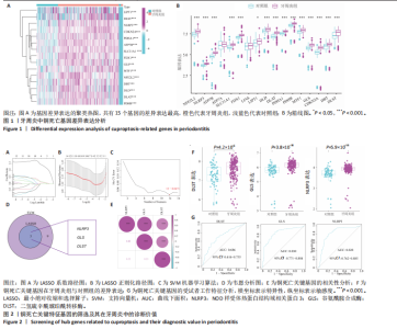
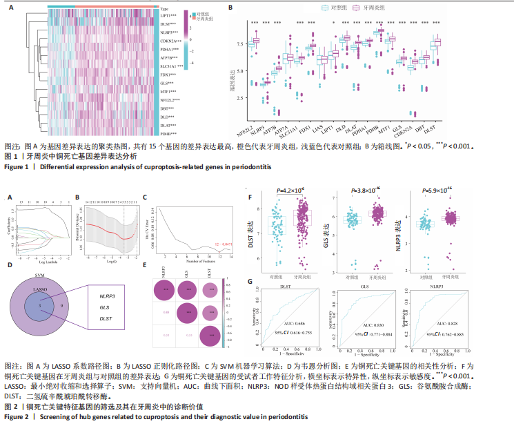
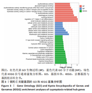
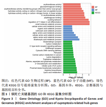
2.1 牙周炎与铜死亡相关差异基因的鉴定 在牙周炎发展过程中,与铜死亡相关的关键分子通路可能会发生改变。为全面探究铜死亡在牙周炎中的调控作用,采用箱线图和聚类热图对17个铜死亡相关基因进行差异分析,旨在找出对牙周炎发生发展有显著影响的关键特征基因。首先,对两个数据集中259个样本的17个铜死亡相关基因进行差异表达分析,结果显示,牙周炎组有15个基因(DLST、DBT、CDKN2A、GLS、MTF1、PDHB、PDHA1、DLAT、DLD、LIPT1、FDX1、SLC31A1、ATP7B、NLRP3、NFE2L2)表达水平显著上调(P < 0.001),见图1A,B,表明这些差异表达基因上调可能在牙周炎的发展中起关键作用。 2.2 关键特征基因的鉴定及其在牙周炎中的诊断意义 通过差异表达分析鉴定出15个铜死亡相关基因。利用最小绝对收缩和选择算子回归、支持向量机-递归特征消除机器学习算法和韦恩分析,进一步筛选出3个特征基因:NOD样受体热蛋白结构域相关蛋白3(NLRP3)、二氢硫辛酰琥珀酰转移酶(DLST)、谷氨酰胺合成酶(GLS),见图2A-D。根据相关性分析结果显示,3个基因间存在显著的正相关关系,其中NOD样受体热蛋白结构域相关蛋白3和谷氨酰胺合成酶的相关性最强(R=0.83),见图2E,这些基因在牙周炎组织中显著上调,见图2F。受试者工作特征曲线分析显示,NOD样受体热蛋白结构域相关蛋白3(曲线下面积=0.828)和谷氨酰胺合成酶(曲线下面积=0.830)具有较好的诊断价值,二氢硫辛酰琥珀酰转移酶(曲线下面积=0.686)具有中等诊断价值,提示它们可能成为牙周炎的诊断生物标志物,见图2G。 2.3 GO功能和KEGG富集分析 为全面阐明关键特征基因的功能机制,对与铜死亡相关的关键特征基因进行了GO功能和KEGG富集分析。基于GO功能分析可知,生物过程主要涉及小分子分解代谢过程、调节Th2细胞的分化以及与泌乳相关的过程;分子功能主要与二羧酸代谢过程、α-氨基酸分解代谢过程和细胞氨基酸分解代谢过程密切相关。KEGG富集分析显示,二氢硫辛酰琥珀酰转移酶基因主要参与硫辛酸代谢和柠檬酸循环通路(三羧酸循环);谷氨酰胺合成酶基因主要参与精氨酸生物合成和近端小管碳酸氢盐再生通路;NOD样受体热蛋白结构域相关蛋白3 基因主要参与百日咳信号通路。基于以上结果提示NOD样受体热蛋白结构域相关蛋白3、二氢硫辛酰琥珀酰转移酶和谷氨酰胺合成酶可能通过上述通路促进牙周炎的发展,见图3。 2.4 与3 个关键特征基因相关的转录因子富集分析 研究结果显示,与二氢硫辛酰琥珀酰转移酶、谷氨酰胺合成酶和NOD样受体热蛋白结构域相关蛋白3相关的转录因子主要分布于脑、血液、睾丸和肌肉等多个组织中,见图4A,这些转录因子主要"
"
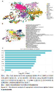
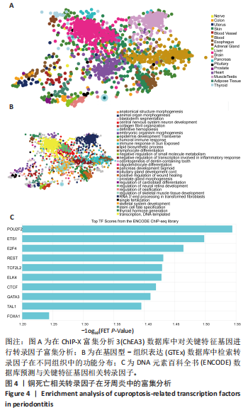
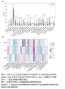
参与转录调控、DNA模板的生成以及动物器官形态的塑造,见图4B。进一步的分析结果显示,在选定的前10个转录因子中,POU结构域2类转录因子2(POU2F2)的频率富集分数最高,见图4C。因此,与二氢硫辛酰琥珀酰转移酶、谷氨酰胺合成酶和NOD样受体热蛋白结构域相关蛋白3相关的转录因子在生物学功能上具有一致性,且POU结构域2类转录因子2与关键特征基因的关联最为密切。 2.5 免疫细胞浸润分析 通过免疫细胞浸润分析进一步阐明参与牙周炎进展的免疫细胞,结果显示在牙周炎组与正常组之间有8种免疫细胞存在显著差异(P < 0.05),包括浆细胞、巨噬细胞、静息NK细胞、 幼稚T细胞、CD4+T细胞、M2巨噬细胞、M0巨噬细胞和CD8+T 细胞,见图5A。此外,浆细胞在这些免疫细胞中的表达水平最高。免疫相关性分析结果显示,NOD样受体热蛋白结构域相关蛋白3、二氢硫辛酰琥珀酰转移酶和谷氨酰胺合成酶的表达与浆细胞的浸润水平呈显著正相关,而与嗜酸性粒细胞和幼稚CD4+T细胞呈负相关(P < 0.05),见图5B。"
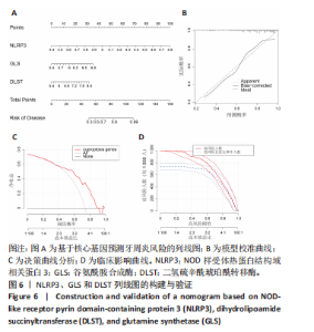
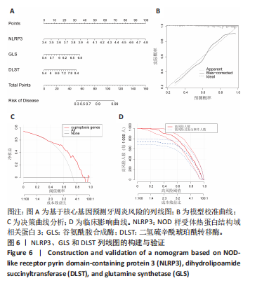
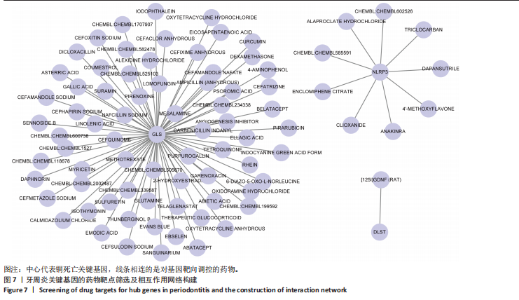
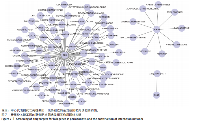
2.6 牙周炎风险评估关键特征基因列线图的构建与验证 基于NOD样受体热蛋白结构域相关蛋白3、谷氨酰胺合成酶和二氢硫辛酰琥珀酰转移酶构建的列线图,将基因表达转化为分数以量化牙周炎风险,并实现个体风险分层,见图6A。校准曲线证实预测风险与实际风险相一致,见图6B。决策曲线显示其净收益优于全民治疗或不治疗策略,见图6C。临床影响曲线有助于明确最佳阈值,见图6D。经验证,这种基于铜死亡基因的列线图具有良好的准确性和适用性,为个性化评估和精准医疗提供支持。 2.7 基因-药物网络构建 研究表明谷氨酰胺合成酶基因受到chembl、羟考酮、盐酸土霉素、头孢西丁钠等多种药物的靶向调控,见图7,提示这些药物可能通过对谷氨酰胺合成酶发挥调控作用,从而影响牙周炎的病理进展。此外,NOD样受体热蛋白结构域相关蛋白3和二氢硫辛酰琥珀酰转移酶基因在该网络中也有各自相关的药物,见图7,但其具体调控模式及对牙周炎的影响仍有待进一步探究。 2.8 牙周炎中与关键基因相关的亚型 通过共识聚类方法将牙周炎分为2个亚型。采用累积分布函数明确达到最大稳定性的k"
"
"
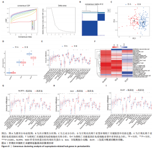
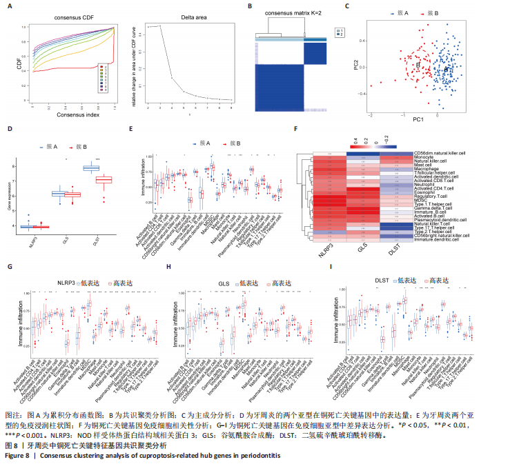
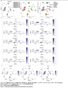
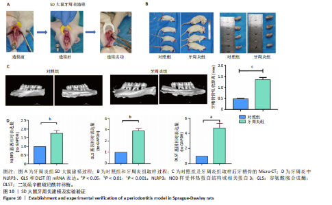
值。基于Delta面积图显示,当k=2时曲线下面积值显著下降,见图8A,B。主成分分析表明,簇A的聚类效果优于簇B,见图8C。此外,簇A相较于簇B,二氢硫辛酰琥珀酰转移酶的表达水平上调,而谷氨酰胺合成酶和NOD样受体热蛋白结构域相关蛋白3的表达则无差异,见图8D。免疫浸润的单样本基因集富集分析揭示了髓系来源抑制细胞在簇A和簇B中的表达水平均达到最高,并存在显著统计学差异(P < 0.05),见图8E。相关性分析显示,谷氨酰胺合成酶(R=0.33)、NOD样受体热蛋白结构域相关蛋白3(R=0.48)、二氢硫辛酰琥珀酰转移酶(R=0.13)与髓系来源抑制细胞呈正相关,见图8F,并在髓系来源抑制细胞中高表达,见图8G-I。 2.9 单细胞分析 基于统一流形逼近与投影图可视化分析细胞类型和基因表达模式,见图9A。免疫细胞聚类显示,IGHG1等标记基因在对应细胞群中高表达,见图9B。NOD样受体热蛋白结构域相关蛋白3、谷氨酰胺合成酶和二氢硫辛酰琥珀酰转移酶在中性粒细胞、浆细胞、上皮细胞和成纤维细胞中表达,见图9C。 2.10 SD大鼠牙周炎构建及体内实验验证 SD大鼠牙周炎模型的造模过程,见图10A;对照组与牙周炎组取样过程,见图10B。Micro-CT扫描图像显示,与对照组比较,牙周炎组上颌第二磨牙牙槽骨高度明显降低。釉牙骨质界至牙槽嵴顶的测量结果显示,牙周炎组的牙槽骨丧失量较对照组明显增加(P < 0.001),见图10C。这些结果表明牙周炎模型的建立是成功的,RT-qPCR结果显示,与对照组相比,牙周炎组牙龈组织中的NOD样受体热蛋白结构域相关蛋白3、二氢硫辛酰琥珀酰转移酶、谷氨酰胺合成酶 mRNA表达显著上升(P < 0.05),见图10D。"
"

"

| [1] AHMAD P, SLOTS J, SIQUEIRA WL. Serum cytokines in periodontal diseases. Periodontol 2000. 2025;98(1):138-180. [2] FU Y, XU T, GUO M, et al. Identification of disulfidptosis- and ferroptosis-related transcripts in periodontitis by bioinformatics analysis and experimental validation. Front Genet. 2024;15:1402663. [3] TRINDADE D, CARVALHO R, MACHADO V, et al. Prevalence of periodontitis in dentate people between 2011 and 2020: A systematic review and meta-analysis of epidemiological studies. J Clin Periodontol. 2023;50(5):604-626. [4] FISCHER RG, LIRA JUNIOR R, RETAMAL-VALDES B, et al. Periodontal disease and its impact on general health in Latin America. Section V: Treatment of periodontitis. Braz Oral Res. 2020;34(supp1 1):e026. [5] HIJRYANA M, MACDOUGALL M, ARIANI N, et al. Impact of Periodontal Disease on the Quality of Life of Older People in Indonesia: A Qualitative Study. JDR Clin Trans Res. 2022;7(4):360-370. [6] CHAPPLE ILC, HIRSCHFELD J, COCKWELL P, et al. Interplay between periodontitis and chronic kidney disease. Nat Rev Nephrol. 2025;21(4):226-240. [7] YE X, WANG Y, TIAN Y, et al. Metformin alleviates junctional epithelium senescence via the AMPK/SIRT1/autophagy pathway in periodontitis induced by hyperglycemia. Heliyon. 2024;10(6):e27478. [8] MICHAUD DS, KELSEY KT, PAPATHANASIOU E, et al. Periodontal disease and risk of all cancers among male never smokers: an updated analysis of the Health Professionals Follow-up Study. Ann Oncol. 2016;27(5):941-947. [9] HAJISHENGALLIS G, LAMONT RJ. Beyond the red complex and into more complexity: the polymicrobial synergy and dysbiosis (PSD) model of periodontal disease etiology. Mol Oral Microbiol. 2012;27(6):409-419. [10] ROBERTS FA, DARVEAU RP. Microbial protection and virulence in periodontal tissue as a function of polymicrobial communities: symbiosis and dysbiosis. Periodontol 2000. 2015;69(1):18-27. [11] XIE J, YANG Y, GAO Y, et al. Cuproptosis: mechanisms and links with cancers. Mol Cancer. 2023;22(1):46. [12] FAN X, CHEN H, JIANG F, et al. Comprehensive analysis of cuproptosis-related genes in immune infiltration in ischemic stroke. Front Neurol. 2023;13:1077178. [13] TSVETKOV P, COY S, PETROVA B, et al. Copper induces cell death by targeting lipoylated TCA cycle proteins. Science. 2022;375(6586):1254-1261. [14] LI W, SHANG Q, YANG D, et al. Abnormal Micronutrient Intake Is Associated with the Risk of Periodontitis: A Dose-response Association Study Based on NHANES 2009-2014. Nutrients. 2022;14(12):2466. [15] PEREIRA TC, CAMPOS MM, BOGO MR. Copper toxicology, oxidative stress and inflammation using zebrafish as experimental model. J Appl Toxicol. 2016;36(7): 876-885. [16] HERMAN M, GOLASIK M, PIEKOSZEWSKI W, et al. Essential and Toxic Metals in Oral Fluid-a Potential Role in the Diagnosis of Periodontal Diseases. Biol Trace Elem Res. 2016;173(2):275-282. [17] ZHENG T, LU F, WU P, et al. Ferroptosis and cuproptosis in periodontitis: recent biological insights and therapeutic advances. Front Immunol. 2025;16:1526961. [18] SUNDARAM G, RAMAKRISHNAN T, PARTHASARATHY H, et al. Evaluation of Micronutrient (Zinc, Magnesium, and Copper) Levels in Serum and Glycemic Status after Nonsurgical Periodontal Therapy in Type 2 Diabetic Patients with Chronic Periodontitis. Contemp Clin Dent. 2017;8(1):26-32. [19] TANG D, CHEN X, KROEMER G. Cuproptosis: a copper-triggered modality of mitochondrial cell death. Cell Res. 2022;32(5):417-418. [20] ZHANG L, TSAI IC, NI Z, et al. Copper Chelation Therapy Attenuates Periodontitis Inflammation through the Cuproptosis/Autophagy/Lysosome Axis. Int J Mol Sci. 2024;25(11):5890. [21] LIU S, GE J, CHU Y, et al. Identification of hub cuproptosis related genes and immune cell infiltration characteristics in periodontitis. Front Immunol. 2023; 14:1164667. [22] YANG Y, XIAO D, LIANG J, et al. Research progress on the expression and prognostic value of copper death related genes in liver cancer. Panminerva Med. 2024;66(3):326-328. [23] LUO W, DU C, HUANG H, et al. The Role of Macrophage Death in Periodontitis: A Review. Inflammation. 2024;47(6):1889-1901. [24] ZHU L, ZHU J, WANG Q, et al. Global, regional, and national burden of 34 cancer groups across 204 countries and territories, 1990-2021, and projections to 2050: a systematic analysis of the Global Burden of Disease Study 2021. Front Oncol. 2025;15:1660125. [25] 朱莎莎,田卫东,郭淑娟.不同类型程序性细胞死亡在牙周炎中的研究进展[J].口腔医学,2024,44(8):624-629. [26] 徐楚蓉,苟格,吴世琪,等.铜代谢障碍和铜死亡在神经退行性疾病和肿瘤发生中的作用[J].生理科学进展,2024,55(5):458-464. [27] LU Z, DING L, ZHANG S, et al. Bioinformatics analysis of copper death gene in diabetic immune infiltration. Medicine (Baltimore). 2023;102(39):e35241. [28] WANG J, LI J, LIU J, et al. Interplay of Ferroptosis and Cuproptosis in Cancer: Dissecting Metal-Driven Mechanisms for Therapeutic Potentials. Cancers (Basel). 2024;16(3):512. [29] SUN X, MAO Y, DAI P, et al. Mitochondrial dysfunction is involved in the aggravation of periodontitis by diabetes. J Clin Periodontol. 2017;44(5):463-471. [30] LOU QM, LAI FF, LI JW, et al. Mechanisms of cuproptosis and its relevance to distinct diseases. Apoptosis. 2024;29(7-8):981-1006. [31] SONG G, PENG G, ZHANG J, et al. Uncovering the potential role of oxidative stress in the development of periodontitis and establishing a stable diagnostic model via combining single-cell and machine learning analysis. Front Immunol. 2023;14:1181467. [32] ROMANO F, CASTIBLANCO A, SPADOTTO F, et al. ICP-Mass-Spectrometry Ionic Profile of Whole Saliva in Patients with Untreated and Treated Periodontitis. Biomedicines. 2020;8(9):354. [33] MASUMOTO J. “The inflammasomes”. Nihon Rinsho Meneki Gakkai Kaishi. 2011;34(5):346-354. [34] CHEN Y, YE X, ESCAMES G, et al. The NLRP3 inflammasome: contributions to inflammation-related diseases. Cell Mol Biol Lett. 2023;28(1):51. [35] 朱博文,陈立妹,郭竹玲.NLRP3炎性小体在牙周疾病中的研究进展[J].国际口腔医学杂志,2019,46(4):450-455. [36] ISOLA G, POLIZZI A, SANTONOCITO S, et al. Periodontitis activates the NLRP3 inflammasome in serum and saliva. J Periodontol. 2022;93(1):135-145. [37] CAO Y, ZHOU B, ZHANG W, et al. DHA Alleviates Excessive Copper-Induced Hepatocyte Injury by Inhibiting Pyroptosis via the ROS/NLRP3/NF-κB Pathway. Biol Trace Elem Res. 2026;204(3): 1483-1495. [38] DE LOS SANTOS-JIMÉNEZ J, CAMPOS-SANDOVAL JA, ALONSO FJ, et al. GLS and GLS2 Glutaminase Isoenzymes in the Antioxidant System of Cancer Cells. Antioxidants (Basel). 2024;13(6):745. [39] SUZUKI S, TANAKA T, POYUROVSKY MV, et al. Phosphate-activated glutaminase (GLS2), a p53-inducible regulator of glutamine metabolism and reactive oxygen species. Proc Natl Acad Sci U S A. 2010;107(16):7461-7466. [40] MARTÍN-RUFIÁN M, NASCIMENTO-GOMES R, HIGUERO A, et al. Both GLS silencing and GLS2 overexpression synergize with oxidative stress against proliferation of glioma cells. J Mol Med (Berl). 2014;92(3):277-290. [41] ANDERSON NM, QIN X, FINAN JM, et al. Metabolic Enzyme DLST Promotes Tumor Aggression and Reveals a Vulnerability to OXPHOS Inhibition in High-Risk Neuroblastoma. Cancer Res. 2021;81(17):4417-4430. [42] DUMONT M, HO DJ, CALINGASAN NY, et al. Mitochondrial dihydrolipoyl succinyltransferase deficiency accelerates amyloid pathology and memory deficit in a transgenic mouse model of amyloid deposition. Free Radic Biol Med. 2009;47(7):1019-1027. [43] MACLEAN A, LEGENDRE F, APPANNA VD. The tricarboxylic acid (TCA) cycle: a malleable metabolic network to counter cellular stress. Crit Rev Biochem Mol Biol. 2023;58(1):81-97. [44] ZHAO G, SUN H, ZHANG T, et al. Copper induce zebrafish retinal developmental defects via triggering stresses and apoptosis. Cell Commun Signal. 2020;18(1):45. [45] TARDITO S, BUSSOLATI O, GACCIOLI F, et al. Non-apoptotic programmed cell death induced by a copper(II) complex in human fibrosarcoma cells. Histochem Cell Biol. 2006;126(4):473-482. [46] COX M, DEKKER N, BOELENS R, et al. NMR studies of the POU-specific DNA-binding domain of Oct-1: sequential 1H and 15N assignments and secondary structure. Biochemistry. 1993;32(23):6032-6040. [47] YANG R, WANG M, ZHANG G, et al. POU2F2 regulates glycolytic reprogramming and glioblastoma progression via PDPK1-dependent activation of PI3K/AKT/mTOR pathway. Cell Death Dis. 2021;12(5):433. [48] MAHANONDA R, CHAMPAIBOON C, SUBBALEKHA K, et al. Human Memory B Cells in Healthy Gingiva, Gingivitis, and Periodontitis. J Immunol. 2016;197(3):715-725. [49] QIU W, GUO R, YU H, et al. Single-cell atlas of human gingiva unveils a NETs-related neutrophil subpopulation regulating periodontal immunity. J Adv Res. 2025;72:287-301. [50] DYAB A, EMNEGARD A, WÄNMAN M, et al. Human gingival fibroblasts are a source of B cell-activating factor during periodontal inflammation. J Periodontol. 2024;95(7):673-681. |
| [1] | Lyu Guoqing, Aizimaitijiang·Rouzi, Xiong Daohai. Irisin inhibits ferroptosis in human articular chondrocytes: roles and mechanisms [J]. Chinese Journal of Tissue Engineering Research, 2026, 30(6): 1359-1367. |
| [2] | Liu Kexin, , Hao Kaimin, Zhuang Wenyue, , Li Zhengyi. Autophagy-related gene expression in pulmonary fibrosis models: bioinformatic analysis and experimental validation [J]. Chinese Journal of Tissue Engineering Research, 2026, 30(5): 1129-1138. |
| [3] | Lian Yong, Xie Zhixing, Lu Jiaxin, Pan Zhaofeng, Chen Qingzhen, Shao Min. Mechanism of high glucose-induced osteoblast cuproptosis [J]. Chinese Journal of Tissue Engineering Research, 2026, 30(36): 9393-9401. |
| [4] | Wang Huaijing, Guo Jinrong, Wan Dongping, Mei Qijie, Yuan Jingzhao, Xu Wenfei, Zeng Chao, Zheng Haijun, Yuan Changshen, Duan Kan. Identification of antimicrobial peptides as key therapeutic targets for necrosis by sodium overload in osteoarthritis using multiple machine learning approaches: cytological validation [J]. Chinese Journal of Tissue Engineering Research, 2026, 30(36): 9413-9422. |
| [5] | Cao Shan, Wang Yanxi, Duan Kaixuan, Qi Xiang, Wang Yuhan. Xiao Ban Tong Mai Fang regulates autophagy via targeting miR-126-3p: bioinformatics analysis for prevention and treatment of atherosclerosis [J]. Chinese Journal of Tissue Engineering Research, 2026, 30(35): 9355-9364. |
| [6] | Yang Huaqun, Abudouainijiang·Abulimiti, Wang Fazheng, Maimaitishawutiaji·Maimaiti, Li Simi, Muhetaer·Maimaitirexiati. Weighted gene co-expression network analysis combined with machine learning identifies autophagy and senescence signature genes in osteoarthritis chondrocytes [J]. Chinese Journal of Tissue Engineering Research, 2026, 30(34): 8889-8898. |
| [7] | Yuan Jingjing, Zhang Xiaomin, Du Pengyang, Wang Weifeng. Mazdutide improves cognitive function in APP/PS1/Tau triple transgenic mice [J]. Chinese Journal of Tissue Engineering Research, 2026, 30(34): 8962-8969. |
| [8] | Mu Wenbo, Wang Yan, Cheng Yao. Association between peri-implantitis and tuberculosis: sample analysis based on GEO and GWAS databases [J]. Chinese Journal of Tissue Engineering Research, 2026, 30(33): 8822-8828. |
| [9] | Zhou Dayu, Dong Na, Geng Ganshu, Zhang Xue. Long non-coding RNA-TNFRSF13C improves periodontal pathomorphology in rat models of periodontitis via target regulation [J]. Chinese Journal of Tissue Engineering Research, 2026, 30(33): 8662-8668. |
| [10] | Yang Yunhong, Guo Lihua, Tang Han, Lin Lvping, Kuang Hongjun, Zhao Hong. Proteomic analysis of the mechanism of moxibustion intervention in a rat model of atopic dermatitis [J]. Chinese Journal of Tissue Engineering Research, 2026, 30(29): 7581-7591. |
| [11] | Li Zhengpeng, Shao Weigang, Zeng Hao, Xiang Kelin, Zhang Botao, Zou Shunyi, Chen Sheng, Qi Wen. Osteoarthritis characteristic genes and prediction of targeted food-medicine homology traditional Chinese medicine: bioinformatics analysis and kinetic simulation [J]. Chinese Journal of Tissue Engineering Research, 2026, 30(29): 7739-7748. |
| [12] | Yan Yuge, Wang Yanxi, Qi Xiang, Cao Shan, Zou Xiaoyan, Liu Yujuan. Screening biomarkers for premature ovarian insufficiency based on cellular senescence and endoplasmic reticulum stress with experimental validation [J]. Chinese Journal of Tissue Engineering Research, 2026, 30(28): 7447-7455. |
| [13] | Zhang Kaijing, Li Chunnian, Li Yizhuo, Xu Shifang, Liu Xinyue. A network meta-analysis of therapeutic effects of different bone repair materials on apical bone defects [J]. Chinese Journal of Tissue Engineering Research, 2026, 30(26): 6946-6951. |
| [14] | Lyu Tianyang, Li Ning, Huang Shuo, Liu Changkui, Guo Yayuan, Hu Kaijin. Preparation and biocompatibility of odanacatib microspheres-gel composite sustained-release carrier [J]. Chinese Journal of Tissue Engineering Research, 2026, 30(26): 6840-6848. |
| [15] | Li Yiwei, Luo Zongming, Rong Yifa, Jiang Kai, Zhang Jiahao, Lu Bowen, Li Gang. Druggable gene and single cell analyses reveal potential therapeutic targets for osteoporosis [J]. Chinese Journal of Tissue Engineering Research, 2026, 30(25): 6654-6660. |
| Viewed | ||||||
|
Full text |
|
|||||
|
Abstract |
|
|||||