Chinese Journal of Tissue Engineering Research ›› 2026, Vol. 30 ›› Issue (33): 8822-8828.doi: 10.12307/2026.378
Previous Articles Next Articles
Association between peri-implantitis and tuberculosis: sample analysis based on GEO and GWAS databases
Mu Wenbo1, Wang Yan1, Cheng Yao2
- 1Department of Prosthodontics, Harbin Stomatological Hospital, Harbin 150090, Heilongjiang Province, China; 2Department of Laboratory Medicine, Beidahuang Group General Hospital, Harbin 150088, Heilongjiang Province, China
-
Received:2025-06-06Revised:2025-09-18Online:2026-11-28Published:2026-06-18 -
Contact:Cheng Yao, MS, Chief technician, Department of Laboratory Medicine, Beidahuang Group General Hospital, Harbin 150088, Heilongjiang Province, China -
About author:Mu Wenbo, MS, Attending physician, Department of Prosthodontics, Harbin Stomatological Hospital, Harbin 150090, Heilongjiang Province, China

CLC Number:
Cite this article
Mu Wenbo, Wang Yan, Cheng Yao. Association between peri-implantitis and tuberculosis: sample analysis based on GEO and GWAS databases[J]. Chinese Journal of Tissue Engineering Research, 2026, 30(33): 8822-8828.
share this article
Add to citation manager EndNote|Reference Manager|ProCite|BibTeX|RefWorks
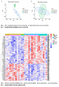
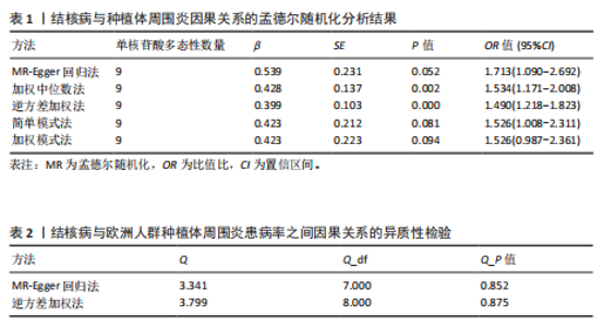
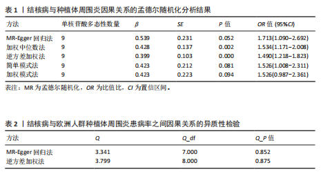
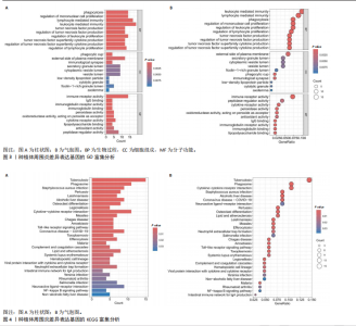
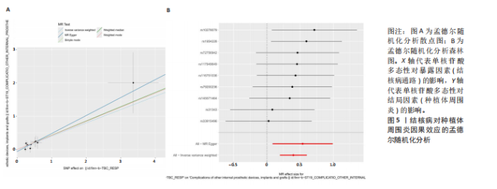
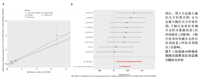
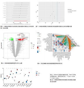
2.1 生物信息学分析结果 将GSE33774、GSE57631和GSE178351整合,批次矫正后批次效应已消除,见图1。将标准化的表达矩阵进行差异基因分析,其中P < 0.05共有2 482个基因,P < 0.05且|log2FC|>0.3共有204个基因。为排除具有统计学差异但表达量变化不大的基因,此次研究采用后者限定差异基因标准。训练组差异分析结果见图2,通过热图直观展示了关键基因差异表达情况。 对训练组进行富集分析后发现,种植体周围炎在GO数据库中主要与白细胞介导免疫、淋巴细胞介导免疫、单核细胞增殖的调控和吞噬作用等生物学功能显著相关,主要与免疫受体活性、肽酶调节剂活性和细胞因子受体活性等分子功能显著相关,见图3。在KEGG的通路富集中,种植体周围炎主要与结核病、吞噬体、细胞因子-细胞因子受体相互作用、金黄色葡萄球菌感染、酒精性肝病、冠状病毒COVID-19等相关,见图4,其中结核病通路差异基因包括TGFB1、CD14、CASP10、FCER1G、IL1B、FCGR2A、FCGR3A、C3、IL10RA、CTSS、IL10RB、CASP3、TLR4、CORO1A、FCGR2B。 2.2 孟德尔随机化分析结果 经过筛选,结核病共纳入9个单核苷酸多态性作为工具变量,分析结果见表1。孟德尔随机化发现,β均> 0,说明肺结核这一暴露因素属于危险因素;OR均> 1,说明结核病的发生与欧洲人群中种植体周围炎的患病率存在因果关系,并且结核病的发生可能是种植体周围炎发展的危险因素。将逆方差加权法作为主要分析方法,结果显示结核病的遗传变异与种植体周围炎患病风险增高相关(β=0.399,OR=1.490,95%CI=1.218-1.823,P < 0.001),见表1,图5。表明结核病患者发生种植体周围炎的风险比较无结核病患者增加了49%。加权中位数法作为补充方法获得了相似结果(β=0.428,OR=1.534,95%CI=1.171-2.008,P≈0.002,见表1,图5。 为了解用不同孟德尔随机化方法估计结核病与种植体周围炎关系的差异,紧接着进行了异质性分析,结果显示,MR-Egger回归法中用于异质性检测的Q=3.341,P=0.852,而在逆方差加权方法中,Q=3.799,P=0.875,说明该研究不存在异质性,见表2。水平多效性检验(MR Egger截距法)结果为Egger截距=-0.059,P=0.520,说明该研究不存在水平多效性。留一法敏感性分析森林图显示,单独去除某个单核苷酸多态性不会对孟德尔随机化的结果产生影响,见图6。观察漏斗图显示基本呈对称状态,见图7,表明单一的单核苷酸多态性作为工具变量时受到的偏倚较小。 2.3 结核病通路差异表达基因与免疫细胞之间的关系 提取结核病通路下的差异基因可以看到大多数差异基因在疾病组中是上调的,与孟德尔随机化结果一致,见图8。对15个差异基因和18个免疫细胞浸润程度的相关性分析发现,多数差异基因均与免疫细胞浸润相关,如图9。单核细胞与CD8+ T细胞之间、活化NK细胞与滤泡辅助性T细胞之间、单核细胞与调节T细胞之间、调节T细胞与CD8+ T细胞之间、中性粒细胞与活化肥大细胞之间具有显著的正调控关系,静息NK细胞与调节T细胞之间、静息NK细胞与活化NK细胞之间、静息NK细胞与单核细胞之间 、滤泡辅助性T细胞与静息CD4+记忆T细胞之间具有显著的负调控关系,多个通路基因共同与静息NK细胞、活化肥大细胞和中性粒细胞之间存在显著的正调控关系,另有多个通路基因共同与CD8+ T细胞、调节T细胞、活化NK细胞和单核细胞之间存在显著的负调控关系。 2.4 验证组差异分析结果 对验证组进行差异分析,结果显示CASP10、FCER1G、IL1B、FCGR2A、FCGR3A、C3、CTSS、CASP3、TLR4、CORO1A在实验组中的表达量显著高于对照组(P < 0.01),IL10RA、FCGR2B在实验组中的表达量显著高于对照组(P < 0.05),见图10。大多数通路基因在实验组中的表达量是上调的,这与孟德尔分析结果一致。"

"
"

"
"

| [1] SCHWARZ F, DERKS J, MONJE A, et al. Peri-implantitis. J Clin Periodontol. 2018; 45(Suppl 20):246-266. [2] HERRERA D, BERGLUNDH T, SCHWARZ F, et al. Prevention and treatment of peri-implant diseases-The EFP S3 level clinical practice guideline. J Clin Periodontol. 2023;50(Suppl 26):4-76. [3] SMEETS R, HENNINGSEN A, JUNG O, et al. Definition, etiology, prevention and treatment of peri-implantitis--a review. Head Face Med. 2014;10:34. [4] RAMS TE, DEGENER JE, VAN WINKELHOFF AJ. Antibiotic resistance in human peri-implantitis microbiota. Clin Oral Implants Res. 2014;25(1):82-90. [5] BESSA LJ, EGAS C, PIRES C, et al. Linking peri-implantitis to microbiome changes in affected implants, healthy implants, and saliva: a cross-sectional pilot study. Front Cell Infect Microbiol. 2025;15:1543100. [6] RENVERT S, POLYZOIS I, CLAFFEY N. Surgical therapy for the control of peri-implantitis. Clin Oral Implants Res. 2012;23 (Suppl 6): 84-94. [7] ITO H, MONOBE K, OKUBO S, et al. Identification of Novel Antimicrobial Compounds Targeting Mycobacterium tuberculosis S-Adenosyl-L-Homocysteine Hydrolase Using Dual Hierarchical In Silico Structure-Based Drug Screening. Molecules. 2024;29(6):1303. [8] Health Organization (WHO). Global tuberculosis report 2024 (2024). [9] LANGE C, AABY P, BEHR MA, et al. 100 years of Mycobacterium bovis bacille Calmette-Guérin. Lancet Infect Dis. 2022;22(1):2-12. [10] PINA P, LEMOS C, DESOUSA S. A buccal mucosa ulcer as the first sign of tuberculosis. J Oral Maxillofac Pathol. 2022;26(3):399-403. [11] MOMBELLI A, DECAILLET F. The characteristics of biofilms in peri-implant disease. J Clin Periodontol. 2011;38(11):203-213. [12] BELIBASAKIS GN. Microbiological and immuno-pathological aspects of peri-implant diseases. Arch Oral Biol. 2014;59(1):66-72. [13] 李志杰,王少果,李跃烘,等.高通量测序研究种植体周围炎龈下微生物多样性[J].四川大学学报(医学版),2015,46(4):568-572. [14] DASILVA ES, FERES M, FIGUEIREDO LC, et al. Microbiological diversity of peri-implantitis biofilm by Sanger sequencing[J]. Clin Oral Implants Res. 2014;25(10):1192-1199. [15] CASADO PL, OTAZU IB, BALDUINO A, et al. Identification of periodontal pathogens in healthy periimplant sites. Implant Dent. 2011;20(3):226-235. [16] 文言,王宇蓝.菌斑生物膜与种植体周围炎相关研究进展[J].口腔疾病防治,2024,32(9):730-736. [17] IUȘAN S, LUCACIU O, PETRESCU N, et al. The Main Bacterial Communities Identified in the Sites Affected by Periimplantitis: A Systematic Review. Microorganisms. 2022;10(6):1232. [18] SAHRMANN P, GILLI F, WIEDEMEIER DB, et al. The Microbiome of Peri-Implantitis: A Systematic Review and Meta-Analysis. Microorganisms. 2020;8(5):661. [19] 唐立新.结核病的病因学分析[J].中国中医药现代远程教育,2011,9(8):95. [20] GAO X, ZHOU J, SUN X, et al. Diversity analysis of subgingival microbial bacteria in peri-implantitis in Uygur population. Medicine (Baltimore). 2018;97(5):9774. [21] MOHANASUNDARAM A, GOHIL NV, ETEKOCHAY MO, et al. Mycobacterium tuberculosis : a new hitchhiker in the etiopathogenesis of periodontitis. Int J Surg. 2024;110(6):3606-3616. [22] RASTOGI T, CHOWDHARY Z, KRISHNA MK, et al. Prevalence of periodontitis in patients with pulmonary disease: A cross-sectional survey in the industrial district of India. J Indian Soc Periodontol. 2019;23(3):269-274. [23] JAVALI MA, PATIL V, AYESHA H. Periodontal disease as the initial oral manifestation of abdominal tuberculosis. Dent Res J (Isfahan). 2012;9(5):634-637. [24] JAIN P, JAIN I. Oral Manifestations of Tuberculosis: Step towards Early Diagnosis. J Clin Diagn Res. 2014;8(12):18-21. [25] DEBATA T, MOHAPATRA A, SAHU SR, et al. Rare case of extrapulmonary tuberculosis masquerading as an intraoral sinus in association with a nonhealing extraction socket in a teenage girl-”A case report with Review of literature”. J Oral Maxillofac Pathol. 2024;28(2):301-306. [26] JAIN S, VIPIN B, KHURANA P. Gingival tuberculosis. J Indian Soc Periodontol. 2009; 13(2):106-108. [27] WEIR MR, THORNTON GF. Extrapulmonary tuberculosis. Experience of a community hospital and review of the literature. Am J Med. 1985;79(4):467-478. [28] LONČAR BRZAK B, HORVAT ALEKSIJEVIĆ L, VINDIŠ E, et al. Osteonecrosis of the Jaw. Dent J (Basel). 2023;11(1):23. [29] EBENEZER J, SAMUEL R, MATHEW GC, et al. Primary oral tuberculosis: report of two cases. Indian J Dent Res. 2006;17(1):41-44. [30] CAPODIFERRO S, LIMONGELLI L, FAVIA G. Oral and Maxillo-Facial Manifestations of Systemic Diseases: An Overview. Medicina (Kaunas). 2021;57(3):271. [31] HAMID R, SHARMA P, SHEIKH RA, et al. Primary oral tuberculosis: A case series of a rare disease. J Oral Maxillofac Pathol. 2020;24(2):332-338. [32] 冯林林,冯彦华.固有免疫细胞在结核分枝杆菌感染中的作用[J].微生物学免疫学进展, 2024,52(5):92-97. [33] FORTUNE SM, SOLACHE A, JAEGER A, et al. Mycobacterium tuberculosis inhibits macrophage responses to IFN-gamma through myeloid differentiation factor 88-dependent and -independent mechanisms. J Immunol. 2004; 172(10):6272-6280. [34] 张贺龙,付琳,刘晓宁,等.TNF-α基因多态性及红细胞沉降率、降钙素原水平对老年骶髂关节结核患者手术预后的影响[J].中国老年学杂志,2025,45(8):1869-1873. [35] RAIMONDO A, BALESTRINO A, LEMBO S. PDE4 Inhibition Reduced Osteoclast Differentiation in Psoriatic Patients. Life (Basel). 2025;15(3):467. [36] QIAN Y, YU Y, YANG F, et al. Jingui Shenqi Wan alleviates bone loss induced by primary osteoporosis by inhibiting osteoblast pyroptosis. J Orthop Surg Res. 2025;20(1):141. [37] WOJDASIEWICZ P, PONIATOWSKI A, SZUKIEWICZ D. The role of inflammatory and anti-inflammatory cytokines in the pathogenesis of osteoarthritis. Mediators Inflamm. 2014;2014:561459. [38] SARAPULTSEV A, GUSEV E, KOMELKOVA M, et al. JAK-STAT signaling in inflammation and stress-related diseases: implications for therapeutic interventions. Mol Biomed. 2023;4(1):40. [39] 尹昭懿,李效宇,王子璇,等.免疫反应在种植体周围炎中的作用研究进展[J]. 现代口腔医学杂志,2022,36(1):46-50. [40] DUTZAN N, KAJIKAWA T, ABUSLEME L, et al. A dysbiotic microbiome triggers TH17 cells to mediate oral mucosal immunopathology in mice and humans. Sci Transl Med. 2018;10(463):0797. [41] FRANCO C, PATRICIA HR, TIMO S, et al. Matrix Metalloproteinases as Regulators of Periodontal Inflammation. Int J Mol Sci. 2017;18(2):440. [42] JIANG N, AN J, YANG K, et al. NLRP3 inflammasome: a new target for prevention and control of osteoporosis? Front Endocrinol. 2021;12:752546. [43] TSUKASAKI M, TAKAYANAGI H. Osteoimmunology: evolving concepts in bone-immune interactions in health and disease[J]. Nat Rev Immunol. 2019; 19(10):626-642. [44] GHIGHI M, LLORENS A, BAROUKH B, et al. Differences between inflammatory and catabolic mediators of peri-implantitis and periodontitis lesions following initial mechanical therapy: An exploratory study. J Periodontal Res. 2018; 53(1):29-39. [45] ESPÉLI M, SMITH KG, CLATWORTHY MR. FcγRIIB and autoimmunity. Immunol Rev. 2016;269(1): 194-211. [46] WEI S, LING D, ZHONG J, et al. Elk1 enhances inflammatory cell infiltration and exacerbates acute lung injury/acute respiratory distress syndrome by suppressing Fcgr2b transcription. Mol Med. 2024;30(1):53. [47] TAKEGAHARA N, KIM H, CHOI Y. Unraveling the intricacies of osteoclast differentiation and maturation: insight into novel therapeutic strategies for bone-destructive diseases. Exp Mol Med. 2024;56(2):264-272. [48] REN F, ZHENG S, LUO H, et al. Fibroblast derived C3 promotes the progression of experimental periodontitis through macrophage M1 polarization and osteoclast differentiation. Int J Oral Sci. 2025;17(1):30. [49] FERNANDES MH, GOMES PS. Bone Cells Dynamics during Peri-Implantitis: a Theoretical Analysis. J Oral Maxillofac Res. 2016;7(3):6. |
| [1] | Lyu Guoqing, Aizimaitijiang·Rouzi, Xiong Daohai. Irisin inhibits ferroptosis in human articular chondrocytes: roles and mechanisms [J]. Chinese Journal of Tissue Engineering Research, 2026, 30(6): 1359-1367. |
| [2] | Wu Zhilin, , He Qin, Wang Pingxi, Shi Xian, Yuan Song, Zhang Jun, Wang Hao . DYRK2: a novel therapeutic target for rheumatoid arthritis combined with osteoporosis based on East Asian and European populations [J]. Chinese Journal of Tissue Engineering Research, 2026, 30(6): 1569-1579. |
| [3] | Liu Hongtao, Wu Xin, Jiang Xinyu, Sha Fei, An Qi, Li Gaobiao. Causal relationship between age-related macular degeneration and deep vein thrombosis: analysis based on genome-wide association study data [J]. Chinese Journal of Tissue Engineering Research, 2026, 30(6): 1602-1608. |
| [4] | Guo Ying, Tian Feng, Wang Chunfang. Potential drug targets for the treatment of rheumatoid arthritis: large sample analysis from European databases [J]. Chinese Journal of Tissue Engineering Research, 2026, 30(6): 1549-1557. |
| [5] | Liu Kexin, , Hao Kaimin, Zhuang Wenyue, , Li Zhengyi. Autophagy-related gene expression in pulmonary fibrosis models: bioinformatic analysis and experimental validation [J]. Chinese Journal of Tissue Engineering Research, 2026, 30(5): 1129-1138. |
| [6] | Gao Zengjie, , Pu Xiang, Li Lailai, Chai Yihui, Huang Hua, Qin Yu. Increased risk of osteoporotic pathological fractures associated with sterol esters: evidence from IEU-GWAS and FinnGen databases [J]. Chinese Journal of Tissue Engineering Research, 2026, 30(5): 1302-1310. |
| [7] | Liu Fengzhi, Dong Yuna, Tian Wenyi, Wang Chunlei, Liang Xiaodong, Bao Lin. Gene-predicted associations between 731 immune cell phenotypes and rheumatoid arthritis [J]. Chinese Journal of Tissue Engineering Research, 2026, 30(5): 1311-1319. |
| [8] | Zhang Cuicui, Chen Huanyu, Yu Qiao, Huang Yuxuan, Yao Gengzhen, Zou Xu. Relationship between plasma proteins and pulmonary arterial hypertension and potential therapeutic targets [J]. Chinese Journal of Tissue Engineering Research, 2026, 30(5): 1331-1340. |
| [9] | Zeng Hao, Sun Pengcheng, Chai Yuan, Huang Yourong, Zhang Chi, Zhang Xiaoyun. Association between thyroid function and osteoporosis: genome-wide data analysis of European populations [J]. Chinese Journal of Tissue Engineering Research, 2026, 30(4): 1019-1027. |
| [10] | Rong Xiangbin, , Zheng Haibo, Mo Xueshen, Hou Kun, Zeng Ping, . Plasma metabolites, immune cells, and hip osteoarthritis: causal inference based on GWAS data from European populations [J]. Chinese Journal of Tissue Engineering Research, 2026, 30(4): 1028-1035. |
| [11] | He Qiwang, , , Chen Bo, Liang Fuchao, Kang Zewei, Zhou Yuan, Ji Anxu, Tang Xialin, . Relationship between Alzheimer’s disease and sarcopenia and body mass index: analysis of GWAS datasets for European populations [J]. Chinese Journal of Tissue Engineering Research, 2026, 30(4): 1036-1046. |
| [12] | Ding Yu, Chen Jingwen, Chen Xiuyan, Shi Huimin, Yang Yudie, Zhou Meiqi, Cui Shuai, . Circulating inflammatory proteins and myocardial hypertrophy: large sample analysis of European populations from GWAS Catalog and FinnGen databases [J]. Chinese Journal of Tissue Engineering Research, 2026, 30(4): 1047-1057. |
| [13] | Wang Huaijing, Guo Jinrong, Wan Dongping, Mei Qijie, Yuan Jingzhao, Xu Wenfei, Zeng Chao, Zheng Haijun, Yuan Changshen, Duan Kan. Identification of antimicrobial peptides as key therapeutic targets for necrosis by sodium overload in osteoarthritis using multiple machine learning approaches: cytological validation [J]. Chinese Journal of Tissue Engineering Research, 2026, 30(36): 9413-9422. |
| [14] | Wang Xiaoxuan, Xiao Lu, Miao Changhong, Yuan Weijie, Guo Dengzhou, Yuan Ziwei. Causal association study between resting-state brain network functional and structural connectivity and autoimmune diseases [J]. Chinese Journal of Tissue Engineering Research, 2026, 30(36): 9640-9648. |
| [15] | Cao Shan, Wang Yanxi, Duan Kaixuan, Qi Xiang, Wang Yuhan. Xiao Ban Tong Mai Fang regulates autophagy via targeting miR-126-3p: bioinformatics analysis for prevention and treatment of atherosclerosis [J]. Chinese Journal of Tissue Engineering Research, 2026, 30(35): 9355-9364. |
| Viewed | ||||||
|
Full text |
|
|||||
|
Abstract |
|
|||||