Chinese Journal of Tissue Engineering Research ›› 2024, Vol. 28 ›› Issue (22): 3539-3547.doi: 10.12307/2024.522
Previous Articles Next Articles
Application of strontium polyphosphate with both radiopaque and osteogenic functions in calcium phosphate cement
Tang Ziniu1, 2, Chu Fengcheng1, 2, Wu Kang1, 2, Zhang Lin1, 2, Bai Yanjie3, Lin Xiao1, 2, Yang Huilin1, 2, Zhou Huan4, Liu Huiling1, 2, Yang Lei1, 2, 4
- 1First Affiliated Hospital of Soochow University, Suzhou 215006, Jiangsu Province, China; 2Institute of Orthopedics, Soochow University, Suzhou 215006, Jiangsu Province, China, 3Department of Chemical Engineering; 4School of Health Sciences and Biomedical Engineering, Hebei University of Technology, Tianjin 300131, China
-
Received:2023-08-21Accepted:2023-10-12Online:2024-08-08Published:2024-01-20 -
Contact:Liu Huiling, Master, Senior experimentalist, First Affiliated Hospital of Soochow University, Suzhou 215006, Jiangsu Province, China; Institute of Orthopedics, Soochow University, Suzhou 215006, Jiangsu Province, China Yang Lei, PhD, Professor, Doctoral supervisor, First Affiliated Hospital of Soochow University, Suzhou 215006, Jiangsu Province, China; Institute of Orthopedics, Soochow University, Suzhou 215006, Jiangsu Province, China; School of Health Sciences and Biomedical Engineering, Hebei University of Technology, Tianjin 300131, China -
About author:Tang Ziniu, Master, First Affiliated Hospital of Soochow University, Suzhou 215006, Jiangsu Province, China; Institute of Orthopedics, Soochow University, Suzhou 215006, Jiangsu Province, China -
Supported by:National Key Research and Development Project, No. 2020YFC1107401 (to YL); National Natural Science Foundation of China, No. 82025025 (to YL); National Natural Science Foundation of China, No. 81802155 (to LHL); National Natural Science Foundation of China, No. 32171321 (to LX); Yunnan Academician Workstations, No. 202205AF150025 (to YL); National Clinical Research Center for Orthopedics, Sports Medicine & Rehabilitation, No. 2021-NCRC-CXJJ-ZH-17 (to YL); Suzhou Key Laboratory of Orthopedics, No. SZS2022017 (to YHL)
CLC Number:
Cite this article
Tang Ziniu, Chu Fengcheng, Wu Kang, Zhang Lin, Bai Yanjie, Lin Xiao, Yang Huilin, Zhou Huan, Liu Huiling, Yang Lei. Application of strontium polyphosphate with both radiopaque and osteogenic functions in calcium phosphate cement[J]. Chinese Journal of Tissue Engineering Research, 2024, 28(22): 3539-3547.
share this article
Add to citation manager EndNote|Reference Manager|ProCite|BibTeX|RefWorks
2.1 SrPP及骨水泥的显影性能 从图2A,B可以看出,皮质骨与SrPP的亮度接近,均显著强于多聚磷酸钠;对于骨水泥而言,CPC亮度较低,CPS的亮度进一步降低,而20%SrPP-CPN的亮度明显提高,与20%BaSO4-CPN的亮度接近,表明20%SrPP-CPN的显影性能较强,且显影能力与添加20%BaSO4骨水泥相似。显影灰度值定量分析显示,皮质骨、SrPP、多聚磷酸钠、CPC、CPS、20%SrPP-CPN、20%BaSO4-CPN的对比值分别为0.74±0.01,0.79±0.01,0.24±0.04,0.78±0.01,0.75±0.01,0.85±0.01,0.84±0.01,见图2C,D。结果证实,20%SrPP-CPN的显影性能可以满足临床应用需求。"

2.2 骨水泥理化性能检测结果 2.2.1 SrPP对骨水泥力学性能与固化时间的影响 各组骨水泥力学性能与固化时间检测结果见表2。20%SrPP-CPN的抗压强度高于CPC、CPS(P < 0.05),前期实验得知20%BaSO4-CPN的抗压强度为(46.19±1.76) MPa[6],20%SrPP-CPN与20%BaSO4-CPN的抗压强度比较差异无显著性意义(P > 0.05)。固液混合后,CPC发生自固化,由于固化速度过快,难以准确测试固化时间;当加入预糊化淀粉后,CPC的固化时间得到延长,但固化时间仍然难以满足手术过程中的操作需要;添加SrPP显著延长了骨水泥固化时间。"

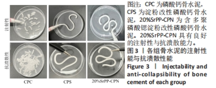
2.2.2 SrPP对骨水泥注射性能与抗溃散性能的影响 骨水泥材料的注射性能是其应用于临床的前提。如图3所示,CPC在推注过程中难以保持连续性,呈现自发节段性断裂,并且易固液分离难以推出,推注效果差;CPS可以保持连续稳定推出,但阻力仍较大,推注效果可;添加了SrPP的骨水泥可以稳定连续推出,推注阻力小,注射效果佳。分别将骨水泥推注到去离子水中,观察骨水泥的溃散效果,发现CPC呈现出不连续节段,并伴有不同程度的溃散;CPS与20%SrPP-CPN均未发生断裂,骨水泥保持稳定连续,表明CPS与SrPP-CPN具有较强的抗溃散能力。"

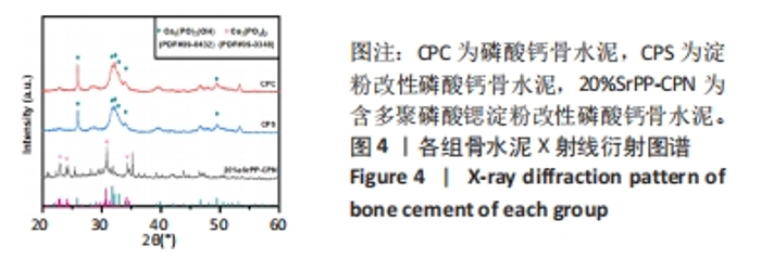
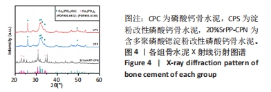
2.2.3 SrPP对骨水泥相成分的影响 通过X射线衍射进一步分析骨水泥的相成分,如图4所示。骨水泥固化后,CPC和CPS位于2θ衍射角32.3°,47°和49°处产生了明显衍射峰,其属于羟基磷灰石的特征峰,表明CPC和CPS在固液混合后发生了水化反应;而20%SrPP-CPN的X射线衍射图仅能观察到磷酸钙衍射峰。以上结果证明SrPP的加入会影响骨水泥的水化反应,20%SrPP-CPN将不再转化为羟基磷灰石。"
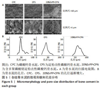
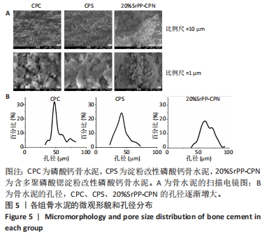
2.2.4 SrPP对骨水泥微观结构的影响 各组骨水泥的微观结构见图5A。CPC横截面可以观察到明显的针尖状晶体结构,添加预糊化淀粉的CPS横截面中同样观察到明显的针尖状晶体结构,结合X射线衍射分析可以确定此晶体为羟基磷灰石,说明CPC经水化反应后生成了羟基磷灰石,并且淀粉的添加并未影响羟基磷灰石的生成[5]。而20%SrPP-CPN的微观结构发生显著改变,未观察到针尖状晶体结构,出现较多微孔结构[28]。骨水泥孔径统计分析结果显示,添加SrPP后骨水泥的孔径进一步增大,见图5B,这将有助于细胞对骨水泥的黏附以及新生钙结节的沉积。"

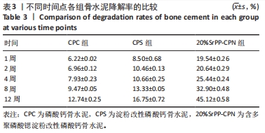
2.2.5 SrPP对骨水泥降解性能及离子释放的影响 各组骨水泥的降解率见表3。随着时间的延长,各组骨水泥的降解率逐渐提高,其中CPC最为稳定,在各个时间段的降解率最低,12周后降解率仅为(12.74±0.25)%;CPS的降解性能得到改善,12周后降解率达到(16.75±0.72)%;20%SrPP-CPN的降解性能在各个时间段得到进一步提升,12周后的降解率达到(45.12±0.58)%。上述结果证明SrPP的加入会加快CPC的降解。"
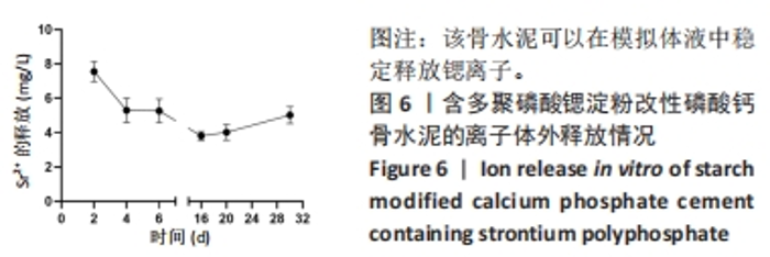
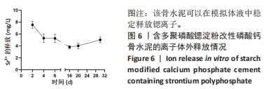
电感耦合等离子检测结果显示,CPC和CPS浸提液中未能检测到Sr2+的存在,20%SrPP-CPN浸提液中能检测到Sr2+,且其浓度保持相对稳定。如图6所示,20%SrPP-CPN在第2,4,6天释放的Sr2+浓度分别为(7.55±0.59),(5.30±0.69),(5.26±0.69) mg/L,在第16,20,30天释放的Sr2+浓度分别为(3.81±0.24),(4.02±0.46),(5.02±0.49) mg/L,表明20%SrPP-CPN可以在模拟体液环境中稳定释放锶离子。"
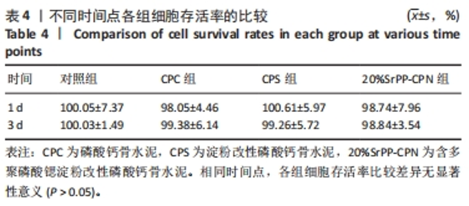
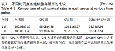
2.3 骨水泥生物相容性检测结果 2.3.1 SrPP对骨水泥调节细胞增殖的影响 CCK-8实验采用不同骨水泥浸提液培养细胞时,相同时间点下各组测得的吸光度值比较差异无显著性意义(P > 0.05),见表4。证明20%SrPP-CPN具有良好的生物相容性,SrPP并未对骨水泥生物相容性产生影响。"
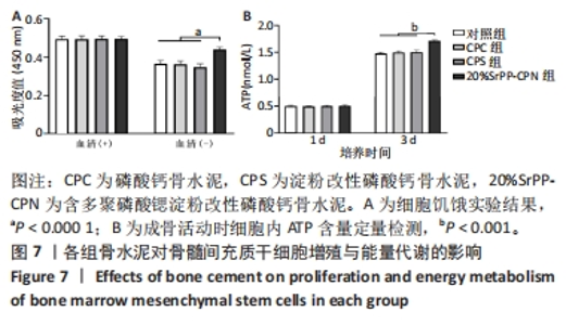
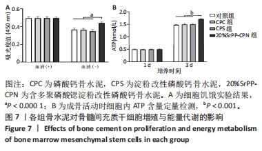
通过饥饿实验进一步验证20%SrPP-CPN浸提液对细胞增殖的影响。培养48 h后,添加胎牛血清时,各组细胞吸光度值比较差异无显著性意义(P > 0.05),表明体积分数10%胎牛血清的供能效果可以满足细胞正常生长;未添加胎牛血清时,各组细胞吸光度值均低于添加胎牛血清时对应组别,并且20%SrPP-CPN组细胞吸光度值高于其他3组(P < 0.000 1),见图7A。实验结果证明,20%SrPP-CPN浸提液能够促进骨髓间充质干细胞的增殖。 2.3.2 SrPP对骨水泥调节细胞能量代谢的影响 培养第1天,对照组、CPC组、CPS组、20%SrPP-CPN组细胞内ATP浓度分别为(0.50±0.02),(0.50±0.01),(0.51±0.01),(0.51±0.02) nmol/L,4组间比较差异无显著性意义(P > 0.05);培养第3天,对照组、CPC组、CPS组、20%SrPP-CPN组细胞内ATP浓度分别为(1.48±0.02),(1.50±0.02),(1.50±0.04),(1.71±0.02) nmol/L,20%SrPP-CPN组高于其他3组(P < 0.001),见图7B。"
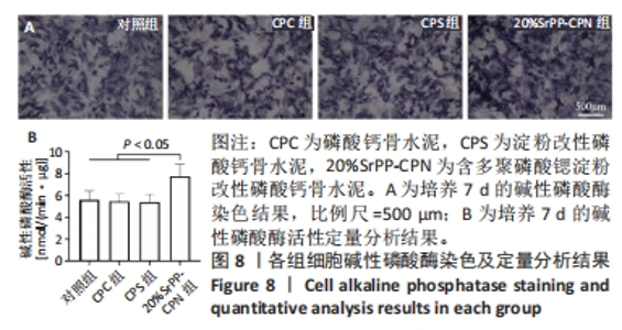
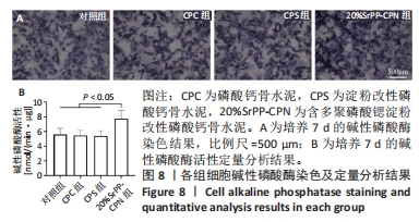
2.4 SrPP对骨水泥体外促进成骨分化的作用 2.4.1 碱性磷酸酶染色结果 从图8A可以观察到各组细胞均呈现阳性染色,证明成骨诱导培养基产生了成骨效能,CPC组、CPS组细胞阳性染色相较于对照组增强的原因可能是浸提液中Ca2+和PO43-浓度升高的作用[23],20%SrPP-CPN组细胞阳性染色增强更加明显,表明SrPP的加入增强了骨水泥促进骨髓间充质干细胞碱性磷酸酶表达的能力。 2.4.2 碱性磷酸酶活性检测结果 成骨诱导培养7 d时,对照组、CPC组、CPS组、20%SrPP-CPN组碱性磷酸酶活性分别为(5.60±0.82),(5.46±0.68),(5.40±0.70),(7.74±1.11) nmol/(min?μg),20%SrPP-CPN组高于其他3组(P < 0.05),见图8B。上述结果表明,SrPP的加入增强了CPC对骨髓间充质干细胞成骨蛋白碱性磷酸酶的诱导表达能力。"
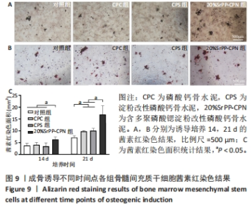
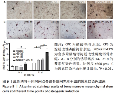
2.4.3 茜素红染色结果 如图9A,B所示,成骨诱导14 d,各组细胞外基质中均观察到茜素红染色;成骨诱导21 d,各组茜素红染色逐渐增多,相较于未添加SrPP组,20%SrPP-CPN组成骨诱导14,21 d的茜素红染色更加明显。Image J统计分析结果显示,成骨诱导14 d,对照组、CPC组、CPS组、20%SrPP-CPN组茜素红染色面积分别为(3.86±0.86),(4.17±0.93),(3.53±1.38),(6.38±1.05) mm2,20%SrPP-CPN组大于其他3组(P < 0.05);成骨诱导21 d,对照组、CPC组、CPS组、20%SrPP-CPN组茜素红染色面积分别为(7.18±0.78),(9.83±0.35),(10.11±0.95),(16.90±3.75) mm2,对照组小于CPC组、CPS组(P < 0.05),CPC组、CPS组小于20%SrPP-CPN组(P < 0.05),见图9C。表明SrPP的添加进一步增强了细胞外基质中的钙沉积。"
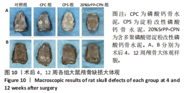
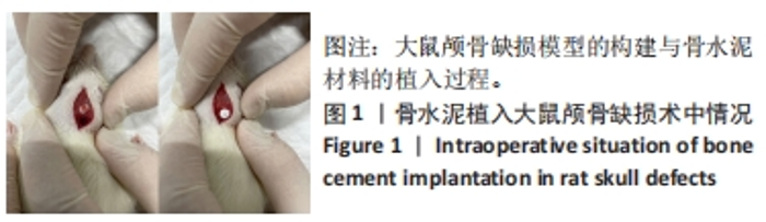
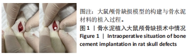
2.5 SrPP对骨水泥体内促进成骨分化的作用 2.5.1 实验动物数量分析 饲养全程观察大鼠状态均正常,无化脓、溃烂、死亡等现象发生,24只大鼠全部进入结果分析。 2.5.2 各组大鼠颅骨大体观 见图10。"

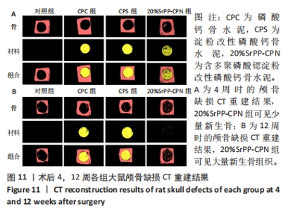
术后4周,对照组颅骨缺损处无明显变化,CPC组与CPS组材料结构完整,20%SrPP-CPN组材料发生降解,缺损部位均未能观察到明显新生骨结构。术后12周,对照组颅骨缺损处仍无明显变化,证明5 mm缺损为大鼠颅骨临界缺损;CPC组材料结构仍保持完整,未发生明显降解,新生骨不明显;CPS组材料出现少量降解,新生骨同样不明显;20%SrPP-CPN组材料降解显著,大体观未见明显骨水泥存在,新生骨结构明显。 2.5.3 各组大鼠颅骨Micro-CT扫描及重建结果 见图11。"
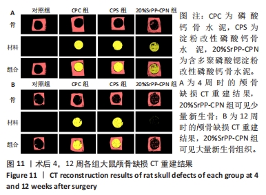
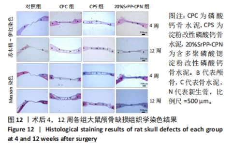
术后4周,对照组缺损处未观察到明显变化,CPC组与CPS组材料未发生明显降解,未见到明显新生骨;20%SrPP-CPN材料出现明显降解,缺损边缘有少量新生骨,并沿着骨水泥降解产生的孔隙长入骨水泥内部。术后12周,对照组缺损处仍未发生明显改变,证明了颅骨5 mm缺损为颅骨的临界缺损;CPC组材料少量降解,仅在缺损边缘产生少量的新生骨;CPS组材料降解较CPC组略微增强,在缺损边缘和降解的骨水泥内部均可观察到少量新生骨;20%SrPP-CPN组材料显著降解,降解率约为80%,新生骨相较于其他组更多,并且散在均匀分布于缺损边缘和降解的骨水泥中。 2.5.4 各组大鼠颅骨标本组织学观察结果 各组大鼠颅骨标本苏木精-伊红与Masson染色见图12。"


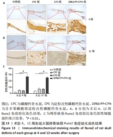
苏木精-伊红染色:术后4周,对照组骨缺损处未发生显著改变,仅有少量纤维组织连接;CPC组骨水泥未见明显变化;CPS组骨水泥发生了少量降解;20%SrPP-CPN组骨水泥降解较为明显,纤维组织均匀分散于降解的骨水泥中。术后12周,对照组骨缺损处仍未发生明显改变;CPC组骨水泥发生部分溃散,较多新生纤维包裹了骨水泥;CPS组骨水泥中存在新生纤维均匀长入;20%SrPP-CPN组中骨水泥降解明显,可观察到致密的纤维组织而未能观察到明显骨水泥存在。 Masson染色:对照组全程未观察到新生骨组织形态,仅观察到新生的纤维组织。术后4周,CPC组由于材料降解缓慢且生物活性较低,因而未能观察到明显新生骨;CPS组骨水泥发生少量降解,极少量新生骨分布于降解的骨水泥中;20%SrPP-CPN组可以观察到少量新生骨分布于增生的纤维组织中。术后12周,CPC组缺损边缘溃散部位产生了少量新生骨,CPS组中少量新生骨散在分布于骨水泥和纤维组织中,20%SrPP-CPN组中有较多新生骨分布于降解的骨水泥中。 各组大鼠颅骨标本Runx2免疫组化染色结果见图13。术后4周,对照组未观察到阳性染色;CPC组骨水泥被染料浸染,未观察到明显的阳性染色细胞;CPS组骨水泥降解部位散在分布少量的阳性染色细胞;20%SrPP-CPN组缺损部位细胞出现较强的阳性染色,均匀分布于新生纤维组织与骨水泥间隙中。术后12周,对照组阳性染色无明显变化;CPC组骨水泥边缘降解部位分布有少量的阳性染色细胞;CPS组骨水泥中均匀分布着纤维组织,少量阳性染色细胞分布其中,且染色程度较浅;20%SrPP-CPN组缺损部位由阳性染色细胞覆盖,阳性染色细胞分布均匀,细胞染色程度较深。Image J统计分析结果显示,术后4周,对照组、CPC组、CPS组、20%SrPP-CPN组Runx2阳性细胞染色面积分别为(2.23±0.39)%,(5.45±1.07)%,(4.66±0.64)%,(27.11±3.36)%,20%SrPP-CPN组大于其他3组(P < 0.01);术后12周,对照组、CPC组、CPS组、20%SrPP-CPN组Runx2阳性细胞染色面积分别为(3.87±1.18)%,(7.53±1.67)%,(7.19±0.39)%,(25.44±1.76)%,20%SrPP-CPN组大于其他3组(P < 0.01)。证实20%SrPP-CPN组表达了更多的成骨相关蛋白。"
| [1] WU T, YANG S, SHI H, et al. Preparation and cytocompatibility of a novel bismuth aluminate/calcium phosphate cement with high radiopacity. J Mater Sci Mater Med. 2018;29(9):149. [2] SNEHA KR, SAILAJA GS. Intrinsically radiopaque biomaterial assortments: a short review on the physical principles, X-ray imageability, and state-of-the-art developments. J Mater Chem B. 2021;9(41):8569-8593. [3] SAS A, HELGASON B, FERGUSON SJ, et al. Mechanical and morphological characterization of PMMA/bone composites in human femoral heads. J Mech Behav Biomed Mater. 2021;115:104247. [4] DEMIR-OĞUZ Ö, BOCCACCINI AR, LOCA D. Injectable bone cements: What benefits the combination of calcium phosphates and bioactive glasses could bring? Bioact Mater. 2023;19:217-236. [5] ZHANG R, LIU H, ZHOU H, et al. Pregelatinized starch as a cohesion promoter improves mechanical property and surgical performance of calcium phosphate bone cement: the effect of starch type. Mater Technol. 2022;37(14):3110-3121. [6] LIU H, ZHANG Z, GAO C, et al. Enhancing effects of radiopaque agent BaSO(4) on mechanical and biocompatibility properties of injectable calcium phosphate composite cement. Mater Sci Eng C Mater Biol Appl. 2020;116:110904. [7] ABERG J, HENRIKSSON HB, ENGQVIST H, et al. Biocompatibility and resorption of a radiopaque premixed calcium phosphate cement. J Biomed Mater Res A. 2012;100(5):1269-1278. [8] SABOKBAR A, FUJIKAWA Y, MURRAY DW, et al. Radio-opaque agents in bone cement increase bone resorption. J Bone Joint Surg Br. 1997;79(1):129-134. [9] LE FERREC M, MELLIER C, BOUKHECHBA F, et al. Design and properties of a novel radiopaque injectable apatitic calcium phosphate cement, suitable for image-guided implantation. J Biomed Mater Res B Appl Biomater. 2018;106(8):2786-2795. [10] HARRISON CJ, HATTON PV, GENTILE P, et al. Nanoscale Strontium-Substituted Hydroxyapatite Pastes and Gels for Bone Tissue Regeneration. Nanomaterials (Basel). 2021;11(6):1161-1180. [11] DAI J, FU Y, CHEN D, et al. A novel and injectable strontium-containing hydroxyapatite bone cement for bone substitution: A systematic evaluation. Mater Sci Eng C Mater Biol Appl. 2021;124:112052. [12] DUMRONGVUTE K, ADEL S, WADA T, et al. Distrontium Cerate as a Radiopaque Component of Hydraulic Endodontic Cement. Materials (Basel). 2021;15(1): 284-294. [13] ARDESHIR, YLAJIMI A, GOLCHIN A, et al. Increased osteogenic differentiation potential of MSCs cultured on nanofibrous structure through activation of Wnt/β-catenin signalling by inorganic polyphosphate. Artif Cells Nanomed Biotechnol. 2018;46(sup3):S943-S949. [14] WANG X, SCHRÖDER HC, MÜLLER WEG. Amorphous polyphosphate, a smart bioinspired nano-/bio-material for bone and cartilage regeneration: towards a new paradigm in tissue engineering. J Mater Chem B. 2018;6(16):2385-2412. [15] LEE NH, KANG MS, KIM TH, et al. Dual actions of osteoclastic-inhibition and osteogenic-stimulation through strontium-releasing bioactive nanoscale cement imply biomaterial-enabled osteoporosis therapy. Biomaterials. 2021;276:121025. [16] CHEN Y, CANELI G, ALMOUSA R, et al. A novel antibacterial zirconia-containing PMMA bone cement. J Mech Behav Biomed Mater. 2022;129:105135. [17] TIAN Y, LIU H, HE L, et al. Calcium phosphate-based composite cement: Impact of starch type and starch pregelatinization on its physicochemical properties and performance in the vertebral fracture surgical models in vitro. J Biomed Mater Res B Appl Biomater. 2021;109(12):2068-2078. [18] LIU H, LIU B, GAO C, et al. Injectable, biomechanically robust, biodegradable and osseointegrative bone cement for percutaneous kyphoplasty and vertebroplasty. Int Orthop. 2018;42(1):125-132. [19] CHEN F, LIU C, MAO Y. Bismuth-doped injectable calcium phosphate cement with improved radiopacity and potent antimicrobial activity for root canal filling. Acta Biomater. 2010;6(8):3199-3207. [20] LU M, ZHANG XS, CHANG L, et al. Preparation, performance and characterization of bioactive bone materials with plasticity. Chin J Tissue Eng Res. 2015;19(21):3323. [21] WU X, TANG Z, WU K, et al. Strontium-calcium phosphate hybrid cement with enhanced osteogenic and angiogenic properties for vascularised bone regeneration. J Mater Chem B. 2021;9(30):5982-5997. [22] CUI X, ZHANG Y, WANG J, et al. Strontium modulates osteogenic activity of bone cement composed of bioactive borosilicate glass particles by activating Wnt/β-catenin signaling pathway. Bioact Mater. 2020;5(2):334-347. [23] HUANG H, LAN LQ, WU JQ, et al.[Effect of bone marrow mesenchymal stem cells on paraquat-induced pulmonary fibrosis in rats]. Zhonghua Lao Dong Wei Sheng Zhi Ye Bing Za Zhi. 2020;38(5):332-338. [24] WU K, YANG Q, ZHANG L, et al. An injectable curcumin-releasing organohydrogel with non-drying property and high mechanical stability at low-temperature for expedited skin wound care. J Mater Sci Technol. 2023;133:123-134. [25] SHIBA T, NISHIMURA D, KAWAZOE Y, et al. Modulation of mitogenic activity of fibroblast growth factors by inorganic polyphosphate. J Biol Chem. 2003;278(29): 26788-26792. [26] LI R, LIN S, ZHU M, et al. Synthetic presentation of noncanonical Wnt5a motif promotes mechanosensing-dependent differentiation of stem cells and regeneration. Sci Adv. 2019;5(10):eaaw3896. [27] LUTOLF MP, WEBER FE, SCHMOEKEL HG, et al. Repair of bone defects using synthetic mimetics of collagenous extracellular matrices. Nat Biotechnol. 2003; 21(5):513-518. [28] OUYANG Y, ZHANG R, CHEN H, et al. Novel, degradable, and cytoactive bone cements based on magnesium polyphosphate and calcium citrate. New J Chem. 2022;46(27):13137-13148. [29] TADDEI P, DI FOGGIA M, ZAMPARINI F, et al. The Influence of the Matrix on the Apatite-Forming Ability of Calcium Containing Polydimethylsiloxane-Based Cements for Endodontics. Molecules. 2022;27(18):5750-5768. [30] WU T, YANG S, LU T, et al. Strontium ranelate simultaneously improves the radiopacity and osteogenesis of calcium phosphate cement. Biomed Mater. 2019;14(3):035005. [31] WON S, KO KH, PARK CJ, et al. Effect of barium silicate filler content on mechanical properties of resin nanoceramics for additive manufacturing. J Adv Prosthodont. 2022;14(5):315-323. [32] MÜLLER WEG, TOLBA E, SCHRÖDER HC, et al. Amorphous polyphosphate-hydroxyapatite: A morphogenetically active substrate for bone-related SaOS-2 cells in vitro. Acta Biomater. 2016;31:358-367. [33] KOBAYASHI K, ANADA T, HANDA T, et al. Osteoconductive property of a mechanical mixture of octacalcium phosphate and amorphous calcium phosphate. ACS Appl Mater Interfaces. 2014;6(24):22602-22611. [34] MÜLLER WE, TOLBA E, FENG Q, et al. Amorphous Ca²⁺ polyphosphate nanoparticles regulate the ATP level in bone-like SaOS-2 cells. J Cell Sci. 2015; 128(11):2202-2207. [35] QUERIDO W, ROSSI AL, FARINA M. The effects of strontium on bone mineral: A review on current knowledge and microanalytical approaches. Micron. 2016;80: 122-134. [36] PING H, WAGERMAIER W, HORBELT N, et al. Mineralization generates megapascal contractile stresses in collagen fibrils. Science. 2022;376(6589):188-192. [37] O’DONNELL MD, HILL RG. Influence of strontium and the importance of glass chemistry and structure when designing bioactive glasses for bone regeneration. Acta Biomater. 2010;6(7):2382-2385. [38] MÜLLER WEG, TOLBA E, SCHRÖDER HC, et al. A new polyphosphate calcium material with morphogenetic activity. Mater Lett. 2015;148:163-166. [39] MÜLLER WEG, TOLBA E, ACKERMANN M, et al. Fabrication of amorphous strontium polyphosphate microparticles that induce mineralization of bone cells in vitro and in vivo. Acta Biomater. 2017;50:89-101. [40] MÜLLER WEG, SCHRÖDER HC, WANG X. Inorganic Polyphosphates As Storage for and Generator of Metabolic Energy in the Extracellular Matrix. Chem Rev. 2019;119(24):12337-12374. [41] WANG X, SCHRÖDER HC, DIEHL-SEIFERT B, et al. Dual effect of inorganic polymeric phosphate/polyphosphate on osteoblasts and osteoclasts in vitro. J Tissue Eng Regen Med. 2013;7(10):767-776. [42] WANG X, SCHRÖDER HC, SCHLOßMACHER U, et al. Inorganic polyphosphates: biologically active biopolymers for biomedical applications. Prog Mol Subcell Biol. 2013;54:261-294. [43] MÜLLER WE, TOLBA E, SCHRÖDER HC, et al. Polyphosphate: A Morphogenetically Active Implant Material Serving as Metabolic Fuel for Bone Regeneration. Macromol Biosci. 2015;15(9):1182-1197. [44] TSAI CH, LIN RM, JU CP, et al. Bioresorption behavior of tetracalcium phosphate-derived calcium phosphate cement implanted in femur of rabbits. Biomaterials. 2008;29(8):984-993. |
| [1] | Liu Zhaohui, Han Xiaoqian, Duan Xin, Guo Pengda, Zhang Yuntao. Salidroside promotes osteogenic differentiation of MC3T3-E1 cells: an in vitro experiment [J]. Chinese Journal of Tissue Engineering Research, 2025, 29(2): 231-237. |
| [2] | Zhou Shijie, Li Muzhe, Yun Li, Zhang Tianchi, Niu Yuanyuan, Zhu Yihua, Zhou Qinfeng, Guo Yang, Ma Yong, Wang Lining. Effect of Wenshen Tongluo Zhitong formula on mouse H-type bone microvascular endothelial cell/bone marrow mesenchymal stem cell co-culture system [J]. Chinese Journal of Tissue Engineering Research, 2025, 29(1): 8-15. |
| [3] | Zheng Qian, Liu Pingping, Gu Yujie, Xie Lei. Effect of ursolic acid on osteogenic differentiation of human periodontal ligament stem cells#br# [J]. Chinese Journal of Tissue Engineering Research, 2025, 29(1): 80-86. |
| [4] | Yang Yufang, Yang Zhishan, Duan Mianmian, Liu Yiheng, Tang Zhenglong, Wang Yu. Application and prospects of erythropoietin in bone tissue engineering [J]. Chinese Journal of Tissue Engineering Research, 2024, 28(9): 1443-1449. |
| [5] | Liu Anhong, Cai Mengmeng, Han Xiao, Wang Zhanhui. Research status on element selection of medical magnesium alloy [J]. Chinese Journal of Tissue Engineering Research, 2024, 28(5): 777-782. |
| [6] | Lan Weiwei, Yu Yaodong, Huang Di, Chen Weiyi. In vitro degradation behavior of Mg-Zn-Ca alloys [J]. Chinese Journal of Tissue Engineering Research, 2024, 28(5): 717-723. |
| [7] | Li Jiaqi, Huang Yuanli, Li Yan, Wang Chunren, Han Qianqian. Mechanism and influencing factors in molecular weight degradation of non-cross-linked hyaluronic acid [J]. Chinese Journal of Tissue Engineering Research, 2024, 28(5): 747-752. |
| [8] | Yang Cheng, Li Yusheng, Jiao Hongzhuo, Shang Man, Liu Qi, Li Linzhen, Fan Fangyang, Zhang Chenglong, Zhang Xiaoyu, Zhang Juntao. Establishment and validation of the Sprague-Dawley rat model of osteoarthritis with kidney deficiency and blood stagnation [J]. Chinese Journal of Tissue Engineering Research, 2024, 28(27): 4273-4280. |
| [9] | Ye Zhikui, Zhang Zhimin, Cui Linna, Jiang Xiaowen. Mechanism by which alendronate promotes rapid mandibular distraction osteogenesis in rabbits [J]. Chinese Journal of Tissue Engineering Research, 2024, 28(23): 3642-3648. |
| [10] | Chu Fuchao, Wang Zhenxin, Zhang Dazhen, Yuan Feng. Osteogenic properties of polyacrylamide-modified gelatin methacryloyl grafted titanium alloy scaffold [J]. Chinese Journal of Tissue Engineering Research, 2024, 28(22): 3472-3477. |
| [11] | An Ran, Shao Guo, Zhang Chunyang. Effect of dura mater on enhancement of cranial osteogenesis in rats [J]. Chinese Journal of Tissue Engineering Research, 2024, 28(22): 3478-3483. |
| [12] | Zheng Heishu, Zhang Yingjuan, Wei Yanhua, Huang Hui, Ma Xiangyu, Liao Hongbing. Antibacterial performance of cerium oxide nanoenzyme against Escherichia coli [J]. Chinese Journal of Tissue Engineering Research, 2024, 28(22): 3496-3501. |
| [13] | Chen Mingxue, Niu Jianhua, Lin Haiyan, Wu Gang, Wan Ben. Physicochemical properties and cytocompatibility of biomimetically precipitated nanocrystalline calcium phosphate granules [J]. Chinese Journal of Tissue Engineering Research, 2024, 28(22): 3502-3508. |
| [14] | Tian Ai, Li Li, Xiao Tianjiao, Kang Jiabing, Zhan Jifan, Wei Yan, Chen Helin. Deferoxamine mesylate improves the repair of jaw bone defects in an ovariectomized rat model of osteoporosis [J]. Chinese Journal of Tissue Engineering Research, 2024, 28(20): 3143-3149. |
| [15] | Lin Tianye, Wu Zhiming, Zhang Wensheng, He Xiaoming, He Mincong, Zhang Qingwen, He Wei, Wei Qiushi, Li Ziqi. Mechanism of compound Shengmai Chenggu capsule in the repair of steroid-induced osteonecrosis of the femoral head [J]. Chinese Journal of Tissue Engineering Research, 2024, 28(2): 200-207. |
| Viewed | ||||||
|
Full text |
|
|||||
|
Abstract |
|
|||||