Chinese Journal of Tissue Engineering Research ›› 2024, Vol. 28 ›› Issue (17): 2631-2636.doi: 10.12307/2024.479
Previous Articles Next Articles
Preparation of heparinized acellular vascular scaffold and hemocompatibility evaluation
Li Xiafei1, Zhao Lingling2, Liang Feng3, Zhang Xuewei2, Zhang Jinjin2, Lin Fei4, Yang Tuo2, Zhao Liang2
- 1College of Medical Engineering, 2College of Life Science and Technology, 3Mingde College, Xinxiang Medical University, Xinxiang 453003, Henan Province, China; 4First Affiliated Hospital of Xinxiang Medical University, Xinxiang 453003, Henan Province, China
-
Received:2023-07-07Accepted:2023-09-15Online:2024-06-18Published:2023-12-14 -
Contact:Zhao Liang, PhD, Associate professor, College of Life Science and Technology, Xinxiang Medical University, Xinxiang 453003, Henan Province, China -
About author:Li Xiafei, Master, Experimentalist, College of Medical Engineering, Xinxiang Medical University, Xinxiang 453003, Henan Province, China -
Supported by:Natural Science Foundation of Henan Province, No. 202300410311 (to ZL); College Student Innovation and Entrepreneurship Training Program Project of Henan Province, No. S202310472023 (to LF); Postgraduate Research and Innovation Support Plan Project of Xinxiang Medical University, No. YJSCX2022108Y (to YT); 2022 Research and Innovation of College Students in Xinxiang Medical University, No. xyxskyz202236 (to ZJJ); Employment and Entrepreneurship Project in Colleges and Universities in Henan Province, No. JYB2023039 (to ZL)
CLC Number:
Cite this article
Li Xiafei, Zhao Lingling, Liang Feng, Zhang Xuewei, Zhang Jinjin, Lin Fei, Yang Tuo, Zhao Liang. Preparation of heparinized acellular vascular scaffold and hemocompatibility evaluation[J]. Chinese Journal of Tissue Engineering Research, 2024, 28(17): 2631-2636.
share this article
Add to citation manager EndNote|Reference Manager|ProCite|BibTeX|RefWorks
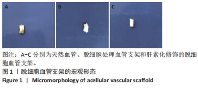
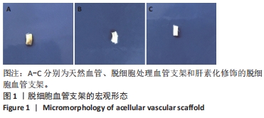
2.1 两组脱细胞血管支架形态观察 2.1.1 表观形态 从大体形态上观察可见天然血管的颜色呈现淡黄色,脱细胞处理血管支架和肝素化修饰的脱细胞血管支架表面均呈白色透明状,血管壁完整(图1)。"
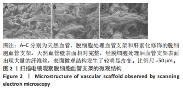
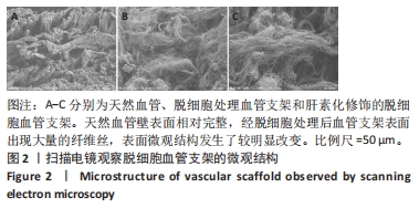
2.1.2 微观形貌 扫描电镜显示天然血管壁表面相对完整,经脱细胞处理后血管支架表面出现大量的纤维丝,表面微观结构发生了较明显改变,表明脱细胞处理对血管的微观结构产生影响,血管中的细胞脱离较完全;与对照组血管支架相比,实验组血管支架微观形貌并未发生较大变化(图2)。"
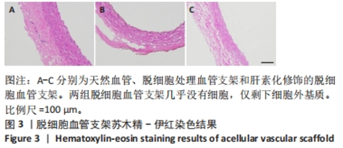
2.1.3 苏木精-伊红染色 天然血管的细胞外基质完整且呈现暗红色,细胞核呈现蓝色,有较多的细胞;两组脱细胞血管支架几乎没有细胞,仅剩下细胞外基质,呈现红色(图3),表明已经去除了细胞。"

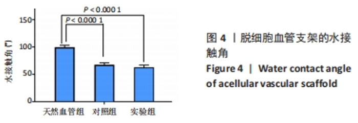
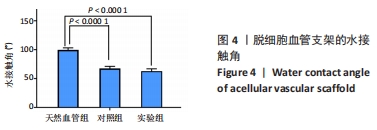
2.2 两组脱细胞血管支架的亲水性 水接触角值表征支架亲水性能强弱,其值越小支架亲水性能越强。对照组和实验组脱细胞血管支架的水接触角分别为(67.50±3.61)° 和(62.67±4.13)°,都明显小于天然血管(P < 0.000 1),两组脱细胞血管支架的水接触角比较差异无显著性意义(P > 0.05),见图4。由此可见,与天然血管相比,脱细胞血管支架的亲水性能更加优良。"
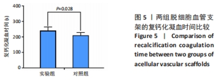
2.3 两组脱细胞血管支架复钙化凝血时间的比较 实验组血管支架的复钙化凝血时间长于对照组[(241±23),(210±19) s,P=0.028],见图5。表明脱细胞血管支架经过肝素化修饰后的抗凝血效果更好。"

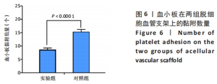
2.4 两组脱细胞血管支架体外血小板黏附的比较 对照组、实验组脱细胞血管支架膜上黏附的血小板平均数量分别为(15.33±0.89),(8.67±0.58)个,组间比较差异有显著性意义(P < 0.000 1),见图6。表明血管支架经Triton-x100/肝素钠处理后的血液相容性能更好。"

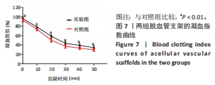
2.5 两组脱细胞血管支架动态凝血时间的比较 实验组和对照组脱细胞血管支架凝血指数在10 min时分别为(78.2±1.39)%,(73.3±2.5)%,组间比较差异有显著性意义(P=0.001 8),在其他各时间点均存在类似情况,见图7。较低的凝血指数值表示较高的凝结率,结果表明实验组脱细胞血管支架具有优良的抗凝作用。"
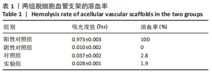
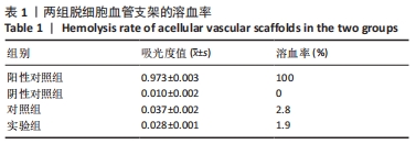
2.6 两组脱细胞血管支架体外溶血实验结果 阳性对照组因红细胞吸水胀破发生溶血。阴性对照组因使用生理盐水红细胞能够维持自己的形态,没有发生溶血。对照组、实验组脱细胞血管支架的溶血率分别为2.8%,1.9%,都满足溶血率低于5%的国家标准(表1)。"

2.7 两组脱细胞血管支架补体激活实验结果 放射免疫测定法检测结果显示,对照组、实验组脱细胞血管支架的补体水平分别为(0.97±0.08),(0.76±0.07) μg/mL,组间比较差异有显著性意义(P=0.000 5)。"

| [1] AWAD N K, NIU H, ALI U, et al. Electrospun fibrous scaffolds for small-diameter blood vessels: a review. Membranes (Basel). 2018;8(1):15. [2] JI J, XU H, LI C, et al. Small-Caliber Tissue-Engineered Vascular Grafts Based on Human-Induced Pluripotent Stem Cells: Progress and Challenges. Tissue Eng Part B Rev. 2023;29(4):441-455. [3] FLIS A, TRÁVNÍČKOVÁ M, KOPER F, et al. Poly(octamethylene citrate) Modified with Glutathione as a Promising Material for Vascular Tissue Engineering. Polymers (Basel). 2023;15(5):1322. [4] PASHNEH-TALA S, MACNEIL S, CLAEYSSENS F. The tissue-engineered vascular graft-past, present, and future. Tissue Eng Part B Rev. 2016; 22(1):68-100. [5] MOHAMMADI F, GOLAFSHAN N, KHARAZIHA M, et al. Chitosan-heparin nanoparticle coating on anodized NiTi for improvement of blood compatibility and biocompatibility. Int J Biol Macromol. 2019;127: 159-168. [6] ZIZHOU R, WANG X, HOUSHYAR S. Review of Polymeric Biomimetic Small-Diameter Vascular Grafts to Tackle Intimal Hyperplasia. ACS Omega. 2022;7:22125-22148. [7] ZHANG F, KING MW. Immunomodulation Strategies for the Successful Regeneration of a Tissue-Engineered Vascular Graft. Adv Healthc Mater. 2022;11:e2200045. [8] OBIWELUOZOR FO, KAYUMOV M, KWAK Y, et al. Rapid remodeling observed at mid-term in-vivo study of a smart reinforced acellular vascular graft implanted on a rat model. J Biol Eng. 2023;17:1. [9] 刘俊,张晓膺.纳米APS外膜肝素化内膜小口径组织工程血管的实验研究[J].上海交通大学学报(医学版),2017,37(3):337-343. [10] NEMENO-GUANZON JG, LEE S, BERG JR, et al. Trends in tissue engineering for blood vessels. J Biomed Biotechnol. 2012;2012: 956345. [11] YAO Y, ZAW AM, ANDERSON DEJ, et al. Fucoidan and topography modification improved in situ endothelialization on acellular synthetic vascular grafts. Bioact Mater. 2023;22:535-550. [12] BAHRAMZADEH E, YILMAZ E, ADALI T. Chitosan-graft-poly(N-hydroxy ethyl acrylamide) copolymers: Synthesis, characterization and preliminary blood compatibility in vitro. Int J Biol Macromol. 2019;123:1257-1266. [13] 夏成勇,刘长建,冉峰,等.等离子体处理脱细胞血管支架的血液相容性[J].中国组织工程研究与临床康复,2009,13(21):4033-4036. [14] DU PC, LI XF, SUN LL, et al. Improved hemocompatibility by modifying acellular blood vessels with bivalirudin and its biocompatibility evaluation. J Biomed Mater Res A. 2022;110(3):635-651. [15] KOPEC K, WOJASINSKI M, EICHLER M, et al. Polydopamine and gelatin coating for rapid endothelialization of vascular scaffolds. Biomater Adv. 2022;134:112544. [16] TARDALKAR K, MARSALE T, BHAMARE N, et al. Heparin Immobilization of Tissue Engineered Xenogeneic Small Diameter Arterial Scaffold Improve Endothelialization. Tissue Eng Regen Med. 2022;19:505-523. [17] YANG F, GUO G, WANG Y. Inflammation-triggered dual release of nitroxide radical and growth factor from heparin mimicking hydrogel-tissue composite as cardiovascular implants for anti-coagulation, endothelialization, anti-inflammation, and anti-calcification. Biomaterials. 2022;289:121761. [18] LV YM, HUANG HM, WANG QL, et al. Acellular porcine aorta matrix as a novel tissue engineered vascular scaffold biocompatibility and mechanical properties. J Cli Reh Tissue Eng Res. 2010;14(47):8921-8925. [19] QIU H, QI P, LIU J, et al. Biomimetic engineering endothelium-like coating on cardiovascular stent through heparin and nitric oxide-generating compound synergistic modification strategy. Biomaterials. 2019;207:10-22. [20] 周莹,肖利吉,姚丽,等.自修复型超疏水材料研究进展[J].材料导报,2019,33(7):159-167. [21] 高云佳,赵庆春,闵鹏,等.脱皮马勃止血有效部位的实验研究[J].解放军药学学报,2010,26(6):82-84. [22] 高娟.壳聚糖止血活性及其作用机理的初步研究[D].无锡:江南大学,2009. [23] 秦静雯,王鸿博,傅佳佳,等.醋纤基载药纳米纤维药物活性评价[J].功能材料,2014,45(14):14077-14080. [24] 欧阳晨曦,李沁,王维慈,等.小口径人工血管血液相容性[J].中国组织工程研究与临床康复,2008,12(6):133-137. [25] LIU HF, LI XM, ZHOU G, et al. Electrospun sulfated silk fibroin nanofibrous scaffolds for vascular tissue engineering. Biomaterials. 2011;32:3784-3793. [26] 陈捷,杨镜秋,刘春晓.灌注法制备全肾脏脱细胞基质支架的体内生物相容性[J].中国组织工程研究,2015,19(16):75-79. [27] 薛正翔,陈登龙,李敏,等.静电纺丝制备小直径血管支架及其血液相容性的研究[J].功能材料,2009(10):129-132. [28] 马慧.羟基磷灰石表面构建压电陶瓷涂层的生物相容性研究[D].乌鲁木齐:新疆医科大学,2015. [29] 贾山山,陈群清,闫玉生.肝素在人工小血管表面改性中的应用[J].中国医学物理学杂志,2018,35(10):126-130. [30] 马巧,宋文静,冀慧雁,等.血管支架材料的应用及研究现状[J].临床医药实践,2018,27(11):57-62. [31] 阮烨.冷空气活动对心脑血管疾病相关指标影响的初步研究[D].兰州:兰州大学,2013. [32] SU H, LIU W, LI X, et al. Cellular energy supply for promoting vascular remodeling of small-diameter vascular grafts: a preliminary study of a new strategy for vascular graft development. Biomater Sci. 2023; 11(9):3197-3213. [33] OMID H, ABDOLLAHI S, BONAKDAR S, et al. Biomimetic vascular tissue engineering by decellularized scaffold and concurrent cyclic tensile and shear stresses. J Mater Sci Mater Med. 2023;34:12. [34] 刘桂阳.皮/芯结构的丝素/聚己内酯纤维及其双层血管再生支架[D].苏州:苏州大学,2015. [35] YAO Y, WANG J, CUI Y, et al. Effect of sustained heparin release from PCL/chitosan hybrid small-diameter vascular grafts on anti-thrombogenic property and endothelialization. Acta Biomater. 2014; 10(6):2739-2749. [36] 赵亮,李霞飞,李成成,等.应用Triton-x100加丹参酚酸B制备脱细胞血管支架及其血液相容性研究[J].中国生物医学工程学报, 2019,38(2):201-207. [37] LIU H, LI X, ZHOU G, et al. Electrospun sulfated silk fibroin nanofibrous scaffolds for vascular tissue engineering. Biomaterials. 2011;32(15): 3784-3793. [38] MONTELIONE N, LORENI F, NENNA A, et al. Tissue Engineering and Targeted Drug Delivery in Cardiovascular Disease: The Role of Polymer Nanocarrier for Statin Therapy. Biomedicines. 2023;11(3):798. [39] LIU Y, YUAN H, LIU Y, et al. Multifunctional nanoparticle-VEGF modification for tissue-engineered vascular graft to promote sustained anti-thrombosis and rapid endothelialization. Front Bioeng Biotechnol. 2023;11:1109058. [40] 王学宁,陈长志,杨岷,等.肝素处理小口径脱细胞异种血管的移植研究[J].中华胸心血管外科杂志,2006,22(5):324-325. |
| [1] | Yang Yufang, Yang Zhishan, Duan Mianmian, Liu Yiheng, Tang Zhenglong, Wang Yu. Application and prospects of erythropoietin in bone tissue engineering [J]. Chinese Journal of Tissue Engineering Research, 2024, 28(9): 1443-1449. |
| [2] | Chen Kaijia, Liu Jingyun, Cao Ning, Sun Jianbo, Zhou Yan, Mei Jianguo, Ren Qiang. Application and prospect of tissue engineering in treatment of osteonecrosis of the femoral head [J]. Chinese Journal of Tissue Engineering Research, 2024, 28(9): 1450-1456. |
| [3] | Mei Jingyi, Liu Jiang, Xiao Cong, Liu Peng, Zhou Haohao, Lin Zhanyi. Proliferation and metabolic patterns of smooth muscle cells during construction of tissue-engineered blood vessels [J]. Chinese Journal of Tissue Engineering Research, 2024, 28(7): 1043-1049. |
| [4] | Wang Shanshan, Shu Qing, Tian Jun. Physical factors promote osteogenic differentiation of stem cells [J]. Chinese Journal of Tissue Engineering Research, 2024, 28(7): 1083-1090. |
| [5] | Shen Ziqing, Xia Tian, Shan Yibo, Zhu Ruijun, Wan Haoxin, Ding Hao, Pan Shu, Zhao Jun. Vascularized tracheal substitutes constructed by exosome-load hydrogel-modified 3D printed scaffolds [J]. Chinese Journal of Tissue Engineering Research, 2024, 28(5): 697-705. |
| [6] | Zhu Liwei, Wang Jiangyue, Bai Ding. Application value of nanocomposite gelatin methacryloyl hydrogels in different bone defect environments [J]. Chinese Journal of Tissue Engineering Research, 2024, 28(5): 753-758. |
| [7] | Chen Xiaofang, Zheng Guoshuang, Li Maoyuan, Yu Weiting. Preparation and application of injectable sodium alginate hydrogels [J]. Chinese Journal of Tissue Engineering Research, 2024, 28(5): 789-794. |
| [8] | Wang Jiani, Chen Junyu. Angiogenesis mechanism of metal ions and their application in bone tissue engineering [J]. Chinese Journal of Tissue Engineering Research, 2024, 28(5): 804-812. |
| [9] | Yang Yuqing, Chen Zhiyu. Role and application of early transient presence of M1 macrophages in bone tissue engineering [J]. Chinese Journal of Tissue Engineering Research, 2024, 28(4): 594-601. |
| [10] | Kong Xiangyu, Wang Xing, Pei Zhiwei, Chang Jiale, Li Siqin, Hao Ting, He Wanxiong, Zhang Baoxin, Jia Yanfei. Biological scaffold materials and printing technology for repairing bone defects [J]. Chinese Journal of Tissue Engineering Research, 2024, 28(3): 479-485. |
| [11] | Xu Jing, Lyu Huixin, Bao Xin, Zhang Yi, Wang Yihan, Zhou Yanmin. Application of near infrared responsive hydrogels in tissue engineering [J]. Chinese Journal of Tissue Engineering Research, 2024, 28(3): 486-492. |
| [12] | Gu Mingxi, Wang Changcheng, Tian Fengde, An Ning, Hao Ruihu, Guo Lin. Preparation and in vitro evaluation of a three-dimensional porous cartilage scaffold made of silk fibroin/gelatin/chitosan [J]. Chinese Journal of Tissue Engineering Research, 2024, 28(3): 366-372. |
| [13] | Wang Xinmin, Yan Wenkai, Song Yahui, Liu Fei. Leukocyte- and platelet-rich fibrin with autologous hamstring tendon for traumatic patella dislocation [J]. Chinese Journal of Tissue Engineering Research, 2024, 28(3): 404-410. |
| [14] | Bi Yujie, Ma Dujun, Peng Liping, Zhou Ziqiong, Zhao Jing, Zhu Houjun, Zhong Qiuhui, Yang Yuxin. Strategy and significance of Chinese medicine combined with medical hydrogel for disease treatment [J]. Chinese Journal of Tissue Engineering Research, 2024, 28(3): 419-425. |
| [15] | Wang Xinyi, Xie Xianrui, Chen Yujie, Wang Xiaoyu, Xu Xiaoqing, Shen Yihong, Mo Xiumei. Electrospun nanofiber scaffolds for soft and hard tissue regeneration [J]. Chinese Journal of Tissue Engineering Research, 2024, 28(3): 426-432. |
| Viewed | ||||||
|
Full text |
|
|||||
|
Abstract |
|
|||||