[1] 李青松,刘少喻,尹宗生.多节段腰椎后路椎体间及后外侧融合椎弓根螺钉内固定治疗老年骨质疏松骨折合并胸腰椎后凸畸形[J].中国组织工程研究,2019,23(4):512-517.
[2] 陈遥,洪正华,洪盾,等.保留中柱经椎弓根开合式截骨术治疗强直性脊柱炎后凸畸形[J].中华骨科杂志,2018,38(22):1349-1356.
[3] 海涌,陈晓明,吴继功,等.后路一期全脊椎截骨术治疗重度僵硬型脊柱侧后凸[J].中国脊柱脊髓杂志,2006,16(3):183-186.
[4] 朱泽章,邱勇.全脊椎截骨术治疗严重脊柱畸形的若干思考[J].中国脊柱脊髓杂志,2016,26(1):19-21.
[5] 陈涛,黎观保,梁科友,等.脊柱-骨盆矢状面平衡及其在脊柱疾病治疗中的作用[J].中国组织工程研究,2013,17(13):2423-2430.
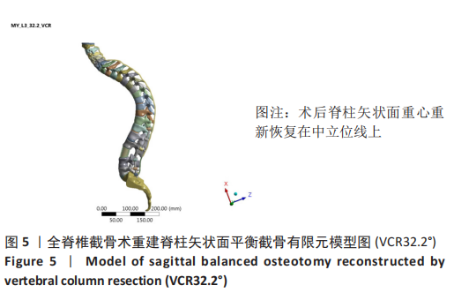
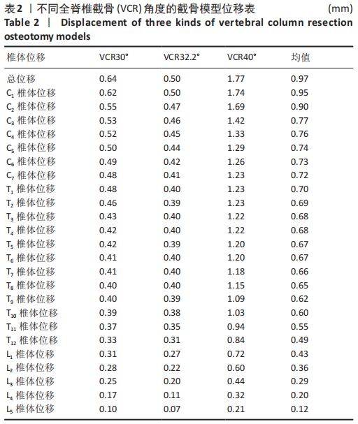
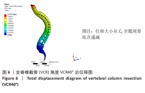
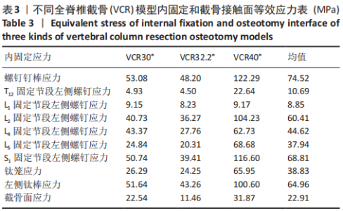
[6] 李辉,高晓玲,马原.不同截骨节段重建ⅢA型强直性脊柱后凸矢状面平衡的生物力学分析[J].中国组织工程研究,2020,24(30): 4824-4828.
[7] 王琦,李辉.颈椎后纵韧带骨化合并椎间融合有限元模型的建立和有效性验证[J].中国组织工程研究,2020,24(33):20-24.
[8] 曾庆馀.关于强直性脊柱炎的早期诊断[J].中华风湿病学杂志,2000, 4(2):69-71.
[9] 金海明,王向阳.脊柱矢状面畸形截骨角度计算方法的研究进展[J].中华骨科杂志,2016,36(5):298-306.
[10] BEHARI S, TUNGERIA A, JAISWAL AK, et al. The ”moustache” sign: Localized intervertebral disc fibrosis and panligamentous ossification in ankylosing spondylitis with kyphosis. Neurol India. 2010;58(5):764-767.
[11] BURSTEIN AH, REILLY DT, MARTENS M. Aging of bone tissue: mechanical properties. J Bone Joint Surg Am. 1976;58(1):82-86.
[12] SHIRAZI-ADL SA, SHRIVASTAVA SC, AHMED AM. Stress analysis of the lumbar disc-body unit in compression a three-dimensional nonlinear finite element study. Spine. 1984;9(2):120-134.
[13] 陈建良.老年骨质疏松性陈旧胸腰椎骨折伴后凸畸形后路截骨矫形的术式选择[J].中国骨伤,2020,33(2):121-126.
[14] 楼宇梁,全仁夫,李伟,等.多节段与单节段截骨治疗陈旧性胸腰椎骨质疏松性骨折伴后凸畸形的疗效比较[J].中华创伤杂志,2019, 35(6):513-519.
[15] 刘祖德,李新锋,臧危平,等.经椎弓根椎体间截骨联合V形截骨术矫正强直性脊柱炎合并重度胸腰椎后凸畸形[J].中华外科杂志, 2009,47(9):681-684.
[16] 袁磊,刘寅昊,曾岩,等.后路全脊椎截骨矫形术治疗重度胸腰椎角状后凸/侧后凸的中远期效果观察[J].中国脊柱脊髓杂志,2020, 30(7):596-603.
[17] YLDZ F. Results of closing wedge osteotomy in the treatment of sagittal imbalance due to ankylosing spondylitis. Acta Orthopaedica Et Traumatologica Turcica. 2016;50(1):63-68.
[18] ZHENG G, SONG K, YAO Z, et al. How to calculate the exact angle for two-level osteotomy in ankylosing spondylitis? Spine. 2016;41(17): E1046-E1052.
[19] 张树文,徐韬,盛伟斌.脊柱矢状面重建术前截骨角度的评估方法及意义[J].中华外科杂志,2020,58(7):551-554.
[20] 夏三强,刘盾,石博,等.SRS-Schwab Ⅳ级截骨术在Ⅰ型先天性脊柱后凸畸形矫形术中的应用[J].中华骨科杂志,2019,39(20): 1268-1274.
[21] 蒋彬,王冰,吕国华,等.腰骶段塌陷型结核后凸畸形后路截骨矫形术后内固定并发症的原因分析[J].中国脊柱脊髓杂志,2019, 29(12):8-15.
[22] WANG T, SONG D, ZHENG G, et al. Staged cervical osteotomy:a new strategy for correcting ankylosing spondylitis thoracolumbar kyphotic deformity with fused cervical spine. J Orthop Surg Res. 2019;14(1):108.
[23] 钱邦平,黄季晨,邱勇,等.截骨矫形术治疗强直性脊柱炎颈胸段畸形的疗效分析[J].中华骨科杂志,2018,38(4):204-211.
[24] 钱邦平,邱勇.强直性脊柱炎颈胸段后凸畸形合并胸腰椎后凸畸形截骨顺序的选择[J].中国脊柱脊髓杂志,2018,28(8):673-674.
[25] 钱邦平,邱勇,潘涛,等.经椎弓根不对称截骨重建强直性脊柱炎胸腰椎侧后凸畸形患者双平面平衡[J].中华骨科杂志,2015,35(4): 341-348.
[26] FABIAN P, DENIS P. Ankylosing spondylitis and axial spondyloarthritis: recent insights and impact of new classification criteria. Ther Adv Musculoskelet Dis. 2018;10(5-6):129-139.
[27] CHIN CM, TZENG ST. Pedicle subtraction osteotomy in lateral decubitus position to correct severe kyphosis in patient with ankylosing spondylitis-Case report. Formosan J Musculoskel Disord. 2019;10(2): 69-73.
[28] KOU J, GUO J, JI X, et al. A posterior-only approach for ankylosing spondylitis (AS) with thoracolumbar pseudoarthrosis: a clinical retrospective study. BMC Musculoskeletal Disorders. 2020;21(1): 370-337
[29] 郑光彬,洪正华,陈遥,等.保留中柱经椎弓根截骨术治疗陈旧性胸腰段骨折伴后凸畸形的疗效[J].中华创伤杂志,2020,36(4): 303-308.
[30] 高博,吴继功,马华松,等.后路三柱截骨矫形术治疗先天性颈胸段脊柱畸形的安全性及并发症分析[J].中国脊柱脊髓杂志,2019, 29(7):604-612.
(责任编辑:WJ,ZN,ZH)
|