[1] 钟品仁,主编.哺乳类实验动物[M].北京:人民卫生出版社,1987: 317.
[2] ARORA T, ZHANG L, PRASAD P. Development of a Subhuman Primate Brain Finite Element Model to Investigate Brain Injury Thresholds Induced by Head Rotation. Stapp Car Crash J. 2019;63:65-82.
[3] GOEL A, KASWA A, SHAH A. Role of atlantoaxial and subaxial spinal instability in pathogenesis of spinal ‘degeneration’ related cervical kyphosis. World Neurosurg. 2017;101:702-709.
[4] WANG XD, FENG MS, HU YC. Establishment and Finite Element Analysis of a Three-dimensional Dynamic Model of Upper Cervical Spine Instability. Orthop Surg. 2019;11(3):500-509.
[5] 邵荣学,全仁夫,王拓,等.新型梯度复合HA/ZrO2组织工程骨支架在猕猴颈椎融合中的应用[J].中华解剖与临床杂志,2017,22(6): 499-509.
[6] BONO CM, MIN W. Avoiding complications in patients with ankylosing spondylitis undergoing spine surgery. Curr Opin Orthop. 2005;16(3): 178-183.
[7] SHIRAZI-ADL A, AHMED AM, SHRIVASTAVA SC, et al. Mechanical response of a lumbar motion segment in axial torque alone and combined with compression. Spine. 1986;11(9):914-927.
[8] SCHMIDT H, HEUER F, DRUMM J, et al. Application of a calibration method provides more realistic results for a finite element model of a lumbar spinal segment. Clin Biomech. 2007;22(4):377-384.
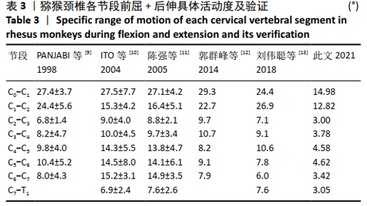
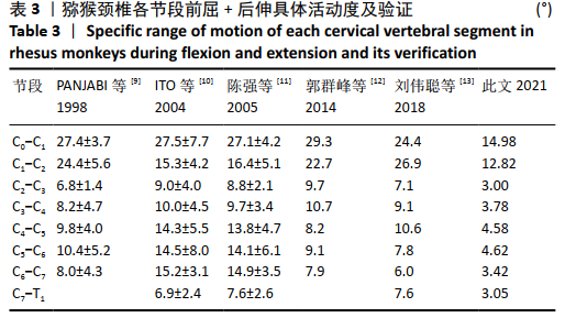
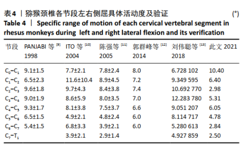
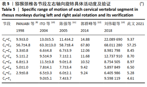
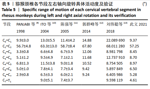
[9] PANJABI MM, CRISCO JJ, VASAVADA A, et al. Mechanical properties of the human cervical spine as shown by three-dimensional load-displacement curves. Spine. 2001;26(24):2692-2700.
[10] ITO S, IVANCIC PC, PANJABI MM, et al. Soft tissue injury threshold during simulated whiplash: a biomechanical investigation. Spine. 2004; 29(9):979-987.
[11] 陈强.挥鞭样损伤的生物力学和临床研究[J].第二军医大学学报, 2005,12(3):631-635.
[12] 郭群峰,陈方经,倪斌,等.带有颅底的全颈椎三维有限元模型的建立及分析[J].中国脊柱脊髓杂志,2014,24(6):550-554.
[13] 刘伟聪,陈雄生,周盛源,等.正常人体C0-T1全颈椎有限元模型的构建及意义[J].中国组织工程研究,2018,22(11):1707-1712.
[14] OLSZKO AV, BELTRAN CM, VASQUEZ KB, et al. Initial analysis of archived non-human primate frontal and rear impact data from the biodynamics data resource. Traffic Injury Prev. 2018;15(7):1165-1169.
[15] 凌泽莎.猕猴椎动脉及横突孔大水的CTA观察及其CSA模型的可行性研究[D].重庆:重庆医科大学,2014.
[16] LEE JH , PARK WM , KIM YH , et al. A Biomechanical Analysis of an Artificial Disc With a Shock-absorbing Core Property by Using Whole-cervical Spine Finite Element Analysis. Spine. 2016; 41(15):893-897.
[17] KIM YH, KHUYAGBAATAR B, KIM K. Recent advances in finite element modeling of the human cervical spine. J Mech Sci Technol. 2018;32(8): 1-10.
[18] BARKER JB, CRONIN DS, NIGHTINGALE RW. Lower Cervical Spine Motion Segment Computational Model Validation: Kinematic and Kinetic Response for Quasi-Static and Dynamic Loading. J Biomech Eng. 2017;139(6):610-613.
[19] LASSWELL TL, CRONIN DS, MEDLEY JB, et al. Incorporating ligament laxity in a finite element model for the upper cervical spine. Spine J. 2017;17(4):1755-1764.
[20] 凌泽莎,贾功伟,谭波涛,等.注射复合rhBMP-2骨水泥制作猕猴椎动脉型颈椎病模型[J].中山大学学报(医学科学版),2016,37(5): 775-780.
[21] CUNNINGHAM AS. Growth and Sexual Dimorphism of the Hyoid Body in Macaca mulatta. Springer US. 2020;28:178-181.
[22] ARORA T, ZHANG L, PRASAD P. Development of a Subhuman Primate Brain Finite Element Model to Investigate Brain Injury Thresholds Induced by Head Rotation. Stapp Car Crash J. 2019;63(3):65-82.
[23] 陈江波,潘希敏,陈应明,等. 磁共振T2 mapping和T1ρ成像研究群养成年恒河猴腰椎间盘的退变过程[J].中国组织工程研究, 2017,21(3):418-422.
[24] 汪韬,党耕町,郭昭庆,等. 自体骨髓基质干细胞与钙磷陶瓷复合体在恒河猴腰椎前路融合中的实验研究[J]. 中华外科杂志,2006, 44(12):843-847.
[25] 宋伟,赵文,魏瑞晗,等.脊髓损伤恒河猴后肢步态数据处理方法的设计[J]. 中国康复理论与实践,2013,19(8):734-738.
[26] 闵少雄,李森,刘成龙,等.恒河猴骨髓间充质干细胞体外培养及诱导分化成骨细胞的实验研究[J]. 中国矫形外科杂志,2011,19(3): 228-232.
[27] 孔杰,王子轩,季爱玉,等.应用微创技术建立恒河猴腰椎间盘早期退变模型[J].中华外科杂志,2008,46(11):835-838.
[28] 黄帆,邓忠良.部分可吸收椎间融合器应用于恒河猴腰椎融合的研究[D].重庆:重庆医科大学,2013.
[29] 凌泽莎,周志明,郑晓,等.实验用猕猴颈部骨骼和血管的影像学及血流动力学分析[J].中国实验动物学报,2015,23(5):500-505.
[30] WATSON DV , GANDHI AA , FREDERICKS DC , et al. Sheep cervical spine biomechanics: a finite element study. Iowa Orthop J. 2014;34:137-143.
[31] MENGONI M, VASILJEVA K, JONES AC, et al. Subject-specific multi-validation of a finite element model of ovine cervical functional spinal units. Biomech. 2016;49:259-266.
[32] 盛孙仁,徐华梓,王向阳,等.猪、小牛与人颈椎的生物力学比较[J].医用生物力学,2010,25(5):380-384.
[33] 薛德明.太行山猕猴寰椎和枢椎的初步研究[J].动物学杂志,2003, 9(2):74-75.
[34] 范春梅,李志雄,周建华.实验猕猴头颈部CT影像学观察[J].畜禽业,2012,10(4):30-32.
[35] 肖莉,杨红,石清明,等.藏酋猴脊柱的解剖学研究[J].西南国防医药,2017,5(10):1037-1040.
[36] SPARREY CJ, SALEGIO EA, CAMISA W, et al. Mechanical Design and Analysis of a Unilateral Cervical Spinal Cord Contusion Injury Model in Non-Human Primates. J Neurotrauma. 2016;33(12):1136-1149.
[37] LING Z, LI L, CHEN Y, et al. Changes of the end plate cartilage are associated with intervertebral disc degeneration:A quantitative magnetic resonance imaging study in rhesus monkeys and humans. J Orthop Translat. 2020;24:23-31. |