Chinese Journal of Tissue Engineering Research ›› 2019, Vol. 23 ›› Issue (29): 4735-4742.doi: 10.3969/j.issn.2095-4344.1781
Previous Articles Next Articles
In vitro embryo culture is the core of embryo engineering technology
Li Nan, Lin Zhong
- Reproductive Medicine Center, Maternal and Child Health Hospital of Liuzhou City, Liuzhou 545001, Guangxi Zhuang Autonomous Region, China
-
Revised:2019-04-02Online:2019-10-18Published:2019-10-18 -
Contact:Lin Zhong, MD, Chief physician, Reproductive Medicine Center, Maternal and Child Health Hospital of Liuzhou City, Liuzhou 545001, Guangxi Zhuang Autonomous Region, China -
About author:Li Nan, MD, Associate researcher, Reproductive Medicine Center, Maternal and Child Health Hospital of Liuzhou City, Liuzhou 545001, Guangxi Zhuang Autonomous Region, China -
Supported by:the Natural Science Foundation of Guangxi Zhuang Autonomous Region, No. 2015GXNSFBA139177 (to LN), 2017GXNSFAA198193 (to LZ) and 2017GXNSFAA198199 (to LN); the Self-Financing Research Project of the Health and Family Planning Commission of Guangxi Zhuang Autonomous Region, No. Z20180027 (to LN); Liuzhou Science and Technology Development Plan, No. 2016G020217 (to LN)
CLC Number:
Cite this article
Li Nan, Lin Zhong . In vitro embryo culture is the core of embryo engineering technology [J]. Chinese Journal of Tissue Engineering Research, 2019, 23(29): 4735-4742.
share this article

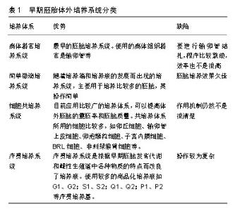
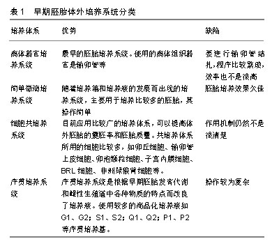
2.1 早期胚胎体外培养系统 目前,胚胎体外培养系统仍无法完全模拟哺乳动物雌性生殖道的物理及环境条件。早期胚胎体外发育的最佳体系不仅受到培养基组成成分的影响,同时还受到多种物理参数的影响(胚胎发育至孵化的环境、饲养细胞层的有无等)。最早在1970年,Bowman等[6]在研究中就发现体外培养的早期胚胎会发生发育阻断现象。体外培养的胚胎经过移植后,会出现大量的胚胎吸收、死亡、流产、死胎、出生后死亡、胎儿过大综合征等问题[7]。目前序贯培养系统的体外胚胎生产的新技术已有了较大的进步,但是仍然无法满足生产及科研的需要,仍然有待进一步完善、优化[8]。体外胚胎培养系统的分类如下: 离体器官培养系统:1949年,Hammond等[9]将生理盐水中加入卵黄,在体外将8-细胞期的BALB/C小鼠胚胎成功培养到囊胚阶段,这打开了早期胚胎体外培养的大门。1962年,Biggers等[10]首次利用离体BALB/C小鼠输卵管膨大部来培养BALB/C小鼠早期胚胎,并且成功获得了囊胚。4年后,Papaioannou等[11]利用离体小鼠输卵管培养系统将小鼠的单细胞胚胎培养至桑椹胚\囊胚阶段,其后采用同样的方法获得大鼠、仓鼠、猪、牛的桑椹胚\囊胚,表明输卵管上皮细胞分泌某些特殊物质能够支持早期胚胎的发育[12-13]。曾经有研究将母兔输卵管作为临时“培养箱”,并获得桑椹胚\囊胚阶段胚胎[14-15],所获得的囊胚经过选择,由胚胎学家装入到移植管中移植到子宫,剩余的囊胚进行冷冻保存,但是要进行输卵管结扎从而避免胚胎的丢失,程序比较繁琐,效率也不是很高。后来随着体外胚胎培养技术的提高而逐渐放弃了该方法。 离体小鼠输卵管培养系统系统在使用CZB基础培养液作为基础培养基时,胚胎发育效果较好。但是更换为其他基础培养基如培养基199、碳酸盐缓冲液、重碳酸盐缓冲液的时候,效果却不尽人意[16]。 简单微滴培养系统:常规培养系统是将胚胎单独在培养液中进行培养,而不添加其他细胞及其分泌因子的一种培养方法,分为微滴法和培养板法[6]。前者是在塑料培养皿中制作30-100 μL的微滴,上面覆石蜡油,将胚胎置于其中培养,一般用于数量较少的胚胎培养; 后者是直接将胚胎置于含500-800 μL 培养液的四孔培养板中培养,一般用于数量较多的胚胎培养[11]。经过胚胎培养液的不断改进和培养条件的逐步完善, 该方法的效率和质量有了很大的提高,因此,成为目前胚胎体外培养的主要方法。 简单微滴培养系统根据所使用的器械的不同分为微滴培养法、平板培养法、微穴培养法。该系统将体外生产的早期胚胎与简单的培养液共同培养。微滴培养法主要针对培养胚胎数量较少的情况,其可创造一个更有利于早期胚胎发育的环境。平板培养法则主要用于培养比较多的胚胎,其操作简单,但效果欠佳,与体内雌性生殖道中的环境相差甚远[17]。近年来随着无透明带核移植技术的发展,在徒手克隆中出现了培养板单孔内进行微穴培养法培养[4]。 细胞共培养系统:体外共培养系统是随着对胚胎发育阻断问题的研究而产生的,该系统主要是指将早期胚胎与辅助细胞或者体细胞在简单培养体系的基础上一起培养,使早期胚胎完成发育。目前用于共培养体系统使用的细胞有卵丘细胞、输卵管上皮细胞、卵泡颗粒细胞、子宫内膜细胞、BRL细胞、非洲绿猴肾细胞等[18]。共培养系统可以提高体外胚胎的囊胚率和胚胎质量,有利于胚胎移植后妊娠。Minami等[12]使用了输卵管上皮细胞与早期羊胚进行共培养。Krisher等[13]将小鼠囊胚与人宫颈癌细胞共培养,发现孵化率有显著提高。目前对共培养的作用机制仍然不是很清楚。Allen等[18]认为胚胎与体细胞的直接接触是主要原因。但是McCaffrey等[19]的试验结果却否定了这个观点。还有研究者认为体细胞主要通过分泌一些有益的细胞因子,如生长因子等,并去除培养液中的重金属物质、细胞毒性物质等,减少过氧化物的产生而发挥作用[20]。研究表明,共培养对克服小鼠早期胚胎发育阻滞作用较为明显,且输卵管上皮细胞共培养的效果优于卵丘细 胞[12]。有研究者建立了小鼠早期胚胎与肿瘤细胞体外共培养模型,结果显示,早期胚胎与肿瘤细胞共培养可显著提高小鼠2-细胞胚胎的体外发育效率[18]。但共培养系统的具体机制仍待研究。而输卵管离体培养因较为烦琐和胚胎发育率不高等原因较少被使用。与体细胞共培养法可以利用体细胞生长过程中产生的有益因子克服发育阻断,从而促进胚胎发育,但随着培养液的不断改良, 实验室更倾向于使用常规培养系统。 序贯培养系统:序贯培养系统是指在对早期胚胎发育特点研究的基础上,针对早期胚胎发育的不同时期对各种物质的不同需求而添加不同的物质到培养基中的连续培养系统[21]。序贯培养系统是在对早期胚胎发育代谢的特点和雌性生殖道中各种物质的认识基础上逐渐发展起来的,其发展对共培养系统等也起到了促进作用。1997年Gardner等[22]利用G1、G2序贯培养基成功地培养了人类的早期胚胎。随着其他科学家的研究及推广,成分确定的序贯培养系统已开始受到了学者们的重视[23]。其他商品化的序贯培养基也得到了开发,如S1、S2;Q1、Q2;P1、P2等序贯培养基[24]。近年来,虽然胚胎培养体系已从静置的微滴法逐渐发展为效果更好的动态微流体培养,这促进了人类辅助生殖技术等的应用和发展[25],但动态培养体系操作较为复杂,限制了其广泛应用,而静置培养体系则因培养效果稳定、易操作而一直占据主导地位,其中微滴法是目前最常用的培养方法。 各种早期胚胎体外培养系统差异见表1。"

| [1]Le Gac S, Nordhoff V. Microfluidics for mammalian embryo culture and selection: where do we stand now? Mol Hum Reprod. 2017;23(4): 213-226.[2]Day BN. Reproductive biotechnologies: current status in porcine reproduction. Anim Reprod Sci. 2000;60-61:161-72.[3]Simopoulou M, Sfakianoudis K, Rapani A, et al. Considerations Regarding Embryo Culture Conditions: From Media to Epigenetics. In Vivo. 2018;32(3):451-460.[4]Wale PL, Gardner DK. The effects of chemical and physical factors on mammalian embryo culture and their importance for the practice of assisted human reproduction. Hum Reprod Update. 2016;22(1):2-22. [5]Chronopoulou E, Harper JC. IVF culture media: past, present and future. Hum Reprod Update. 2015;21(1):39-55. [6]Bowman P, McLaren A. Cleavage rate of mouse embryos in vivo and in vitro. J Embryol Exp Morphol. 1970;24(1):203-207.[7]Sinclair KD, Young LE, Wilmut I, et al. In-utero overgrowth in ruminants following embryo culture: lessons from mice and a warning to men. Hum Reprod. 2000;15 Suppl 5:68-86.[8]Otsuki J, Nagai Y, Matsuyama Y, et al. The redox state of recombinant human serum albumin and its optimal concentration for mouse embryo culture. Syst Biol Reprod Med. 2013;59(1):48-52.[9]Hammond J Jr. Recovery and culture of tubal mouse ova. Nature. 1949;163(4131):28.[10]Biggers JD, Gwatkin RB, Brinster RL. Development of mouse embryos in organ cultures of fallopian tubes on a chemically defined medium. Nature. 1962;194:747-749.[11]Papaioannou VE, Ebert KM. Development of fertilized embryos transferred to oviducts of immature mice. J Reprod Fertil. 1986;76(2): 603-608.[12]Minami N, Bavister BD, Iritani A. Development of hamster two-cell embryos in the isolated mouse oviduct in organ culture system. Gamete Res. 1988;19(3):235-240.[13]Krisher RL, Petters RM, Johnson BH, et al. Development of porcine embryos from the one-cell stage to blastocyst in mouse oviducts maintained in organ culture. J Exp Zool. 1989;249(2):235-239.[14]Sirard MA, Lambert RD, Guay P, et al. In vivo and in vitro development of in vitro fertilized bovie follicular oocytes obtained by laparoscopy. Theriogenology. 1985;23(1):230.[15]Ueda SKM, Shioya Y, Hanada A. Culture of in vitro fertilized bovine embyros in a chemically defined medium(BO-medium). Jpn J Anim Reprod. 1988;34:5.[16]Sharif HVE. Development of early bovine embyros in mouse oviducts maintained in organ culture. Theriogenology. 1991;35:270.[17]Leese HJ. The formation and function of oviduct fluid. J Reprod Fertil. 1988;82(2):843-856.[18]Allen RL, Wright RW Jr. In vitro development of porcine embryos in coculture with endometrial cell monolayers or culture supernatants. J Anim Sci. 1984;59(6):1657-1661.[19]McCaffrey C, McEvoy TG, Diskin MG, et al. Successful co-culture of 1-4-cell cattle ova to the morula or blastocyst stage. J Reprod Fertil. 1991;92(1):119-124.[20]Batista M, Torres A, Diniz P, et al. Development of a bovine luteal cell in vitro culture system suitable for co-culture with early embryos. In Vitro Cell Dev Biol Anim. 2012;48(9):583-592. [21]Lin S, Li R, Zheng X, et al. Influence of embryo culture medium on incidence of ectopic pregnancy in in vitro fertilization. Fertil Steril. 2015;104(6):1442-1445.[22]Gardner DK, Lane M. Culture and selection of viable blastocysts: a feasible proposition for human IVF? Hum Reprod Update. 1997;3(4): 367-382.[23]Sirard MA. The influence of in vitro fertilization and embryo culture on the embryo epigenetic constituents and the possible consequences in the bovine model. J Dev Orig Health Dis. 2017;8(4):411-417. [24]López-Pelayo I, Gutiérrez-Romero JM, Armada AIM, et al. Comparison of two commercial embryo culture media (SAGE-1 step single medium vs. G1-PLUSTM/G2-PLUSTM sequential media): Influence on in vitro fertilization outcomes and human embryo quality. JBRA Assist Reprod. 2018;22(2):128-133. [25]Kamath MS, Mascarenhas M, Kirubakaran R, et al. Use of embryo culture supernatant to improve clinical outcomes in assisted reproductive technology: a systematic review and meta-analysis. Hum Fertil (Camb). 2018;21(2):90-97. [26]Brinster RL. Studies on the development of mouse embryos in vitro. IV. Interaction of energy sources. J Reprod Fertil. 1965;10(2):227-240.[27]Schini SA, Bavister BD. Two-cell block to development of cultured hamster embryos is caused by phosphate and glucose. Biol Reprod. 1988;39(5):1183-1192.[28]Brown JJ, Whittingham DG. The dynamic provision of different energy substrates improves development of one-cell random-bred mouse embryos in vitro. J Reprod Fertil. 1992;95(2):503-511.[29]Brackett BG, Zuelke KA. Analysis of factors involved in the in vitro production of bovine embyros. Theriogenology. 1993;39(1):43-64.[30]Petters RM, Johnson BH, Reed ML, et al. Glucose, glutamine and inorganic phosphate in early development of the pig embryo in vitro. J Reprod Fertil. 1990;89(1):269-275.[31]Khurana NK, Niemann H. Energy metabolism in preimplantation bovine embryos derived in vitro or in vivo. Biol Reprod. 2000;62(4): 847-856.[32]Lane M, Gardner DK. Lactate regulates pyruvate uptake and metabolism in the preimplantation mouse embryo. Biol Reprod. 2000;62(1):16-22.[33]Johnson MH, Nasr-Esfahani MH. Radical solutions and cultural problems: could free oxygen radicals be responsible for the impaired development of preimplantation mammalian embryos in vitro? Bioessays.1994;16(1):31-38.[34]Schini SA, Bavister BD. Two-cell block to development of cultured hamster embryos is caused by phosphate and glucose. Biol Reprod. 1988;39(5):1183-1192.[35]Chatot CL, Ziomek CA, Bavister BD, et al. An improved culture medium supports development of random-bred 1-cell mouse embryos in vitro. J Reprod Fertil.1989;86(2):679-688.[36]Lane M, Gardner DK. Increase in postimplantation development of cultured mouse embryos by amino acids and induction of fetal retardation and exencephaly by ammonium ions. J Reprod Fertil. 1994;102(2):305-312.[37]Ludwig TE, Squirrell JM, Palmenberg AC, et al. Relationship between development, metabolism, and mitochondrial organization in 2-cell hamster embryos in the presence of low levels of phosphate. Biol Reprod. 2001;65(6):1648-1654.[38]Bedzhov I, Graham SJ, Leung CY, et al. Developmental plasticity, cell fate specification and morphogenesis in the early mouse embryo. Philos Trans R Soc Lond B Biol Sci. 2014;369(1657):20130538. [39]Kirkegaard K, Ahlström A, Ingerslev HJ, et al. Choosing the best embryo by time lapse versus standard morphology. Fertil Steril. 2015;103(2):323-332. [40]Rose-Hellekant TA, Libersky-Williamson EA, Bavister BD. Energy substrates and amino acids provided during in vitro maturation of bovine oocytes alter acquisition of developmental competence. Zygote. 1998;6(4):285-294.[41]Bavister BD, Arlotto T. Influence of single amino acids on the development of hamster one-cell embryos in vitro. Mol Reprod Dev. 1990;25(1):45-51.[42]Spindle AI, Pedersen RA. Hatching, attachment, and outgrowth of mouse blastocysts in vitro: fixed nitrogen requirements. J Exp Zool. 1973;186(3):305-318.[43]Swain JE, Carrell D, Cobo A, et al. Optimizing the culture environment and embryo manipulation to help maintain embryo developmental potential. Fertil Steril. 2016;105(3):571-587.[44]Olson SE, Seidel GE Jr. Culture of in vitro-produced bovine embryos with vitamin E improves development in vitro and after transfer to recipients. Biol Reprod. 2000;62(2):248-252.[45]Kane MT. The effects of water-soluble vitamins on the expansion of rabbit blastocysts in vitro. J Exp Zool. 1988;245(2):220-223.[46]Carolan C, Lonergan P, Van Langendonckt A, et al. Factors affecting bovine embryo development in synthetic oviduct fluid following oocyte maturation and fertilization in vitro. Theriogenology.1995;43(6): 1115-1128.[47]Sagirkaya H, Misirlioglu M, Kaya A, et al. Developmental potential of bovine oocytes cultured in different maturation and culture conditions. Anim Reprod Sci. 2007;101(3-4):225-240. [48]Li XX, Cao PH, Han WX, et al. Non-invasive metabolomic profiling of culture media of ICSI- and IVF-derived early developmental cattle embryos via Raman spectroscopy. Anim Reprod Sci. 2018;196:99-110. [49]Youssef MM, Mantikou E, van Wely M, et al. Culture media for human pre-implantation embryos in assisted reproductive technology cycles. Cochrane Database Syst Rev. 2015;(11):CD007876. [50]Mattioli M, Galeati G, Barboni B, et al. Concentration of cyclic AMP during the maturation of pig oocytes in vivo and in vitro. J Reprod Fertil. 1994;100(2):403-409.[51]Nureddin A, Epsaro E, Kiessling AA. Purines inhibit the development of mouse embryos in vitro. J Reprod Fertil. 1990;90(2):455-464.[52]Loutradis D, John D, Kiessling AA. Hypoxanthine causes a 2-cell block in random-bred mouse embryos. Biol Reprod. 1987;37(2):311-316.[53]Bouillon C, Léandri R, Desch L, et al. Does Embryo Culture Medium Influence the Health and Development of Children Born after In Vitro Fertilization? PLoS One. 2016;11(3):e0150857.[54]Kane MT, Carney EW, Ellington JE. The role of nutrients, peptide growth factors and co-culture cells in development of preimplantation embryos in vitro. Theriogenology. 1992;38(2):297-313.[55]Flood MR, Gage TL, Bunch TD. Effect of various growth-promoting factors on preimplantation bovine embryo development in vitro. Theriogenology. 1993;39(4):823-833.[56]Li X, Xu Y, Fu J, et al. Non-invasive metabolomic profiling of embryo culture media and morphology grading to predict implantation outcome in frozen-thawed embryo transfer cycles. J Assist Reprod Genet. 2015; 32(11):1597-1605. [57]Rieger D, Luciano AM, Modina S, et al. The effects of epidermal growth factor and insulin-like growth factor I on the metabolic activity, nuclear maturation and subsequent development of cattle oocytes in vitro. J Reprod Fertil. 1998;112(1):123-130.[58]Swain JE. Optimal human embryo culture. Semin Reprod Med. 2015; 33(2):103-117.[59]Shi F, Li H, Wang E, et al. Melatonin reduces two-cell block via nonreceptor pathway in mice. J Cell Biochem. 2018;119(11):9380-9393. [60]Mishra A, Reddy IJ, Gupta P, et al. Developmental regulation and modulation of apoptotic genes expression in sheep oocytes and embryos cultured in vitro with L-carnitine. Reprod Domest Anim. 2016;51(6):1020-1029. [61]Takahashi M, Honda T, Hatoya S, et al. Efficacy of mechanical micro-vibration in the development of bovine embryos during in vitro maturation and culture. J Vet Med Sci. 2018;80(3):532-535.[62]Highet AR, Bianco-Miotto T, Pringle KG, et al. A novel embryo culture media supplement that improves pregnancy rates in mice. Reproduction. 2017;153(3):327-340. [63]Borland RM, Biggers JD, Lechene CP, et al. Elemental composition of fluid in the human Fallopian tube. J Reprod Fertil. 1980;58(2):479-482.[64]Duan XG, Huang ZQ, Hao CG, et al. The role of propofol on mouse oocyte meiotic maturation and early embryo development. Zygote. 2018;26(4):261-269. [65]Gardner DK, Lane M. Amino acids and ammonium regulate mouse embryo development in culture. Biol Reprod. 1993;48(2):377-385.[66]Nakamura H, Hosono T, Kumasawa K, et al. Vaginal bioelectrical impedance determines uterine receptivity in mice. Hum Reprod. 2018;33(12):2241-2248. [67]Roblero LS, Guadarrama A, Ortiz ME, et al. High potassium concentration and the cumulus corona oocyte complex stimulate the fertilizing capacity of human spermatozoa. Fertil Steril. 1990;54(2): 328-332.[68]Swain JE. Optimizing the culture environment in the IVF laboratory: impact of pH and buffer capacity on gamete and embryo quality. Reprod Biomed Online. 2010;21(1):6-16. [69]Amir AA, Kelly JM, Kleemann DO, et al. Phyto-oestrogens affect fertilisation and embryo development in vitro in sheep. Reprod Fertil Dev. 2018. doi: 10.1071/RD16481. [Epub ahead of print][70]Graves CN, Biggers JD. Carbon dioxide fixation by mouse embryos prior to implantation. Science. 1970;167(3924):1506-1508.[71]Azambuja RM, Kraemer DC, Westhusin ME. Effect of low temperatures on in-vitro matured bovine oocytes. Theriogenology. 1998;49(6):1155-1164.[72]Latorre-Pellicer A, Moreno-Loshuertos R, Lechuga-Vieco AV, et al. Corrigendum: Mitochondrial and nuclear DNA matching shapes metabolism and healthy ageing. Nature. 2017;542(7639):124.[73]Hu K, Yu Y. Metabolite availability as a window to view the early embryo microenvironment in vivo. Mol Reprod Dev. 2017;84(10):1027-1038. [74]Wondimu SF, von der Ecken S, Ahrens R, et al. Integration of digital microfluidics with whispering-gallery mode sensors for label-free detection of biomolecules. Lab Chip. 2017;17(10):1740-1748. [75]Lee YS, Thouas GA, Gardner DK. Developmental kinetics of cleavage stage mouse embryos are related to their subsequent carbohydrate and amino acid utilization at the blastocyst stage. Hum Reprod. 2015; 30(3):543-552. [76]Wheeler MB, Rubessa M. Integration of microfluidics in animal in vitro embryo production. Mol Hum Reprod. 2017;23(4):269. |
| [1] | Wu Xun, Meng Juanhong, Zhang Jianyun, Wang Liang. Concentrated growth factors in the repair of a full-thickness condylar cartilage defect in a rabbit [J]. Chinese Journal of Tissue Engineering Research, 2021, 25(8): 1166-1171. |
| [2] | Hou Jingying, Yu Menglei, Guo Tianzhu, Long Huibao, Wu Hao. Hypoxia preconditioning promotes bone marrow mesenchymal stem cells survival and vascularization through the activation of HIF-1α/MALAT1/VEGFA pathway [J]. Chinese Journal of Tissue Engineering Research, 2021, 25(7): 985-990. |
| [3] | Li Cai, Zhao Ting, Tan Ge, Zheng Yulin, Zhang Ruonan, Wu Yan, Tang Junming. Platelet-derived growth factor-BB promotes proliferation, differentiation and migration of skeletal muscle myoblast [J]. Chinese Journal of Tissue Engineering Research, 2021, 25(7): 1050-1055. |
| [4] | Luo Xuanxiang, Jing Li, Pan Bin, Feng Hu. Effect of mecobalamine combined with mouse nerve growth factor on nerve function recovery after cervical spondylotic myelopathy surgery [J]. Chinese Journal of Tissue Engineering Research, 2021, 25(5): 719-722. |
| [5] | Nie Huijuan, Huang Zhichun. The role of Hedgehog signaling pathway in transforming growth factor beta1-induced myofibroblast transdifferentiation [J]. Chinese Journal of Tissue Engineering Research, 2021, 25(5): 754-760. |
| [6] | Zhang Zhenkun, Li Zhe, Li Ya, Wang Yingying, Wang Yaping, Zhou Xinkui, Ma Shanshan, Guan Fangxia. Application of alginate based hydrogels/dressings in wound healing: sustained, dynamic and sequential release [J]. Chinese Journal of Tissue Engineering Research, 2021, 25(4): 638-643. |
| [7] | Hao Xiaona, Zhang Yingjie, Li Yuyun, Xu Tao. Bone marrow mesenchymal stem cells overexpressing prolyl oligopeptidase on the repair of liver fibrosis in rat models [J]. Chinese Journal of Tissue Engineering Research, 2021, 25(25): 3988-3993. |
| [8] | Jiang Tao, Ma Lei, Li Zhiqiang, Shou Xi, Duan Mingjun, Wu Shuo, Ma Chuang, Wei Qin. Platelet-derived growth factor BB induces bone marrow mesenchymal stem cells to differentiate into vascular endothelial cells [J]. Chinese Journal of Tissue Engineering Research, 2021, 25(25): 3937-3942. |
| [9] | Liu Jinwei, Chen Yunzhen, Wan Chunyou. Changes of osteogenic growth factors in the broken end of bone nonunion under stress [J]. Chinese Journal of Tissue Engineering Research, 2021, 25(23): 3619-3624. |
| [10] | Zhou Wu, Wang Binping, Wang Yawen, Cheng Yanan, Huang Xieshan. Transforming growth factor beta combined with bone morphogenetic protein-2 induces the proliferation and differentiation of mouse MC3T3-E1 cells [J]. Chinese Journal of Tissue Engineering Research, 2021, 25(23): 3630-3635. |
| [11] | Xie Yang, Lü Zhiyu, Zhang Shujiang, Long Ting, Li Zuoxiao. Effects of recombinant adeno-associated virus mediated nerve growth factor gene transfection on oligodendrocyte apoptosis and myelination in experimental autoimmune encephalomyelitis mice [J]. Chinese Journal of Tissue Engineering Research, 2021, 25(23): 3678-3683. |
| [12] | Yang Li, Li Xueli, Song Jinghui, Yu Huiqian, Wang Weixia. Effect of cryptotanshinone on hypertrophic scar of rabbit ear and its related mechanism [J]. Chinese Journal of Tissue Engineering Research, 2021, 25(20): 3150-3155. |
| [13] | Wei Qin, Zhang Xue, Ma Lei, Li Zhiqiang, Shou Xi, Duan Mingjun, Wu Shuo, Jia Qiyu, Ma Chuang. Platelet-derived growth factor-BB induces the differentiation of rat bone marrow mesenchymal stem cells into osteoblasts [J]. Chinese Journal of Tissue Engineering Research, 2021, 25(19): 2953-2957. |
| [14] | Zhang Shengmin, Cao Changhong, Liu Chao. Adipose-derived stem cells integrated with concentrated growth factors prevent bisphosphonate-related osteonecrosis of the jaws in SD rats [J]. Chinese Journal of Tissue Engineering Research, 2021, 25(19): 2982-2987. |
| [15] | Ailimaierdan·Ainiwaer, Wang Ling, Gu Li, Dilidaer•Taxifulati, Wang Shan, Yin Hongbin. Effect of transforming growth factor-beta3 on the proliferation and osteogenic capability of osteoblasts [J]. Chinese Journal of Tissue Engineering Research, 2021, 25(17): 2664-2669. |
| Viewed | ||||||
|
Full text |
|
|||||
|
Abstract |
|
|||||