Chinese Journal of Tissue Engineering Research ›› 2017, Vol. 21 ›› Issue (5): 707-712.doi: 10.3969/j.issn.2095-4344.2017.05.009
Previous Articles Next Articles
Effect of human amniotic mesenchymal stem cells transplantation via tail vein on neurological function recovery from ischemic reperfusion injury
Zhao Yu
- Department of Neurosurgery, Tianjin Fifth Central Hospital, Tianjin 300450, China
-
Online:2017-02-18Published:2017-03-20 -
About author:Zhao Yu, Physician, Department of Neurosurgery, Tianjin Fifth Central Hospital, Tianjin 300450, China -
Supported by:the Science and Technology Fund of Tianjin Health Department, No. 2014KZ020
CLC Number:
Cite this article
Zhao Yu. Effect of human amniotic mesenchymal stem cells transplantation via tail vein on neurological function recovery from ischemic reperfusion injury[J]. Chinese Journal of Tissue Engineering Research, 2017, 21(5): 707-712.
share this article
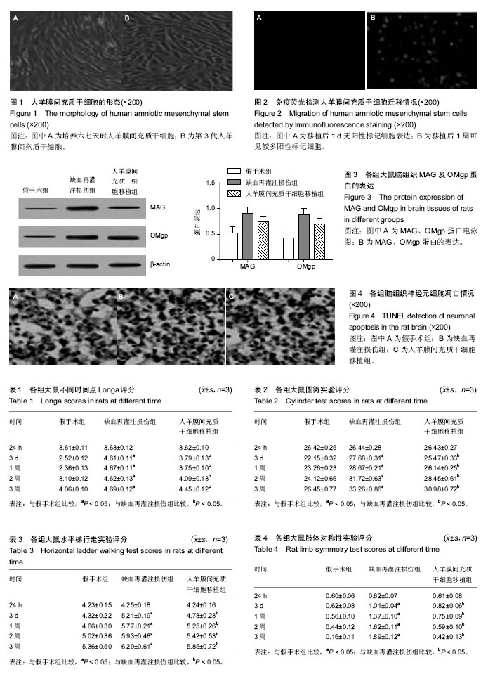
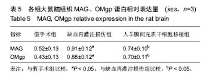
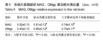
2.1 人羊膜间充质干细胞的形态 人羊膜间充质干细胞复苏后重悬于新鲜的细胞培养液中,除少数细胞贴壁死亡外,多数细胞生长良好,培养六七天即可传代,培养14 d可见呈平形排列或漩涡状集落生长,细胞排列紧密(图1A);第3代细胞生长迅速,纯度高、形态单一(图1B)。实验选取第3代细胞进行移植。 2.2 移植细胞迁移和分布情况 免疫荧光检测显示,细胞移植后第1天在损伤区未观察到阳性细胞,移植后1周在损伤区周围可见红色阳性标记细胞,并且随移植时间的延长,移植细胞表达逐渐增多,见图2。 2.3 实验动物数量分析 实验共纳入60只SD大鼠,均进入结果分析。 2.4 行为学评分 移植后不同时间点进行Longa行为学评分、圆筒实验评分、水平梯行走实验评分、肢体对称性实验评分。结果显示:在移植后24 h各组行为学评分差异无显著性意义;移植后3 d及1,2,3周,细胞移植组较缺血再灌注损伤组神经受损得到明显改善(P < 0.05);对人羊膜间充质干细胞移植组不同时间点进行比较发现,随着时间的延长,大鼠神经功能亦得到明显改善(P < 0.05),而缺血再灌注组不同时间点行为学评分比较,差异无显著性意义(P > 0.05),见表1-4。 2.5 各组大鼠脑组织MAG及OMgp蛋白的表达变化 与假手术组比较,缺血再灌注组MAG及OMgp蛋白的表达量均明显升高;与缺血再灌注损伤组比较,人羊膜间充质干细胞移植组MAG及OMgp蛋白的表达量均显著降低,差异有显著性意义(P < 0.05),见表5和图3。 2.6 TUNEL染色观察神经元凋亡情况 与假手术组相比,缺血再灌注损伤组凋亡细胞数增多;人羊膜间充质干细胞移植组凋亡细胞数目较缺血再灌注损伤组下降,见图4。"
"
| [1] Jia Q, Liu LP, Wang YJ. Stroke in China. Clin Exp Pharmacol Physiol. 2010;37(2):259-264. [2] Pandey AK, Bhattacharya P, Shukla SC,et al.Resveratrol inhibits matrix metalloproteinases to attenuate neuronal damage in cerebral ischemia: a molecular docking study exploring possible neuroprotection. Neural Regen Res. 2015; 10(4): 568-575.[3] Zhao D, Liu J, Wang W, et al. Epidemiological transition of stroke in China: twenty-one-year observational study from the Sino-MONICA-Beijing Project. Stroke. 2008;39(6):1668-1674.[4] 蒋建平,李静,陈永顺,等.冰七片对大鼠局灶性脑缺血再灌注损伤的保护作用研究[J]. 中外医学研究,2016, 14(1):148-149. [5] Gao HJ,Liu PF,Li PW,et al.Ligustrazine monomer against cerebral ischemia/reperfusion injury. Neural Regen Res. 2015;10(5): 832-840.[6] Wu PF, Zhang Z, Wang F, et al. Natural compounds from traditional medicinal herbs in the treatment of cerebral ischemia/reperfusion injury.Acta Pharmacol Sin. 2010;31(12): 1523-1531.[7] Martino G, Pluchino S, Bonfanti L, et al. Brain regeneration in physiology and pathology: the immune signature driving therapeutic plasticity of neural stem cells. Physiol Rev. 2011; 91(4):1281-1304.[8] Cafferty WB, Duffy P, Huebner E, et al. MAG and OMgp synergize with Nogo-A to restrict axonal growth and neurological recovery after spinal cord trauma. J Neurosci. 2010;30(20):6825-6837.[9] Uccelli A, Benvenuto F, Laroni A, et al. Neuroprotective features of mesenchymal stem cells. Best Pract Res Clin Haematol. 2011;24(1):59-64.[10] Andres RH, Horie N, Slikker W, et al. Human neural stem cells enhance structural plasticity and axonal transport in the ischaemic brain.Brain. 2011;134(Pt 6):1777-1789.[11] Zhu JM, Zhao YY, Chen SD, et al. Functional recovery after transplantation of neural stem cells modified by brain-derived neurotrophic factor in rats with cerebral ischaemia. J Int Med Res. 2011;39(2):488-498.[12] Hosseini SM, Farahmandnia M, Razi Z,et al.12 hours after cerebral ischemia is the optimal time for bone marrow mesenchymal stem cell transplantation. Neural Regen Res. 2015;10(6): 904-908.[13] Ba?enková D, Rosocha J, Tóthová T, et al. Isolation and basic characterization of human term amnion and chorion mesenchymal stromal cells. Cytotherapy. 2011;13(9):1047- 1056.[14] Lu Y, Hui GZ,Wu ZY,et al.Transformation of human amniotic epithelial cells into neuron-like cells in the microenvironment of traumatic brain injury in vivo and in vitro. Neural Regen Res. 2011; 6(10):744-749.[15] Díaz-Prado S, Muiños-López E, Hermida-Gómez T, et al. Human amniotic membrane as an alternative source of stem cells for regenerative medicine. Differentiation. 2011;81(3): 162-171.[16] Huang H, Liu N, Wang JH, et al. The effects of adipose-derived stem cells transplantation on the expression of TGF-β1 in rat brain after cerebral ischemia. Xi Bao Yu Fen Zi Mian Yi Xue Za Zhi. 2011;27(8):872-875.[17] Sapin V, Souteyrand G, Bonnin N, et al. Therapeutic use of amniotic membranes and their derivate cells. Gynecol Obstet Fertil. 2011;39(6):388-390.[18] Sun F, Xie L, Mao X, et al. Effect of a contralateral lesion on neurological recovery from stroke in rats. Restor Neurol Neurosci. 2012;30(6):491-495.[19] Pignataro G, Meller R, Inoue K, et al. In vivo and in vitro characterization of a novel neuroprotective strategy for stroke: ischemic postconditioning. J Cereb Blood Flow Metab. 2008; 28(2):232-241.[20] Zhang Q, Yuan W, Wang G, et al. The protective effects of a phosphodiesterase 5 inhibitor, sildenafil, on postresuscitation cardiac dysfunction of cardiac arrest: metabolic evidence from microdialysis. Crit Care. 2014;18(6):641.[21] 王灿,王琳琳,苗明三.脑脉舒康胶囊对沙土鼠脑缺血再灌注损伤的影响[J].中华中医药杂志,2014,29(7):2352-2355.[22] Miao MS,Guo L,Li RQ,et al.Radix Ilicis Pubescentis total flavonoids combined with mobilization of bone marrow stem cells to protect against cerebral ischemia/reperfusion injury. Neural Regen Res. 2016;11(2): 278-284.[23] 刘灵芝.巴戟天醇提物对离体大鼠缺血再灌注损伤心肌能量代谢的影响[J].中国医药导报,2012,9(28):10-11。[24] 张向前,王雪侠.巴戟天醇提物对大鼠肾缺血再灌注损伤的保护作用[J].中国医药导报,2013,10(5):22-24.[25] 赵刚,王伟民,杨帅,等.白黎芦醇对大鼠脑缺血再灌注损伤的保护作用[J].中国老年学杂志,2014,27(21):6149-6150.[26] 肖爱娇,陈日新,康明非,等.热敏灸对脑缺血再灌注损伤大鼠SOD、MDA的影响[J].天津医药,2014,27(1):51-53.[27] Peng B, Guo QL, He ZJ, et al. Remote ischemic postconditioning protects the brain from global cerebral ischemia/reperfusion injury by up-regulating endothelial nitric oxide synthase through the PI3K/Akt pathway. Brain Res. 2012;1445:92-102.[28] Wang WB,Yang LF,He QS,et al.Mechanisms of electroacupuncture effects on acute cerebral ischemia/ reperfusion injury: possible association with upregulation of transforming growth factor beta 1. Neural Regen Res. 2016; 11(7): 1099-1101.[29] Xie D, Zhang EL, Li JM, et al. Design, synthesis and anti-platelet aggregation activities of ligustrazine- tetrahydroisoquinoline derivatives. Yao Xue Xue Bao. 2015; 50(3):326-331. [30] Hu J, Lang Y, Cao Y, et al. The Neuroprotective Effect of Tetramethylpyrazine Against Contusive Spinal Cord Injury by Activating PGC-1α in Rats. Neurochem Res. 2015;40(7): 1393-1401.[31] Li N, Deng XG, Zhang SH, et al. Effects of different concentrations of tetramethylpyrazine, an active constituent of Chinese herb, on human corneal epithelial cell damaged by hydrogen peroxide. Int J Ophthalmol. 2014;7(6):947-951.[32] 马淑媛,高芳.川穹嗪注射液治疗急性脑梗死临床疗效观察[J].牡丹江医学院学报,2008, 29(6): 34-35.[33] 余奇劲,杨洁,陈娟. 缺血前和缺血后联合应用丙泊酚对兔脊髓缺血再灌注损伤的保护作用[J].中华临床医师杂志,2011, 5(20): 5955-5959.[34] Moon JH, Lee JR, Jee BC, et al. Successful vitrification of human amnion-derived mesenchymal stem cells. Hum Reprod. 2008;23(8):1760-1770.[35] Lei L,Zhou RX. Migration and differentiation of bone marrow-derived multipotent adult progenitor cells through tail vein injection in a rat model of cerebral ischemia. Neural Regen Res. 2009; 4(2):118-122.[36] Pan FH, Ding XS, Ding HX, et al. Stem cell transplantation for treatment of cerebral ischemia in rats: effects of human umbilical cord blood stem cells and human neural stem cells. Neural Regen Res. 2010; 5(7):485-490.[37] 郝永岗,张正春,薛群,等.骨髓间充质干细胞移植治疗脑出血的实验研究[J].中国急救医学,2009,29(7):615-618. |
| [1] | Yao Xiaoling, Peng Jiancheng, Xu Yuerong, Yang Zhidong, Zhang Shuncong. Variable-angle zero-notch anterior interbody fusion system in the treatment of cervical spondylotic myelopathy: 30-month follow-up [J]. Chinese Journal of Tissue Engineering Research, 2022, 26(9): 1377-1382. |
| [2] | Wang Shuo, Liu Wenying, Lü Chaofan, Li Jiacong, Geng Yi, Zhao Yungang. Cardioprotective effect of 3-nitro-N-methyl salicylamide on the isolated rat heart under cold ischemia preservation [J]. Chinese Journal of Tissue Engineering Research, 2022, 26(8): 1194-1201. |
| [3] | Li Zhiyi, He Pengcheng, Bian Tianyue, Xiao Yuxia, Gao Lu, Liu Huasheng. Bibliometric and visualized analysis of ferroptosis mechanism research [J]. Chinese Journal of Tissue Engineering Research, 2022, 26(8): 1202-1209. |
| [4] | Yang Shenglin, Pu Xingwei, Luo Chunshan, Yang Jianwen. Neuroprotective effects of tetrandrine preconditioning in rabbits with spinal cord ischemia-reperfusion injury [J]. Chinese Journal of Tissue Engineering Research, 2022, 26(8): 1223-1227. |
| [5] | An Weizheng, He Xiao, Ren Shuai, Liu Jianyu. Potential of muscle-derived stem cells in peripheral nerve regeneration [J]. Chinese Journal of Tissue Engineering Research, 2022, 26(7): 1130-1136. |
| [6] | Zhang Jinglin, Leng Min, Zhu Boheng, Wang Hong. Mechanism and application of stem cell-derived exosomes in promoting diabetic wound healing [J]. Chinese Journal of Tissue Engineering Research, 2022, 26(7): 1113-1118. |
| [7] | Dong Miaomiao, Lai Han, Li Manling, Xu Xiuhong, Luo Meng, Wang Wenhao, Zhou Guoping. Effect of electroacupuncture on expression of nucleotide binding oligomerization domain-like receptor protein 3/cysteinyl aspartate specific proteinase 1 in rats with cerebral ischemia/reperfusion injury [J]. Chinese Journal of Tissue Engineering Research, 2022, 26(5): 749-755. |
| [8] | He Yunying, Li Lingjie, Zhang Shuqi, Li Yuzhou, Yang Sheng, Ji Ping. Method of constructing cell spheroids based on agarose and polyacrylic molds [J]. Chinese Journal of Tissue Engineering Research, 2022, 26(4): 553-559. |
| [9] | He Guanyu, Xu Baoshan, Du Lilong, Zhang Tongxing, Huo Zhenxin, Shen Li. Biomimetic orientated microchannel annulus fibrosus scaffold constructed by silk fibroin [J]. Chinese Journal of Tissue Engineering Research, 2022, 26(4): 560-566. |
| [10] | Chen Xiaoxu, Luo Yaxin, Bi Haoran, Yang Kun. Preparation and application of acellular scaffold in tissue engineering and regenerative medicine [J]. Chinese Journal of Tissue Engineering Research, 2022, 26(4): 591-596. |
| [11] | Kang Kunlong, Wang Xintao. Research hotspot of biological scaffold materials promoting osteogenic differentiation of bone marrow mesenchymal stem cells [J]. Chinese Journal of Tissue Engineering Research, 2022, 26(4): 597-603. |
| [12] | Shen Jiahua, Fu Yong. Application of graphene-based nanomaterials in stem cells [J]. Chinese Journal of Tissue Engineering Research, 2022, 26(4): 604-609. |
| [13] | Zhang Tong, Cai Jinchi, Yuan Zhifa, Zhao Haiyan, Han Xingwen, Wang Wenji. Hyaluronic acid-based composite hydrogel in cartilage injury caused by osteoarthritis: application and mechanism [J]. Chinese Journal of Tissue Engineering Research, 2022, 26(4): 617-625. |
| [14] | Li Hui, Chen Lianglong. Application and characteristics of bone graft materials in the treatment of spinal tuberculosis [J]. Chinese Journal of Tissue Engineering Research, 2022, 26(4): 626-630. |
| [15] | Gao Cangjian, Yang Zhen, Liu Shuyun, Li Hao, Fu Liwei, Zhao Tianyuan, Chen Wei, Liao Zhiyao, Li Pinxue, Sui Xiang, Guo Quanyi. Electrospinning for rotator cuff repair [J]. Chinese Journal of Tissue Engineering Research, 2022, 26(4): 637-642. |
| Viewed | ||||||
|
Full text |
|
|||||
|
Abstract |
|
|||||