Chinese Journal of Tissue Engineering Research ›› 2017, Vol. 21 ›› Issue (36): 5886-5891.doi: 10.3969/j.issn.2095-4344.2017.36.025
Previous Articles Next Articles
Endoplasmic reticulum stress and apoptosis of chondrocytes
Xiong Fei1, Wei Yi-shan2
- 1Inner Mongolia Medical University, Hohhot 010059, Inner Mongolia Autonomous Region, China; 2Department of Pediatric Orthopedics, the Second Affiliated Hospital of Inner Mongolia Medical University, Hohhot 010059, Inner Mongolia Autonomous Region, China
-
Received:2017-07-30Online:2017-12-28Published:2018-01-04 -
Contact:Wei Yi-shan, M.D., Chief physician, Department of Pediatric Orthopedics, the Second Affiliated Hospital of Inner Mongolia Medical University, Hohhot 010059, Inner Mongolia Autonomous Region, China -
About author:Xiong Fei, Studying for master’s degree, Inner Mongolia Medical University, Hohhot 010059, Inner Mongolia Autonomous Region, China
CLC Number:
Cite this article
Xiong Fei1, Wei Yi-shan2. Endoplasmic reticulum stress and apoptosis of chondrocytes[J]. Chinese Journal of Tissue Engineering Research, 2017, 21(36): 5886-5891.
share this article
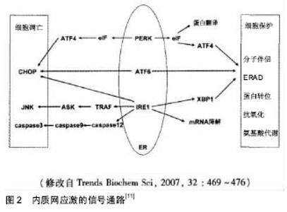
2.1 内质网应激与细胞凋亡 遗传或环境损伤会引起细胞内钙稳态失衡、氧化应激、营养缺乏、糖基化抑制和蛋白质错误折叠,从而破环内质网功能,内质网应激随之形成[2-5],细胞中会出现未折叠蛋白反应,然后内质网超载反应会随之出现,最后会减少蛋白质的合成,同时蛋白质会出现正确的折叠,应激过度也会启动对应的凋亡分子[6-7]。 2.1.1 未折叠蛋白反应和细胞凋亡 蛋白激酶样内质网激酶(PERK)及转录激活因子6(ATF6)和1型内质网转膜蛋白激酶(IRE-1),它们是内质网应激的感受蛋白质,能够联结GRP78/BIP构成平稳的复合物,介导未折叠蛋白反应[8]。当内质网应激没有发生时,它们处于结合的状态,该状态没有活性,当内质网应激发生时,积聚的未折叠蛋白能够让GRP78/BIP从3种跨膜的蛋白上分离开来,转而去连接未折叠蛋白。感受蛋白分离后会被活化并导致未折叠蛋白反应的产生[3,9-10],未折叠蛋白反应可以庇护由内质网应激所激发的细胞损害,修复细胞功能,有停歇初期的蛋白质组成,内质网的分子伴侣和折叠有关的酶的转录激活和内质网有关降解,为了提高不正确蛋白质折叠的处理,增进内质网对积蓄的蛋白质处置,有利于维护细胞的一般功能和细胞的生存。 PERK、ATF6和IRE-l信号不光是可以发动ERS的生存路线,惨重或长期的内质网应激毁损了内质网的功能时,内质网应激所带领的凋亡的信号线路照样能够被这3个信号通路启动(图2)[11],致使细胞产生凋亡,然后去掉受伤的细胞.目前认为,激活CHOP/GADD153基因的转录路线;C-Jun氨基酸末端激酶(C-Jun N-terminal kinase,JNK) 的激活通路;内质网中Caspase-12被激活。"
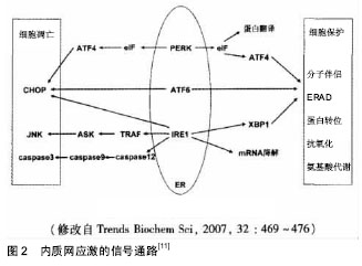
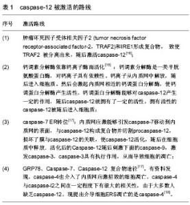
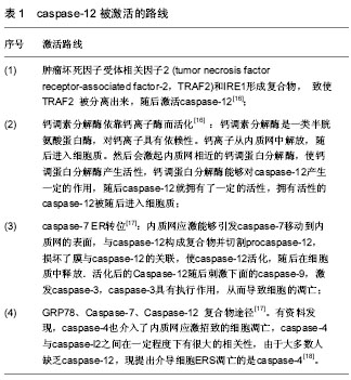
CHOP/GADD153基因的激活转录:CHOP/ GADD153内质网应激特异的转录因子,CHOP从属于转录因子 CCAAT/增强子结合蛋白(C/EBP)家族,经常结合转录因子CCAAT/增强子结合蛋白(C/EBP)家族的其他成员,然后会导致二聚体的出现,在一般的情况下,基本不会有CHOP的表达。当出现一个内质网的应激时,通过激活IRE1、PERK和 ATF-6能够产生大量的CHOP。在内质网应激反应的跨膜蛋白IRE1 和ATF-6激活后进入细胞核,连接ERS反应元件,驱动CHOP转录与表达,最后致使凋亡。ATF4在PERK/elF2α的下游,AARE域在CHOP启动子上,ATF4能够与AARE 域联合,随后出现CHOP的表达,在CHOP的表达中PERK与elF2α及其ATF4三者形成的复合物是必不可少的。有研究说明,升高CHOP,将能够对抗细胞凋亡的蛋白物Bcl-2的表达起下降作 用[12],对细胞内的谷胱甘肽的产生有抑制的作用,也对促成反馈性氧中介物(的产生有着抑制的作用,进而发生细胞凋亡。 c-Jun氨基端激酶激活途径:应激条件下,IRE1活化后能够召集C-JUN氨基末端激酶(C-Jun N-terminal kinase,JNK)和肿瘤坏死因子受体有关因子(TRAF2),肿瘤坏死因子受体有关因子刺激细胞凋亡信号激酶1 (ASK1),继而发生IRE-1、TRAF2和ASK1构成的三聚体,然后引起C-JUN氨基末端激酶,并诱导细胞凋亡[13-14]。 Caspase -12的激活:细胞凋亡中内质网的应激的关键分子是caspase-12,只有在内质网应激发生的时候才能被激活,而在线粒体或死亡受体凋亡路线中不被激活[15]。当内质网产生应激时, caspase-12能够经过以下的路线被激活(表1)。"
| [1] Gorman AM,Healy SJ,Jäger R,et al.Stress managementat theER:regulators of ER stress-inducedapoptosis.Pharmacol Ther.2012;134(3):306-316.[2] Paschen W,Frandsen A. Endoplasmic reticulum dysfunction acommon denominator for cell injury in acute and degenerativediseases of the brain.J Neurochem.2001;79(4) : 719-725.[3] Schroder M,Kaufman RJ. ER stress and the unfolded proteinresponse. Mutat Res.2005;569(1-2):29-63.[4] Zhang HY,Wang ZG,Lu XH,et al.Endoplasmic reticulumstress: relevance and therapeutics in central nervous systemdiseases.Mol Neurobiol.2015;51(3) : 1343-1352.[5] Breckenridge DG,Germain M,Mathai JP,et al.Regulation ofapoptosis by endoplasmic reticulum pathways. Oncogene.2003;22(53):8608-8618.[6] Volmer R,van der Ploeg K,Ron D.Membrane lipid saturation activates endoplasmic reticulum unfolded protein responsetransducers through their transmembrane domains. Proc Natl AcadSci USA.2013;110(12) : 4628-4633.[7] Hetz C,Chevet E,Harding HP.Targeting the unfolded proteinresponse in disease.Nat Rev Drug Discov.2013;12(9):703-719.[8] Szegezdi E,Logue S,Gorman A,et al.Mediators of endoplasmicreticulum stress-induced apoptosis. EMBO Rep.2006;7 (9) :880-885.[9] Shen J,Chen X,Hendershot L,et al. ER stress regulation ofATF6 localization by dissociation of BiP /GRP78 binding andunmasking of Golgi localization signals.Dev Cell.2002;3(1):99-111.[10] Bertolotti A,Zhang Y,Hendershot LM,et al. Dynamicinteraction of BiP and ER stress transducers in the unfoldedproteinresponse. Nat Cell Biol.2000;2: 326-332.[11] 滕旭,齐永芬,唐朝枢.内质网应激与心脏疾病[J].生理科学进展,2009, 40(2):106-110.[12] Fu HY,Okada K,Liao Y,et al.Ablation of C/EBP homologousproteinatteuates ER-mediatedapoptosis and cardiacdysfunction induced by pressureoverload. Circulation. 2010;122(4):361-369.[13] Yoneda T,Imaizumi K,Oono K,et al. Activation of Caspase-12,an endoplastic reticulum (ER) resident caspase,throughtumor necrosis factor receptor-associated factor 2-dependentmechanism in response to the ER stress. J Biol Chem.2001;276(17):13935-13940.[14] Tabas I,Ron D.Integrating the mechanisms of apoptosis inducedby endoplasmic reticulum stress.Nat Cell Biol. 2011;13(3):184-190.[15] 张凯强,张颖,顾何锋,等. 内质网应激与细胞凋亡研究进展[J]. 口腔医学,2013,33(6):415-417.[16] 张红菊, 赵忠新.大鼠脑缺血后内质网伴侣蛋白GRP78 和GRP94及胱冬酶12表达的改变[J]. 中国脑血管病杂志,2006, 3(4):164-166.[17] 刘洪亮,崔玉山. Caspase-12在内质网应激中的激活途径及与疾病关系研究进展[J]. 环境卫生学杂志,2011,1(4):42-43.[18] 杨琼,吴永全.内质网应激与心血管疾病[J]. 心脏杂志, 2015, 27(1):99-101.[19] 周婷,刘铁夫.内质网应激与肝脏疾病[J]. 现代生物医学进展, 2012,12(30):5982-5984.[20] Taniguchi N, Carames B, Ronfani L, et al. Aging-related loss ofthe chromatin protein HMGB2 in articular cartilage is linked toreduced cellularity and osteoarthritis. Proc Natl Acad Sci USA.2009;106(4):1181-1186.[21] Johnson EO, Charchandi A, Babis GC, et al. Apoptosis inosteoarthritis: morphology, mechanisms, and potential meansfor therapeutic intervention. J Surg Orthop Adv.2008;17(3):147-152.[22] Kawaguchi H. Mechanism of molecular backgrounds of osteoarthritis.Nihon Rinsho.2014;72(10):1729-1733.[23] Saito S, Murakoshi K, Kotake S, et al. Granzyme B inducesapoptosis of chondrocytes with natural killer cell-like cytotoxicityin rheumatoid arthritis. J Rheumatol.2008;35(10): 1932-1943.[24] Becker SJ, Teunis T, Blauth J, et al. Medical services and associated costs vary widely among surgeons treating patients with hand osteoarthritis. Clin Orthop Relat Res. 2015;473(3):1111-1117.[25] Palmieri B, Lodi D, Capone S. Osteoarthritis and degenerativejoint disease: local treatment options update. Acta Biomed.2010;81(2):94-100.[26] Ryu JH, Shin Y, Huh YH, et al. Hypoxia-inducible factor-2alpharegulates Fas-mediated chondrocyte apoptosisduringosteoarthritic cartilage destruction. Cell Death Differ. 2012;19(3):440-450.[27] Yang L, Carlson SG, McBurney D, et al. Multiple signalsinduce endoplasmic reticulum stress in both primary andimmortalized chondrocytes resulting in loss of differentiation,impaired cell growth, and apoptosis. J Biol Chem.2005;280(35): 31156-31165.[28] Oliver BL, Cronin CG, Zhang-Benoit Y, et al. Divergent stressresponses to IL-1beta, nitric oxide, and tunicamycin bychondrocytes. J Cell Physiol. 2005;204(1): 45-50.[29] Nugent AE, Speicher DM, Gradisar I, et al. Advancedosteoarthritis in humans is associated with altered collagen VIexpression and upregulation of ER-stress markers Grp78 andbag-1. J Histochem Cytochem. 2009; 57(10):923-931.[30] Yamabe S, Hirose J, Uehara Y, et al. Intracellular accumulationof advanced glycation end products induces apoptosis viaendoplasmic reticulum stress in chondrocytes. FEBS J.2013;280(7):1617-1629.[31] Palmieri B, Lodi D, Capone S. Osteoarthritis and degenerativejoint disease: local treatment options update. Acta Biomed.2010;81(2):94-100.[32] Ron D, Walter P. Signal integration in the endoplasmicreticulum unfolded protein response. Nat Rev Mol Cell Biol.2007;8(7): 519-529.[33] Tsang KY, Chan D, Cheslett D, et al. Surviving endoplasmicreticulum stress is coupled to altered chondrocyte differentiation and function. PLoS Biol. 2007; 5(3): e44.[34] van Meurs JB, Uitterlinden AG. Osteoarthritis year 2012 inreview: genetics and genomics. OsteoarthritisCartilage. 2012;20(12):1470-1476.[35] Thomas CM, Fuller CJ, Whittles CE, et al.Chondrocyte deathby apoptosis is associated with the initiation andseverity ofarticular cartilage degradation. Int J Rheum Dis.2011;14(2):191-198.[36] Li H, Zhang XY, Wu TJ, et al. Endoplasmic reticulum stressregulates rat mandibular cartilage thinning undercompressivemechanical stress. J Biol Chem.2013; 288(25): 18172-18183.[37] Rasheed Z, Haqqi TM. Endoplasmic reticulum stress inducesthe expression of COX-2 through activation of eIF2alpha,p38-MAPK and NF-kappaB in advanced glycation endproducts stimulated human chondrocytes. Biochim Biophys Acta. 2012;1823(12): 2179-2189.[38] Takada K, Hirose J, Senba K, et al. Enhanced apoptotic andreduced protective response in chondrocytes followingendoplasmic reticulum stress in osteoarthritic cartilage.Int JExp Pathol.2011;92(4): 232-242.[39] 李载权,周爱儒,唐朝枢.内质网应激反应分子机理研究进展[J].中国生物化学与分子生物学报, 2004, 20(3): 283-288.[40] Feng J, Li S, Chen H. Tanshinone IIA ameliorates apoptosis of cardiomyocytes induced by endoplasmic reticulum stress. Experimental biology and medicine (Maywood, NJ).2016, 241(18): 2042-2048.[41] Guo R, Wu Z, Jiang J,et al. New mechanism of lipotoxicity in diabetic cardiomyopathy: Deficiency of Endogenous H2S Production and ER stress. Mech Ageing Dev. 2017;162: 46-52.[42] Li B, Tian J, Sun Y,et al. Activation of NADPH oxidase mediates increased endoplasmic reticulum stress and left ventricular remodeling after myocardial infarction in rabbits. Biochim Biophys Acta. 2015;1852(5):805-815.[43] Lin Y,Zhang X,Wang L,et al.Polyamine depletion attenuates isoproterenol-induced hypertrophy and endoplasmic reticulum stress in cardiomyocytes. Cell Physiol Biochem. 2014;34(5):1455-1465.[44] Lin Y, Zhang X, Xiao W,et al. Endoplasmic Reticulum Stress is Involved in DFMO Attenuating Isoproterenol-Induced Cardiac Hypertrophy in Rats. Cell Physiol Biochem. 2016; 38(4):1553-1562.[45] Liu X, Kwak D, Lu Z,et al. Endoplasmic reticulum stress sensor protein kinase R-like endoplasmic reticulum kinase (PERK) protects against pressure overload-induced heart failure and lung remodeling. Hypertension (Dallas, Tex : 1979).2014;64(4): 738-744.[46] Meng C, Yuan CH, Zhang CC,et al. Ophiopogonin D protects cardiomyocytes against doxorubicin-induced injury through suppressing endoplasmic reticulum stress. Yao Xue Xue Bao. 2014;49(8):1117-123.[47] Sreedhar R, Giridharan VV, Arumugam S,et al. Role of MAPK-mediated endoplasmic reticulum stress signaling in the heart during aging in senescence-accelerated prone mice. BioFactors (Oxford, England).2016;42(4): 368-375.[48] Tse G, Yan BP, Chan YW,et al. Reactive Oxygen Species, Endoplasmic Reticulum Stress and Mitochondrial Dysfunction: The Link with Cardiac Arrhythmogenesis. Front Physiol. 2016;7:313.[49] Xu Z, Zhao Y, Zhong P,et al. EGFR inhibition attenuates diabetic nephropathy through decreasing ROS and endoplasmic reticulum stress. Oncotarget.2017;8(20): 32655-32667.[50] Zhang B, He P, Lu Y,et al. HSF1 Relieves Amyloid-beta-Induced Cardiomyocytes Apoptosis. Cell biochem biophys. 2015;72(2): 579-587.[51] Zhang GG, Cai HQ, Li YH,et al. Ghrelin protects heart against ERS-induced injury and apoptosis by activating AMP-activated protein kinase. Peptides.2013;48:156-165.[52] Zhang Q, Lu L, Liang T,et al. MAPK pathway regulated the cardiomyocyte apoptosis in mice with post-infarction heart failure . Bratislavske lekarske listy, 2017;118(6): 339-346.[53] Zhang Z,Zhao L,Zhou Y,et al.Taurine ameliorated homocysteine-induced H9C2 cardiomyocyte apoptosis by modulating endoplasmic reticulum stress. Apoptosis. 2017; 22(5):647-661.[54] Ahn C,An BS,Jeung EB.Streptozotocin induces endoplasmic reticulum stress and apoptosis via disruption of calcium homeostasis in mouse pancreas. Mol Cell Endocrinol. 2015; 412:302-308.[55] Ali BR.Is cystic fibrosis-related diabetes an apoptotic consequence of ER stress in pancreatic cells?. Med Hypotheses. 2009; 72(1):55-57.[56] Carrera Boada CA, Martinez-Moreno JM. Pathophysiology of diabetes mellitus type 2: beyond the duo "insulin resistance-secretion deficit" .Nutricion hospitalaria.2013; 28 Suppl 2:78-87.[57] Clemens DL, Schneider KJ, Arkfeld CK,et al. Alcoholic pancreatitis: New insights into the pathogenesis and treatment. World J Gastrointest Pathophysiol. 2016; 7(1): 48-58.[58] Martinez-Useros J, Georgiev-Hristov T, Borrero-Palacios A,et al. Identification of Poor-outcome Biliopancreatic Carcinoma Patients With Two-marker Signature Based on ATF6alpha and p-p38 "STARD Compliant". Medicine (Baltimore). 2015;94(45):e1972.[59] Zhao L, Guo H, Chen H,et al. Effect of Liraglutide on endoplasmic reticulum stress in diabetes. Biochem Biophys Res Commun. 2013 ;441(1):133-138. |
| [1] | Yao Xiaoling, Peng Jiancheng, Xu Yuerong, Yang Zhidong, Zhang Shuncong. Variable-angle zero-notch anterior interbody fusion system in the treatment of cervical spondylotic myelopathy: 30-month follow-up [J]. Chinese Journal of Tissue Engineering Research, 2022, 26(9): 1377-1382. |
| [2] | An Weizheng, He Xiao, Ren Shuai, Liu Jianyu. Potential of muscle-derived stem cells in peripheral nerve regeneration [J]. Chinese Journal of Tissue Engineering Research, 2022, 26(7): 1130-1136. |
| [3] | Wen Dandan, Li Qiang, Shen Caiqi, Ji Zhe, Jin Peisheng. Nocardia rubra cell wall skeleton for extemal use improves the viability of adipogenic mesenchymal stem cells and promotes diabetes wound repair [J]. Chinese Journal of Tissue Engineering Research, 2022, 26(7): 1038-1044. |
| [4] | Zhang Yujie, Yang Jiandong, Cai Jun, Zhu Shoulei, Tian Yuan. Mechanism by which allicin inhibits proliferation and promotes apoptosis of rat vascular endothelial cells [J]. Chinese Journal of Tissue Engineering Research, 2022, 26(7): 1080-1084. |
| [5] | Zhang Jinglin, Leng Min, Zhu Boheng, Wang Hong. Mechanism and application of stem cell-derived exosomes in promoting diabetic wound healing [J]. Chinese Journal of Tissue Engineering Research, 2022, 26(7): 1113-1118. |
| [6] | Deng Shuang, Pu Rui, Chen Ziyang, Zhang Jianchao, Yuan Lingyan . Effects of exercise preconditioning on myocardial protection and apoptosis in a mouse model of myocardial remodeling due to early stress overload [J]. Chinese Journal of Tissue Engineering Research, 2022, 26(5): 717-723. |
| [7] | He Yunying, Li Lingjie, Zhang Shuqi, Li Yuzhou, Yang Sheng, Ji Ping. Method of constructing cell spheroids based on agarose and polyacrylic molds [J]. Chinese Journal of Tissue Engineering Research, 2022, 26(4): 553-559. |
| [8] | He Guanyu, Xu Baoshan, Du Lilong, Zhang Tongxing, Huo Zhenxin, Shen Li. Biomimetic orientated microchannel annulus fibrosus scaffold constructed by silk fibroin [J]. Chinese Journal of Tissue Engineering Research, 2022, 26(4): 560-566. |
| [9] | Chen Xiaoxu, Luo Yaxin, Bi Haoran, Yang Kun. Preparation and application of acellular scaffold in tissue engineering and regenerative medicine [J]. Chinese Journal of Tissue Engineering Research, 2022, 26(4): 591-596. |
| [10] | Kang Kunlong, Wang Xintao. Research hotspot of biological scaffold materials promoting osteogenic differentiation of bone marrow mesenchymal stem cells [J]. Chinese Journal of Tissue Engineering Research, 2022, 26(4): 597-603. |
| [11] | Shen Jiahua, Fu Yong. Application of graphene-based nanomaterials in stem cells [J]. Chinese Journal of Tissue Engineering Research, 2022, 26(4): 604-609. |
| [12] | Zhang Tong, Cai Jinchi, Yuan Zhifa, Zhao Haiyan, Han Xingwen, Wang Wenji. Hyaluronic acid-based composite hydrogel in cartilage injury caused by osteoarthritis: application and mechanism [J]. Chinese Journal of Tissue Engineering Research, 2022, 26(4): 617-625. |
| [13] | Li Hui, Chen Lianglong. Application and characteristics of bone graft materials in the treatment of spinal tuberculosis [J]. Chinese Journal of Tissue Engineering Research, 2022, 26(4): 626-630. |
| [14] | Gao Cangjian, Yang Zhen, Liu Shuyun, Li Hao, Fu Liwei, Zhao Tianyuan, Chen Wei, Liao Zhiyao, Li Pinxue, Sui Xiang, Guo Quanyi. Electrospinning for rotator cuff repair [J]. Chinese Journal of Tissue Engineering Research, 2022, 26(4): 637-642. |
| [15] | Guan Jian, Jia Yanfei, Zhang Baoxin , Zhao Guozhong. Application of 4D bioprinting in tissue engineering [J]. Chinese Journal of Tissue Engineering Research, 2022, 26(3): 446-455. |
| Viewed | ||||||
|
Full text |
|
|||||
|
Abstract |
|
|||||