Chinese Journal of Tissue Engineering Research ›› 2017, Vol. 21 ›› Issue (27): 4348-4353.doi: 10.3969/j.issn.2095-4344.2017.27.015
Previous Articles Next Articles
Anatomical characters of the proximal tibial osteotomy in healthy Mongolia populations
Zheng Jin-yang1, Ren Zhi-yong1, Zhang Guo-liang2, Mori Gele2, Puri Busurong2, Li Qiang2, Wang Yue-wen2
- 1Orthopedic Center, Sunshine Union Hospital of Weifang, Weifang 261061, Shandong Province, China; 2Department of Orthopedics, the Affiliated Hospital of Inner Mongolia Medical University, Hohhot 010050, Inner Mongolia Autonomous Region, China
-
Online:2017-09-28Published:2017-10-24 -
Contact:Wang Yue-wen, Professor, Master’s supervisor, Department of Orthopedics, the Affiliated Hospital of Inner Mongolia Medical University, Hohhot 010050, Inner Mongolia Autonomous Region, China -
About author:Zheng Jin-yang, Master, Orthopedic Center, Sunshine Union Hospital of Weifang, Weifang 261061, Shandong Province, China -
Supported by:the Medical Health Research Project of Health and Family Planning Commission of Inner Mongolia Autonomous Region, No. 201301054; the Research Project of Universities in Inner Mongolia Autonomous Region, No. NJ09112
CLC Number:
Cite this article
Zheng Jin-yang, Ren Zhi-yong, Zhang Guo-liang, Mori Gele, Puri Busurong, Li Qiang, Wang Yue-wen. Anatomical characters of the proximal tibial osteotomy in healthy Mongolia populations [J]. Chinese Journal of Tissue Engineering Research, 2017, 21(27): 4348-4353.
share this article
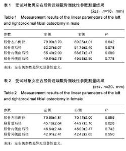
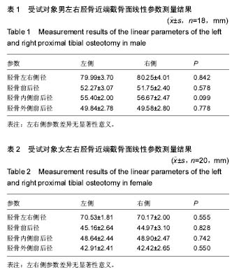
2.1 受试者数量分析 健康蒙古族成人志愿者38例,均行膝关节CT扫描,数据保存完整,无毁损及缺失,全部CT数据用于结果分析。 2.2 受试对象同性别间左、右胫骨近端截骨面各线性参数比较 蒙古族人(男/女)左、右侧胫骨近端截骨面各线性参数差异无显著性意义(P > 0.05;表1,2)。"

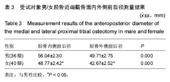
2.3 受试对象同性别间内、外侧胫骨近端截骨面各线性参数比较 本组蒙古族人(男/女)内、外侧胫骨近端截骨面对应的各线性参数差异有显著性意义(P < 0.05;表3)。"
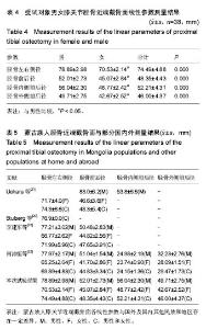
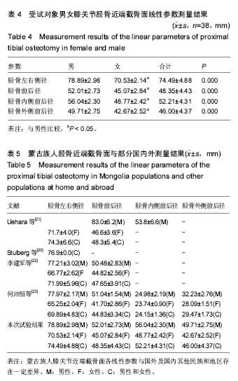
2.4 受试对象男女胫骨近端截骨面各线性参数比较 本组蒙古族人男女胫骨近端截骨面对应的各线性参数差异有显著性意义(P < 0.05;表4)。 2.5 试验结果与既往文献比较 蒙古族人膝关节近端截骨面各线性参数与国外及国内其他民族和地区存在一定差异(表5)。"
| [1] Galante J. Overaw of current attempts to eliminate methyl-methacylate. J Bone. 1980;60:A181-A189.[2] 阿布力克木,王成伟,曲铁兵,等.维吾尔族人正常胫骨平台线性参数测量及特性分析[J].中国骨与关节外科,2009,3(2):225-228.[3] 谷长增,朱新晨,季明华,等.维吾尔族正常膝关节参数的测量结果及其意义[J].创伤外科杂志,2015,1(17):46-49.[4] 张国梁,吕铁刚,赵建民,等.正常蒙古族人股骨外旋角的测量及临床意义[J].内蒙古医学院学报,2012,34(4):316-320.[5] 李强,赵建民,王跃文,等.蒙古族人正常髌骨三维测量[J].内蒙古医学院学报,2012,5(34):349-355.[6] 黄健,张志峰,张元智,等.汉族与蒙古族股骨后髁角及股骨面率的数字化测量对比研究[J].中华关节外科杂志(电子版),2015,1(9): 67-70.[7] Moreland JR, Bassett LW, Hanker GJ. Radiograghic analysis of the axial alignment of the lower extremity. J Bone Joint Surg (Am). 1987;69:745-749.[8] Kwak DS, Surendran S, Pengatteeri YH. Morphometry of the proximal tibia to design the tibial component of total knee arthroplasty for the Korean population. Knee. 2007;14:295-300. [9] Li GA, Suggs J, Hanson G, et al. Three-dimensional tibiofemoral articular contact kinematics of a cruciate-retaining total knee arthroplasty. J Bone Joint Surg Am. 2006;88: 395-402. [10] Siu D, Rudan J, Wevers HW, et al. Femoral articular shape and Geometry A three-dimensional computerized analysis of the knee. J Arthroplasty. 1996;11(2):166-173.[11] Liau JJ, Cheng CK, Huang CH, et al. The effect of malalignment on stresses in polyethylene component of total knee prosthses a finit element analysis. Clin Biomech. 2002; 17(2):140-146.[12] Godest AC, Beaugonin M, Haug E, et al. Simulation of a knee joint replacement during a gait cycle using explicit finite element analysis. J Biomech. 2002;35(2):267-275.[13] Myers H, Davis A, Lazicki R, et al. Sex differences in rectus femoris morphology across different knee flexion positions. Int J Sports Phys Ther. 2013;8(2):84-90.[14] Liebs TR, Kloos SA, Herzberg W, et al. The significance of an asymmetric extension gap on routine radiographs after total knee replacement: a new sign and its clinical significance. Bone Joint J. 2013;95-B(4):472-477. [15] Burkhart TA, Asa B, Payne MW, et al. Anatomy of the proximal tibiofibular joint and interosseous membrane, and their contributions to joint kinematics in below-knee amputations. J Anat. 2015;226(2):143-149. [16] 徐勃兴,王伟,张力,等.三维数字化重建在中国人胫骨近端形态学研究中的应用[J].中国组织工程研究,2012,16(44):8218-8222. [17] Moore SR, Milz S, Knothe Tate ML. Periosteal thickness and cellularity in mid-diaphyseal cross-sections from human femora and tibiae of aged donors. J Anat. 2014;224(2): 142-149.[18] 郭胜杰,周一新,邵宏翊,等.正常髌骨解剖形态学的CT研究[J].中华骨科杂志,2013,33(3):273-277.[19] 王涛,王坤正,王伟,等.西北地区中老年人正常膝关节影像学测量及临床意义[J].西安交通大学学报,2010,31(2):224-226.[20] Baldwin JL, House CK. Anatomic dimensions of the patella measured during total knee arthroplasty. J Arthroplasty. 2005;20(2):250-257. [21] Kim TK, Chung BJ, Kang YG, et al. Clinical implications of anthropometric patellar dimensions for TKA in Asians. Clin Orthop Relat Res. 2009;467(4):1007-1014.[22] Johnson AJ, Costa CR, Mont MA, et al. Do we need gender-specific total joint arthroplasty? Clin Orthop Relat Res. 2011;469:1852-1858.[23] Merchant AC, Arendt EA, Dye SF, et al. The female knee: anatomic variations and the female-specific total knee design. Clin Orthop Relat Res. 2008;466:3059-3065.[24] Varadarajan KM, Gill TJ, Freiberg AA, et al. Gender differences in trochlear groove orientation and rotational Kinematics of human knee.J Orthop Res. 2009;27:871-878.[25] Yue B, Varadarajan KM, Ai S, et al. Differences of knee anthropometry between chinese and white men and women. J Arthroplasty. 2011;26:124-130.[26] 黄菲,曲铁兵.关于当前人工膝关节假体设计的焦点与展望[J].中华关节外科杂志(电子版), 2012,4(6):617-624.[27] Stulberg BN, Dombrowski RM, Fromson M, et al. Computed tomography analysis of proximal tibial coverage. Clin Orthop Relat Res. 1995;(311):148-156.[28] Uehara K, Kadoya Y, Kobayashi A, et al. Anthropometry of the proximal tibia to design a total knee prosthesis for the Japanese population. J Arthroplasty. 2002;17(8):1028-1032.[29] 李建军,冯天天,许伟华,等.国人膝关节数字化骨形态学研究[J].华中科技大学学报(医学版),2013,5(42):610-613.[30] 何沛恒,徐栋梁,左建伟,等.保留前后交叉韧带人工全膝关节假体的胫骨近端解剖测量[J].中国组织工程研究,2013,26(17):4775-4781. [31] 刘峰,李士春,云才.胫骨平台线性几何参数在CT三维重建中的测量及其意义[J].中国临床解剖学杂志,2013,31(3):265-268.[32] Rao C, Fitzpatrick CK, Rullkoetter PJ, et al. A statistical finite element model of the knee accounting for shape and alignment variability. Med Eng Phys. 2013;35(10):1450-1456.[33] 丁小明,廖丹琼,庄跃宏,等.中国福建东南部成人女性胫骨近端表面及断面几何参数三维数字化测量及临床意义[J].中国组织工程研究,2016,20(35):5250-5255. [34] Cointry GR, Nocciolino L, Ireland A, et al. Structural differences in cortical shell properties between upper and lower human fibula as described by pQCT serial scans. A biomechanical interpretation. Bone. 2016. doi: 10.1016/j.bone.2016.06.007.[35] Gupta G, Ahmad S, Zahid M, et al. Management of traumatic tibial diaphyseal bone defect by "induced-membrane technique". Indian J Orthop. 2016;50(3):290-296. [36] Chávez-Martínez P, Ortega-Palma A, Castrejón-Caballero JL, et al. Equations to estimate fetal age at the moment of death in the Mexican population. Forensic Sci Int. 2016. doi: 10.1016/j.forsciint.2016.04.011.[37] Liu Q, Miao J, Lin B, et al. Development of a new type elastic syndesmosis hook plate. Zhongguo Yi Liao Qi Xie Za Zhi. 2016;40(1):33-34. [38] Vasanad GH, Antin SM, Akkimaradi RC, et al. The role of fibular fixation in distal tibial fractures. J Clin Diagn Res. 2016;10(4):RC12-RC14. [39] Tevanov I, Enescu DM, B?l?nescu R, et al. Negative pressure wound therapy (npwt) to treat complex defect of the leg after electrical burn. Chirurgia (Bucur). 2016;111(2):175-179.[40] D'Amato Gutiérrez M, Palacio Díaz FA. A case report of a patient with FATCO syndrome:fibular aplasia, tibial campomelia and oligosyndactyly. Arc Argent Pediatr. 2016; 114(3):e167-e170.[41] Tamayo Y Ortiz M, Téllez-Rojo MM, et al. Longitudinal associations of age and prenatal lead exposure on cortisol secretion of 12-24 month-old infants from Mexico City. Environ Health. 2016;15:41. [42] Lorbergs AL, Noseworthy MD, Adachi JD, et al. Fat Infiltration in the Leg is Associated with Bone Geometry and Physical Function in Healthy Older Women. Calcif Tissue Int. 2015; 97(4):353-363.[43] Robison CI, Rice M, Makagon MM, et al. Duck gait: Relationship to hip angle, bone ash, bone density, and morphology. Poult Sci. 2015;94(5):1060-1067. [44] Zou S, Chen T, Wang Y, et al. Mesenchymal stem cells overexpressing Ihh promote bone repair. J Orthop Surg Res. 2014;9:102. [45] Shea KG, Polousky JD, Jacobs JC Jr, et al. Anatomical dissection and CT imaging of the posterior cruciate and lateral collateral ligaments in skeletally immature cadaver knees. J Bone Joint Surg Am. 2014;96(9):753-759. [46] Ambers A, Gill-King H, Dirkmaat D, et al. Autosomal and Y-STR analysis of degraded DNA from the 120-year-old skeletal remains of Ezekiel Harper. Forensic Sci Int Genet. 2014;9:33-41. [47] Mounasamy V, Desai P. Peri-implant fracture of the distal tibia after intra-medullary nailing of a tibial fracture: a report of two cases. Eur J Orthop Surg Traumatol. 2013;23 Suppl 2: S279-S283. [48] Suk M, Daigl M, Buckley RE, et al. Teftom: a promising general trauma expectation/outcome measure-results of a validation study on pan-american ankle and distal tibia trauma patients. ISRN Orthop. 2013;2013:801784.[49] Gilbert CC, Goble ED, Kingston JD, et al. Partial skeleton of Theropithecus brumpti (Primates, Cercopithecidae) from the Chemeron Formation of the Tugen Hills, Kenya. J Hum Evol. 2011;61(4):347-362.[50] Ahn SJ, Leesungbok R, Lee SW. Histomorphometric analysis and removal torque of small diameter implants with alternative surface treatments and different designs. J Oral Implantol. 2010;36(4):263-272. [51] Walker TW, Modayil PC, Cascarini L, et al. Retrospective review of donor site complications after harvest of cancellous bone from the anteriomedial tibia. Br J Oral Maxillofac Surg. 2009;47(1):20-22. [52] Swords MP, Nemec S. Osteotomy for salvage of the arthritic ankle. Foot Ankle Clin. 2007;12(1):1-13. [53] Cooperstein R, Haneline M, Young M. Mathematical modeling of the socalled Allis test: a field study in orthopedic confusion. Chiropr Osteopat. 2007;15:3.[54] Banks S, Bellemans J, Nozaki H, et al. Knee motions during maximum flexion in fixed and mobile-bearing arthroplasties. Clin Orthop Relat Res. 2003;(410):131-138.[55] Khan U, Pickford M. Use of an islanded fasciocutaneous flap in the lower limb following distraction callotasis. Br J Plast Surg. 2000;53(8):705-706.[56] DeSalle R, Giddings LV, Kaneshiro KY. Mitochondrial DNA variability in natural populations of Hawaiian Drosophila. II. Genetic and phylogenetic relationships of natural populations of D. silvestris and D. heteroneura. Heredity (Edinb). 1986;56 ( Pt 1):87-96.[57] Carson HL. Evolution of Drosophila on the newer Hawaiian volcanoes. Heredity (Edinb). 1982;48(Pt 1):3-25. [58] Carson HL, Bryant PJ. Change in a secondary sexual character as evidence of incipient speciation in Drosophila silvestris. Proc Natl Acad Sci U S A. 1979;76(4):1929-1932. |
| [1] | Wang Jianping, Zhang Xiaohui, Yu Jinwei, Wei Shaoliang, Zhang Xinmin, Xu Xingxin, Qu Haijun. Application of knee joint motion analysis in machanism based on three-dimensional image registration and coordinate transformation [J]. Chinese Journal of Tissue Engineering Research, 2022, 26(在线): 1-5. |
| [2] | Xu Xinzhong, Wu Zhonghan, Yu Shuisheng, Zhao Yao, Xu Chungui, Zhang Xin, Zheng Meige, Jing Juehua. Biomechanical analysis of different ways of inserting Steinmann Pins into the femoral head [J]. Chinese Journal of Tissue Engineering Research, 2022, 26(9): 1313-1317. |
| [3] | Wei Guoqiang, Li Yunfeng, Wang Yi, Niu Xiaofen, Che Lifang, Wang Haiyan, Li Zhijun, Shi Guopeng, Bai Ling, Mo Kai, Zhang Chenchen, Xu Yangyang, Li Xiaohe. Biomechanical analysis of non-uniform material femur under different loads [J]. Chinese Journal of Tissue Engineering Research, 2022, 26(9): 1318-1322. |
| [4] | Yao Xiaoling, Peng Jiancheng, Xu Yuerong, Yang Zhidong, Zhang Shuncong. Variable-angle zero-notch anterior interbody fusion system in the treatment of cervical spondylotic myelopathy: 30-month follow-up [J]. Chinese Journal of Tissue Engineering Research, 2022, 26(9): 1377-1382. |
| [5] | Wu Bingshuang, Wang Zhi, Tang Yi, Tang Xiaoyu, Li Qi. Anterior cruciate ligament reconstruction: from enthesis to tendon-to-bone healing [J]. Chinese Journal of Tissue Engineering Research, 2022, 26(8): 1293-1298. |
| [6] | An Weizheng, He Xiao, Ren Shuai, Liu Jianyu. Potential of muscle-derived stem cells in peripheral nerve regeneration [J]. Chinese Journal of Tissue Engineering Research, 2022, 26(7): 1130-1136. |
| [7] | Zhang Jinglin, Leng Min, Zhu Boheng, Wang Hong. Mechanism and application of stem cell-derived exosomes in promoting diabetic wound healing [J]. Chinese Journal of Tissue Engineering Research, 2022, 26(7): 1113-1118. |
| [8] | Shao Yangyang, Zhang Junxia, Jiang Meijiao, Liu Zelong, Gao Kun, Yu Shuhan. Kinematics characteristics of lower limb joints of young men running wearing knee pads [J]. Chinese Journal of Tissue Engineering Research, 2022, 26(6): 832-837. |
| [9] | Li Shuo, Su Peng, Zhang Li, Wu Qiulong, Hu Xiangyu, Lai Yuliang. Positive effect of supracondylar femoral osteotomy on the correction of knee varus based on three-dimensional reconstruction and finite element analysis [J]. Chinese Journal of Tissue Engineering Research, 2022, 26(6): 858-863. |
| [10] | Zhou Jianguo, Liu Shiwei, Yuan Changhong, Bi Shengrong, Yang Guoping, Hu Weiquan, Liu Hui, Qian Rui. Total knee arthroplasty with posterior cruciate ligament retaining prosthesis in the treatment of knee osteoarthritis with knee valgus deformity [J]. Chinese Journal of Tissue Engineering Research, 2022, 26(6): 892-897. |
| [11] | Wei Xing, Liu Shufang, Mao Ning. Roles and values of blood flow restriction training in the rehabilitation of knee joint diseases [J]. Chinese Journal of Tissue Engineering Research, 2022, 26(5): 774-779. |
| [12] | Chen Xiaoxu, Luo Yaxin, Bi Haoran, Yang Kun. Preparation and application of acellular scaffold in tissue engineering and regenerative medicine [J]. Chinese Journal of Tissue Engineering Research, 2022, 26(4): 591-596. |
| [13] | Kang Kunlong, Wang Xintao. Research hotspot of biological scaffold materials promoting osteogenic differentiation of bone marrow mesenchymal stem cells [J]. Chinese Journal of Tissue Engineering Research, 2022, 26(4): 597-603. |
| [14] | Shen Jiahua, Fu Yong. Application of graphene-based nanomaterials in stem cells [J]. Chinese Journal of Tissue Engineering Research, 2022, 26(4): 604-609. |
| [15] | Zhang Tong, Cai Jinchi, Yuan Zhifa, Zhao Haiyan, Han Xingwen, Wang Wenji. Hyaluronic acid-based composite hydrogel in cartilage injury caused by osteoarthritis: application and mechanism [J]. Chinese Journal of Tissue Engineering Research, 2022, 26(4): 617-625. |
| Viewed | ||||||
|
Full text |
|
|||||
|
Abstract |
|
|||||