Chinese Journal of Tissue Engineering Research ›› 2016, Vol. 20 ›› Issue (41): 6183-6189.doi: 10.3969/j.issn.2095-4344.2016.41.016
Previous Articles Next Articles
Transplantation of erythropoietin gene-modified endothelial progenitor cells to treat lower extremity artery occlusion: a magnetically-labeled MRI evaluation
Xu Guang-yu1, Tian Su-hong2, Zhou Shi-qi2
- 1CT/MRI Room, Tangshan Union Hospital, Tangshan 063000, Hebei Province, China
2Department of Interventional Radiology, Affiliated Hospital of North China University of Technology, Tangshan 063000, Hebei Province, China
-
Revised:2016-08-19Online:2016-10-07Published:2016-10-07 -
Contact:Zhou Shi-qi, Attending physician, Department of Interventional Radiology, Affiliated Hospital of North China University of Technology, Tangshan 063000, Hebei Province, China -
About author:Xu Guang-yu, Attending physician, CT/MRI Room, Tangshan Union Hospital, Tangshan 063000, Hebei Province, China
CLC Number:
Cite this article
Xu Guang-yu, Tian Su-hong, Zhou Shi-qi. Transplantation of erythropoietin gene-modified endothelial progenitor cells to treat lower extremity artery occlusion: a magnetically-labeled MRI evaluation[J]. Chinese Journal of Tissue Engineering Research, 2016, 20(41): 6183-6189.
share this article
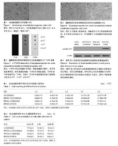
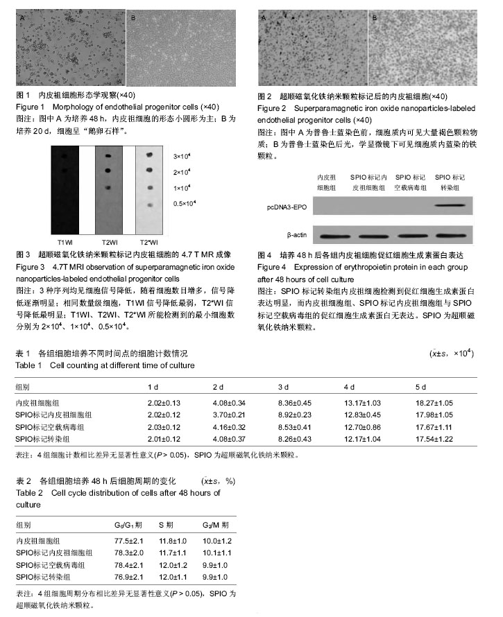
2.1 内皮祖细胞形态学观察 培养48 h后,内皮祖细胞开始贴壁生长,细胞形态主要为小圆形,之后可见小的细胞集落形成,细胞体积逐渐增大、拉长,呈长梭状或线状,其他混杂细胞大部分逐渐脱落;培养10 d左右,可见索条样结构形成;培养20 d左右,细胞长满培养瓶底壁,形成典型“鹅卵石样”外观(图1A,B)。细胞按1∶2比例传代培养4-6 d长满瓶底。流式细胞术检测内皮祖细胞免疫表型。培养至第7天的内皮祖细胞分析细胞表面标志物CD31、CD34、VEGFR-2的阳性表达率分别为53.68%、11.62%和64.82%,空白对照管结果均小于1%。 2.2 内皮祖细胞的SPIO标记率测定 SPIO标记内皮祖细胞后,鲁士蓝染色前细胞质内可见大量褐色颗粒物质,即标记的SPIO颗粒(图2A);行普鲁士蓝染色后,光学显微镜下可见细胞胞质内蓝染的铁颗粒(图2B),镜下随机计数5个视野显示标记率达97%。 2.3 体外MR成像 分别对不同数量级标记内皮祖细胞行4.7T MR,T1WI、T2WI、T2*WI扫描,3种序列均见细胞信号降低,随着细胞数目增多,信号降低逐渐明显;相同数量级细胞,T1WI信号降低最弱,T2*WI信号降低最明显;T1WI、T2WI、T2*WI所能检测到的最小细胞数分别为2×10⁴、1×10⁴、0.5×10⁴(图3)。 2.4 CCK-8检测结果 普通内皮祖细胞、SPIO标记内皮祖细胞组、SPIO标记空载病毒组、SPIO标记转染组的A490值分别为0.20±0.05、0.19±0.03、0.18±0.03与0.21±0.04,3组细胞与普通内皮祖细胞A490值比较差异无显著性意义(P > 0.05),表明SPIO标记的内皮祖细胞体外促红细胞生成素基因修饰后对内皮祖细胞的增殖无明显影响。 2.5 细胞生长曲线 3组标记细胞与普通内皮祖细胞生长曲线比较差异无显著性意义(P > 0.05),表明SPIO标记的内皮祖细胞体外促红细胞生成素基因修饰后对内皮祖细胞的增殖无明显影响,见表1。 2.6 细胞周期分布 3组标记细胞与普通内皮祖细胞的细胞周期分布比较差异无显著性意义(P > 0.05),表明SPIO标记的内皮祖细胞体外促红细胞生成素基因修饰后对内皮祖细胞的细胞周期分布无明显影响,见表2。 2.7 Western blot检测结果 pcDNA3-EPO重组质粒稳定转染促红细胞生成素基因在转染48 h后,SPIO标记转染组内皮祖细胞检测到促红细胞生成素蛋白表达明显,而内皮祖细胞组、SPIO标记内皮祖细胞组与SPIO标记空载病毒组的促红细胞生成素蛋白无表达,说明了促红细胞生成素基因已稳定整合入SPIO标记转染组内皮祖细胞中,且可以行目的蛋白的稳定表达,见图4。"
| [1] Zhao XH,Huang L.The characteristics of endothelial progenitor cell biology and its effects on the cardiovascular system.Chin J Interven Card.2005; 13(6):405-407. [2] 范思均,何守志.血管内皮生长因子A的选择性剪接与眼内新生血管的生成[J].中华眼科杂志,2011,47(4):373- 377. [3] Kawamoto A,Losordo DW.Endothelial progenitor cells for cardiovascular regeneration.Trends Cardiovasc Med.2008;18(1):33-37. [4] Chade AR,Zhu X,Lavi R,et al.Endothelial progenitor cells restore renal function in chronic experimental renovaseular disease. Circulation. 2009;119(4): 547-557. [5] Patschan D,Krupineza K,Patschan S,et al.Dynamics of mobilization and homing of endothelial progenitor cells after acute renal ischemia:modulation by ischemic pre. conditioning.Am J Physiol Renal Physiol.2006; 291(1): F176-185. [6] Lu Y,Gong Y,Lian J,et al.Expansion of Endothelial Progenitor Cells in High Density Dot Culture of Rat Bone Marrow Cells.PLoS One.2014;9(9):e107127. [7] Ching YH,Sutton TL,Pierpont YN,et al.The use of growth factors and other humeral agents to accelerate and enhance burn wound healing.Eplasty. 2011; 11(e41):429-449. [8] Bao P, Kodra A, Tomic-Canic M, et al.The role of vascular endothelial growth factor in would healing.J Surg Res.2009;153(2):347-358. [9] Patschan D,Krupincza K,Patschan S,et al.Dynamics of mobilization and homing of endothelial pmgenitor ceUs after acute renal ischemia:modulation by ischemi. preconditioning.Am J Physiol Renal Physiol. 2006;291: 176-185. [10] atenaude A,Parker J,Kglsan A.Involvement of endothelial pro-genitor cells in tumor vascularization. Micmvasc Res.2010;79(3):217-223. [11] Ambms JT,HeⅡ℃m-Fresneda I,Borau 0G,et al. Ischemic preconditioning in solid organ an transplantation: fmm experimental to clinics.Tmnsplant Int.2007;20:219-229. [12] 蒋素华,邹建洲,刘红,等.通过短时间预缺血改变肾小管上皮细胞命运诱导肾脏缺血耐受[J].中华肾脏病杂志,2011, 27(3):198-202. [13] Li M, Nishimura H, wakura A,et al.Endothelial pmgenitor cells are rapidly recmited to myocardium and mediate pmtective e bct of ischemic preconditioning via ”imported” nitric oxide synthase activity.Circulation.2005;111(9):1114-1120. [14] Lee PS,Poh KK.Endothlia progenitor cells in cardiovascular diseases.World J Stem Cells 2014; 6(3):355. [15] Hu CH,Li ZW,DU ZM,et al.Human umbilical cord- derived endothelial progenitorcells promote growth cytokines-mediated neorevascularization in rat myocardial infarction.Chin Med J(Engl).2009;122(5): 548. [16] Sambataro M,Seganfreddo E,Canal F,et al.Prognostic significance of circulating and endothelial progenitor cell markers in type 2 diabetic foot.Int J Vasc Med. 2014;2014:589412. [17] Menne J,Park JK,Shushakova N,et al.Continuous erythropoietin receptor activation affects different pathways of diabetic renal injury.J AIn Soc Nephrol. 2007;18:2046-2053. [18] Leyland-Jones B,Semiglazov V,Pawlicki M,et al. Maintaining normal hemo-globin levels with epoetin alfa in mainly nonanemic patients with metastatic breast cancer receiving first-line chemotherapy: a survival study.J Clin Oncol. 2005;23(25):5960-5972. [19] 陈波,赵卫红,吴剑卿.促红细胞生成素对肺腺癌A549细胞生长和凋亡作用的实验研究[J].中国肺癌杂志,2009, 12(9):956-960. [20] Bohlius J,Wilson J,Seidenfeld J,et al.Recombinant human erythropoietins and cancer patients: updated meta-analysis of 57 studies including 9 353 patients.J Nati Cancer Inst.2006;98(10):708-714. [21] 朱蓓,赵卫红,吴剑卿.构建促红细胞生成素表达载体及其在肾小管上皮细胞中的表达[J].中国血液净化,2009, 8(4):211-214. [22] 麦筱莉,韩 冰,范海健,等.体外标记内皮祖细胞小鼠缺血性脑梗死模型局部移植后向病灶侧迁移的MRI实验研究[J].医学影像学杂志,2015,25(1):143-147. [23] 康涛,李晓强,盂庆友,等.新型超顺磁性氧化铁标记血管内皮祖细胞的实验研究[J].中国血管外科杂志(电子版), 2012,4(2):108-111. [24] Liu G,Xia C,Wang Z,et al.Magnetic resonance imaging probes for labeling of chondrocyte cells.J Mater Sci Mater Med.2011;22(3):601-606. [25] ampetaki A,Kirton JP,Xu Q.Vascular repair by endothelial pro-genitor ceils.Cardiovasc Res. 2008; 78(3):413-421. [26] Jianguo W,Tianhang L,Hong Z,et al.Optimization of culture conditions for endothelial progenitor cells from porcine bone malTOW in vitro.Cell Pmlif.2010; 43(4): 418-426. [27] ariucci S,Rovati B,Bencardino K,et al.Flow cytometric detec. tion of circulating endothelial cells and endothelial progenitor cells in healthy subjects.Int J Lab Hematol.2010;32(1 Ptl):e4048. [28] Estes ML,Mund JA,Mead LE,et al.Application of polychmmatic flow cytometry to identify novel subsets of circulating cells with an-giogenic potential.Cytometry A.2010;77(9):831-839. [29] Richardson MR,Yoder MC.Endothelial progenitor cells:quo vadis.J Mol Cell Cardiol,2011,50(2):266-272. [30] Yang N,Li D,Jiao P,et al.The characteristics of endothelial pro-genitor cells derived from mononuclear cells of rat bone marrowin different culture conditions. Cytotechnology.2011;63(3):217-226. [31] Amsalem Y,Mardor Y,Feinberg MS,et al.Iron-oxide labeling and outcome of transplanted mesenchymal stem cells in the infarcted myocardium. Circulation 2007;116(11 Suppl):138-145. [32] Hauger O,Frost EE,van Heeswijk R,et al.MR evaluation of the gIomeruIar homing of magneticalIy labeled mesenchymal stem cells in a rat model of nephropathy.Radiology.2006;238(1):200-210. [33] Ju SH,Teng GJ.Lu HH,et al.In vivo MR tracking of mesenchymal stem ceils in rat liver after intrasplenic transpIantation.Radiology,2007,245(1):206-215. [34] Arbab AS,Pandit SD,Anderson SA,et al.Magnetic resonance imaging and confocal microscopy studies of magnetically labeled endothellal progenitor ceIIs trafficking to sites of tumor angiogenesis.Stem Cells. 2006;24(3):671-678. [35] Henning TD,Gawande R,Khurana A,et al.Magnetic resonance imaging of ferumoxide 1abeled mesenchymal stem cells in cartiIage defects: in vitro and in vivo investigations.Mol Imaging. 2012;11(3): 197-209. [36] Khurana A,Nejadnik H,Gawande R,et al.Intravenous ferumoxytol allows noninvasive MR imaging monitoring of macrophage migration into stem cell transpIants.Radiology.2012;264(3):803-811. [37] Khurana A,Chapelin F,Beck G,et al.Iron administration before stem celI harvest enables MR imaging tracking after transplantation.Radiology.2013;269(1):186-197. [38] Liu W,Frank Jh.Detection and quantification of magnetically la-beled cells by cellularMRI.Eur J Radiol. 2009;70(2):258-264. [39] Ai H.Layer-by-layer capsules for magnetic resonance imaging and dmg delivery.Adv Drug Deliv Rev.2011; 63(9):772-788. |
| [1] | Pu Rui, Chen Ziyang, Yuan Lingyan. Characteristics and effects of exosomes from different cell sources in cardioprotection [J]. Chinese Journal of Tissue Engineering Research, 2021, 25(在线): 1-. |
| [2] | Min Youjiang, Yao Haihua, Sun Jie, Zhou Xuan, Yu Hang, Sun Qianpu, Hong Ensi. Effect of “three-tong acupuncture” on brain function of patients with spinal cord injury based on magnetic resonance technology [J]. Chinese Journal of Tissue Engineering Research, 2021, 25(在线): 1-8. |
| [3] | Lin Qingfan, Xie Yixin, Chen Wanqing, Ye Zhenzhong, Chen Youfang. Human placenta-derived mesenchymal stem cell conditioned medium can upregulate BeWo cell viability and zonula occludens expression under hypoxia [J]. Chinese Journal of Tissue Engineering Research, 2021, 25(在线): 4970-4975. |
| [4] | Zhang Tongtong, Wang Zhonghua, Wen Jie, Song Yuxin, Liu Lin. Application of three-dimensional printing model in surgical resection and reconstruction of cervical tumor [J]. Chinese Journal of Tissue Engineering Research, 2021, 25(9): 1335-1339. |
| [5] | Zhang Xiumei, Zhai Yunkai, Zhao Jie, Zhao Meng. Research hotspots of organoid models in recent 10 years: a search in domestic and foreign databases [J]. Chinese Journal of Tissue Engineering Research, 2021, 25(8): 1249-1255. |
| [6] | Hou Jingying, Yu Menglei, Guo Tianzhu, Long Huibao, Wu Hao. Hypoxia preconditioning promotes bone marrow mesenchymal stem cells survival and vascularization through the activation of HIF-1α/MALAT1/VEGFA pathway [J]. Chinese Journal of Tissue Engineering Research, 2021, 25(7): 985-990. |
| [7] | Shi Yangyang, Qin Yingfei, Wu Fuling, He Xiao, Zhang Xuejing. Pretreatment of placental mesenchymal stem cells to prevent bronchiolitis in mice [J]. Chinese Journal of Tissue Engineering Research, 2021, 25(7): 991-995. |
| [8] | Liang Xueqi, Guo Lijiao, Chen Hejie, Wu Jie, Sun Yaqi, Xing Zhikun, Zou Hailiang, Chen Xueling, Wu Xiangwei. Alveolar echinococcosis protoscolices inhibits the differentiation of bone marrow mesenchymal stem cells into fibroblasts [J]. Chinese Journal of Tissue Engineering Research, 2021, 25(7): 996-1001. |
| [9] | Fan Quanbao, Luo Huina, Wang Bingyun, Chen Shengfeng, Cui Lianxu, Jiang Wenkang, Zhao Mingming, Wang Jingjing, Luo Dongzhang, Chen Zhisheng, Bai Yinshan, Liu Canying, Zhang Hui. Biological characteristics of canine adipose-derived mesenchymal stem cells cultured in hypoxia [J]. Chinese Journal of Tissue Engineering Research, 2021, 25(7): 1002-1007. |
| [10] | Geng Yao, Yin Zhiliang, Li Xingping, Xiao Dongqin, Hou Weiguang. Role of hsa-miRNA-223-3p in regulating osteogenic differentiation of human bone marrow mesenchymal stem cells [J]. Chinese Journal of Tissue Engineering Research, 2021, 25(7): 1008-1013. |
| [11] | Lun Zhigang, Jin Jing, Wang Tianyan, Li Aimin. Effect of peroxiredoxin 6 on proliferation and differentiation of bone marrow mesenchymal stem cells into neural lineage in vitro [J]. Chinese Journal of Tissue Engineering Research, 2021, 25(7): 1014-1018. |
| [12] | Zhu Xuefen, Huang Cheng, Ding Jian, Dai Yongping, Liu Yuanbing, Le Lixiang, Wang Liangliang, Yang Jiandong. Mechanism of bone marrow mesenchymal stem cells differentiation into functional neurons induced by glial cell line derived neurotrophic factor [J]. Chinese Journal of Tissue Engineering Research, 2021, 25(7): 1019-1025. |
| [13] | Duan Liyun, Cao Xiaocang. Human placenta mesenchymal stem cells-derived extracellular vesicles regulate collagen deposition in intestinal mucosa of mice with colitis [J]. Chinese Journal of Tissue Engineering Research, 2021, 25(7): 1026-1031. |
| [14] | Pei Lili, Sun Guicai, Wang Di. Salvianolic acid B inhibits oxidative damage of bone marrow mesenchymal stem cells and promotes differentiation into cardiomyocytes [J]. Chinese Journal of Tissue Engineering Research, 2021, 25(7): 1032-1036. |
| [15] | Guan Qian, Luan Zuo, Ye Dou, Yang Yinxiang, Wang Zhaoyan, Wang Qian, Yao Ruiqin. Morphological changes in human oligodendrocyte progenitor cells during passage [J]. Chinese Journal of Tissue Engineering Research, 2021, 25(7): 1045-1049. |
| Viewed | ||||||
|
Full text |
|
|||||
|
Abstract |
|
|||||