Chinese Journal of Tissue Engineering Research ›› 2014, Vol. 18 ›› Issue (47): 7566-7572.doi: 10.3969/j.issn.2095-4344.2014.47.005
Previous Articles Next Articles
Calcium phosphate bone cement and biodegradable mesh-like microporous balloon for vertebroplasty
Xie Zhi-yong1, Liu Xun-wei1, Zhong Jian2, Wei Dai-xu2, Ye Yong1, Du Yan-xia1, Sun Gang1
- 1Department of Medical Image, Jinan Military General Hospital, Jinan 250031, Shandong Province, China
2National Engineering Research Center for Nanotechnology (NERCN), Shanghai 200241, China
-
Revised:2014-09-19Online:2014-11-19Published:2014-11-19 -
Contact:Sun Gang, M.D., Chief physician, Professor, Doctoral supervisor, Department of Medical Image, Jinan Military General Hospital, Jinan 250031, Shandong Province, China -
About author:Xie Zhi-yong, Associate chief physician, Department of Medical Image, Jinan Military General Hospital, Jinan 250031, Shandong Province, China -
Supported by:the National High Technology Research and Development Program of China (863 Program), No. 2013AA032203; the National Natural Science Foundation of China, No. 51073173
CLC Number:
Cite this article
Xie Zhi-yong, Liu Xun-wei, Zhong Jian, Wei Dai-xu, Ye Yong, Du Yan-xia, Sun Gang . Calcium phosphate bone cement and biodegradable mesh-like microporous balloon for vertebroplasty[J]. Chinese Journal of Tissue Engineering Research, 2014, 18(47): 7566-7572.
share this article
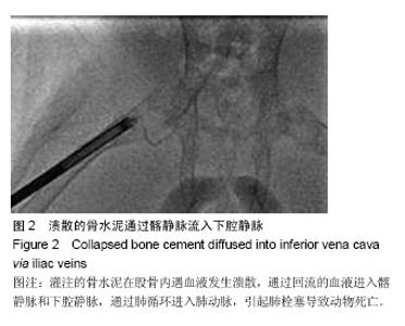
2.1 实验动物数量分析 实验用新西兰兔48只,实验术中2只因股骨骨折放弃入组,对照组灌注磷酸钙骨水泥时因骨水泥渗漏致肺栓塞死亡3只,补充实验动物5只,术后动物饲养情况同术前,术后及观察期未出现动物感染及死亡,最终新西兰兔48只进入结果分析。 2.2 球囊膨胀及骨水泥渗漏的观察 实验组在灌注磷酸钙骨水泥过程中球囊逐渐膨胀,呈椭圆球形,在X射线下未见明确骨水泥渗出,球囊与输送装置结合紧密,无球囊脱离现象发生,球囊松解后,球囊位置无移动。对照组骨水泥直接进入骨小梁间隙,3只因骨水泥溃散渗漏入髂静脉,通过肺循环进入肺动脉,引起肺栓塞导致动物死亡(图2),发生率为11%。"


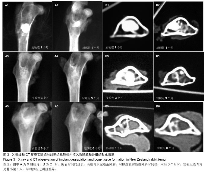
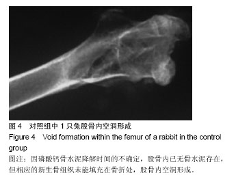
2.3 影像学检查 术后1个月时,实验组兔股骨内骨水泥形态呈球形,对照组形态不规整;3个月后,实验组形态仍保持,股骨内见骨小梁长入,对照组骨水泥降解速度较实验组快,骨小梁形态恢复;6个月后,骨水泥基本降解,股骨内见粗大骨小梁,见图3。 术后1个月时,对照组有1只股骨内骨水泥完全降解,新骨组织未形成的情况发生,查对照组其余动物,磷酸钙骨水泥仍存在于股骨内,本只动物进一步证实磷酸钙骨水泥降解时间的不确定性[25],见图4。"

"
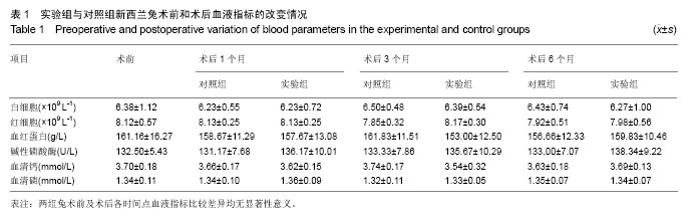
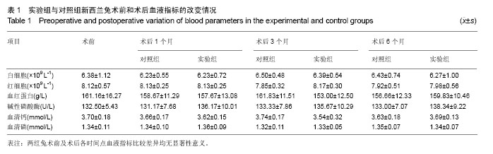
2.4 血液指标检测 在术前、术后1,3,6个月时,两组血细胞、血清碱性磷酸酶和钙磷指标手术前后组间和组内比较差异均无显著性意义,见表1。"
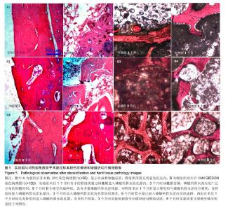
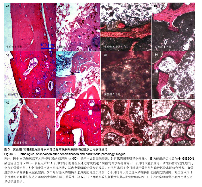
2.5 组织学检测 标本在脱钙过程中磷酸钙骨水泥丢失,因此未能显示出新生骨组织与磷酸钙骨水泥及球囊的关系,脱钙苏木精-伊红染色病理图片只是显示出成骨细胞活跃,骨组织周围无明显免疫反应。硬组织切片显示:①实验组:术后1个月时有小的骨组织通过球囊膜进入磷酸钙骨水泥孔隙内;3个月时球囊膜变薄,磷酸钙骨水泥内见广泛分布的骨髓组织;6个月时骨小梁交织成网状,其内少量磷酸钙骨水泥残留。②对照组:术后1个月时显示骨组织与磷酸钙骨水泥结合紧密,有骨组织探入磷酸钙骨水泥孔隙内,3个月时进入磷酸钙骨水泥内的骨组织增多,6个月时骨小梁已进入磷酸钙骨水泥内交织成网。两组在术后1个月时均见有骨组织进入磷酸钙骨水泥孔隙,差异性不明显;3个月时实验组新骨生长情况较对照组活跃,6个月时实验组骨小梁增生情况明显优于对照组,见图5。"
| [1] Tzermiadianos MN,Renner SM,Phillips FM,et al.Altered discpressure profile after an osteoporotic vertebral fracture is a risk factor for adjacent vertebral body fracture.Eur Spine J. 2008;17:1522-1530. [2] Sun YC,Teng MM,Yuan WS,et al. Risk of post-vertebroplasty fracture in adjacent vertebral bodies appears correlated with the morphologic extent of bone cement. J Chin Med Assoc. 2011;74(8):357-362. [3] Frankel BM,Monroe T,Wang C.Percutaneous vertebral augmentation: an elevation in adjacent-level fracture risk in kyphoplasty as compared with vertebroplasty. Spine J. 2007; 7(5):575-582. [4] Zou J,Mei X,Zhu X,et al.The long-term incidence of subsequent vertebral body fracture after vertebral augmentation therapy: a systemic review and meta-analysis. Pain Physician.2012;15(4):E515-522. [5] Brodano GB,Amendola L,Martikos K,et al.Vertebroplasty: benefits are more than risks in selected and evidence-based informed patients. A retrospective study of 59 cases. Eur Spine J.2011;20(8):1265-1271. [6] Bula P,Lein T,Strassberger C,et al.Balloon kyphoplasty in the treatment of osteoporotic vertebral fractures: indications- treatment strategy-complications. Z Orthop Unfall. 2010; 148(6):646-656. [7] Sun G,Jin P,Li M,et al.Height restoration and wedge anglecorrection effects of percutaneous vertebroplasty: association with intraosseous clefts.Eur Radiol. 2011;21: 2597-2603. [8] Wagner AL,Baskurt E.Refracture with cement extrusion following percutaneous vertebroplasty of a large interbody cleft.AJR Am J Roentgenol.2006;27:230-231. [9] Voormolen MH,Lohle PN,Juttmann JR,et al.The risk of new osteoporotic vertebral compression fractures in the year after percutaneous vertebroplasty. J Vasc Interv Radiol. 2006; 17(1):71-76. [10] Togawa D,Kovacic JJ,Bauer TW,et al.Radiographic and histologic findings of vertebral augmentation using polymethylmethacrylate in the primate spine: percutaneous vertebroplasty versus kyphoplasty.Spine.2006;31:E4-10. [11] 周鑫,夏群,苗军,等.磷酸钙骨水泥改性研究进展[J].中国矫形外科杂志,2008,16(1):55-57. [12] Lieberman IH,Togawa D,et al.Vertebroplasty and kyphoplasty: filler materials .Spine J.2005;11:305S-316S. [13] van de Watering FC,van den Beucken JJ,Walboomers XF,et al.Calcium phosphate/poly(D,L-lactic-co-glycolic acid) composite bone substitute materials: evaluation of temporal degradation and bone ingrowth in a rat critical-sized cranial defect.Clin Oral Implants Res.2012;23:151-159. [14] Aberg J,Henriksson HB,Engqvist H,et al. Biocompatibility and resorption of a radiopaque premixed calcium phosphate cement.J Biomed Mater Res A.2012;100: 1269-1278. [15] Cherng A,Takagi S,Chow LC.Effects of hydroxypropyl methylcellulose and other gelling agents on the handling properties of calcium phosphate cement. J Biomed Mater Res.1997;35(3):273-277. [16] Ling Chen, Hong Xiang, Xiao Xi Li, et al. Improvement of the anti-washout performance of calcium phosphate cement by use of modified starch. Key Engineering Materials. 2007; 336-338:1628-1631. [17] Qian G,Dong Y,Yang W,et al.Injectable calcium phosphate cement and fibrin sealant recombined human bone morphogenetic protein-2 composite in vertebroplasty: an animal study.Bosn J Basic Med Sci.2012;12(4):231-235. [18] Schumacher M,Hen A,Rohnke M,et al.A novel and easy-to-prepare strontium(II) modified calcium phosphate bone cement with enhanced mechanical properties. Acta Biomater.2013;9(7):7536-7544. [19] Christel T,Kuhlmann M,Vorndran E,et al.Dual setting α-tricalcium phosphate cements.J Mater Sci Mater Med. 2013;24(3):573-581. [20] Tarsuslugil SM,O'Hara RM,Dunne NJ,et al.Development of calcium phosphate cement for the augmentation of traumatically fractured porcine specimens using vertebroplasty. J Biomech.2013;46(4):711-715. [21] Sun G,Wei D,Liu X,et al.Novel biodegradable electrospun nanofibrous P(DLLA-CL) balloons for the treatment of vertebral compression fractures.Nanomedicine. 2013;9(6): 829-838. [22] 彭湘涛,刘训伟,李敏,等.可降解高分子网状球囊及磷酸钙骨水泥椎体填充后的力学变化[J].中国组织工程研究,2013,17(51): 8795-8800. [23] 刘训伟,钟建,彭湘涛,等.联合钙盐骨水泥及可降解网状微孔球囊椎体成形的体外实验[J].中国组织工程研究,2014,18(12): 1817-1823. [24] 中华人民共和国科学技术部.关于善待实验动物的指导性意见. 2006-09-30. [25] Galovich LA,Perez-Higueras A,Altonaga JR,et al. Biomechanical, histological and histomorphometric analyses of calcium phosphate cement compared to PMMA for vertebral augmentation in a validated animal model. Eur Spine J.2011;20(Suppl 3):S376-S382. [26] Galibert P,Deramond H,Rosat P,et al.Preliminary note on the treatment of vertebral angioma by percutaneous acrylic vertebroplasty. Neurochirurgie.1987;33(2):166-168. [27] Rao RD,Singrakhia MD.Painful osteoporotic vertebral fracture. Pathogenesis, evaluation, and roles of vertebroplasty and kyphoplasty in its management. J Bone Joint Surg Am. 2003; 85-A(10):2010-2022. [28] Foo LS,Yeo W,Fook S,et al.Results, experience and technical points learnt with use of the SKy Bone Expander kyphoplasty system for osteoporotic vertebral compression fractures: a prospective study of 40 patients with a minimum of 12 months of follow-up.Eur Spine J.2007;16(11):1944-1950. [29] 孙钢,金鹏,易玉海,等.用国产器械行椎体后凸成形术治疗老年骨质疏松脊柱压缩骨折的价值[J].中华放射学杂志,2006,40(10): 1089-1092. [30] Zheng Z,Luk KD,Kuang G,et al. Vertebral augmentation with a novel Vessel-X bone void filling container system and bioactive bone cement.Spine (Phila Pa 1976). 2007;32(19): 2076-2082. [31] Panagiotis K1,Konstantinos V,Vasilios V,et al.Is Kiva implant advantageous to balloon kyphoplasty in treating osteolytic metastasis to the spine? Comparison of 2 percutaneous minimal invasive spine techniques: a prospective randomized controlled short-term study.Spine (Phila Pa 1976). 2014; 39(4):E231- E239. |
| [1] | Zou Shouping, Lu Daoyun, Ye Li. Minimally invasive percutaneous pedicle screw technique for thoracolumbar fractures: biomechanical changes of the spine during 6-month follow-up [J]. Chinese Journal of Tissue Engineering Research, 2021, 25(24): 3865-3869. |
| [2] | Wang Ziao, Song Wenhui, Liu Changwen . Short-segment fixation of thoracolumbar burst fractures: method modification and strategies to reduce failure [J]. Chinese Journal of Tissue Engineering Research, 2021, 25(24): 3902-3907. |
| [3] | Liu Gang, Zhang Baolu, Shi Jie, Bao Dingsu, Zeng Shengqiang, Deng Kai, Liu Yang, Wang Guoyou, Fu Shijie. Difference between proximal humeral locking plate and cannulated screw fixation in the treatment of Mutch type II fracture of greater tuberosity of humerus [J]. Chinese Journal of Tissue Engineering Research, 2021, 25(18): 2863-2868. |
| [4] | Xie Jian, Su Jiansheng. Advantages and characteristics of electrospun aligned nanofibers as scaffolds for tissue engineering [J]. Chinese Journal of Tissue Engineering Research, 2021, 25(16): 2575-2581. |
| [5] | Han Shichong, Li Chang, Xing Haiyang, Ge Wenlong, Wang Gang . Finite element analysis of two internal fixation methods for treating extra-articular proximal tibial fractures [J]. Chinese Journal of Tissue Engineering Research, 2021, 25(15): 2329-2333. |
| [6] | Jing Wanli, Zhang Tao, Teng Donghui, Shi Tao, Zhou Qiang. Poor outcomes of bone filling mesh container vertebroplasty for the treatment of osteoporotic vertebral compression fractures with vertebral body wall incompetence [J]. Chinese Journal of Tissue Engineering Research, 2021, 25(10): 1522-1527. |
| [7] | Zhang Zhiwei, Li Li, Huang Ziyu, Wu Duoyi, Gan Farong, Ye Baofei, Zhang Yan, Zhang Taibiao, Hu Wanjun. Percutaneous vertebroplasty through unilateral and bilateral pedicle approaches and unilateral pedicle extrapedicle approach for the treatment of thoracolumbar vertebral compression fractures: bone cement perfusion volume and cement leakage rate [J]. Chinese Journal of Tissue Engineering Research, 2020, 24(9): 1353-1358. |
| [8] | Liu Yan, Ge Hongqing, Guan Hua, Chen Wenzhi. Finite element analysis of different fixation methods for poor medial column support proximal humeral fracture [J]. Chinese Journal of Tissue Engineering Research, 2020, 24(9): 1384-1389. |
| [9] | Wang Liang, Huang Zhaozhao, Yu Jiaona, Gu Weidong, Wang Ren, Qian Zhiyi. Biomechanical characteristics of bridge-link type combined internal fixation system with mixed-rod versus double-rod in the treatment of femoral and tibial fractures [J]. Chinese Journal of Tissue Engineering Research, 2020, 24(6): 888-892. |
| [10] | Li Kaiming, Wang Shangquan, Li Linghui, Zhu Liguo, Zhang Qing, Xie Rui. Bone filling bag vertebroplasty and percutaneous kyphoplasty for the treatment of thoracolumbar osteoporotic compression fractures: a meta-analysis of improving Cobb angle and reducing bone cement leakage [J]. Chinese Journal of Tissue Engineering Research, 2020, 24(4): 650-656. |
| [11] | Weng Nengyuan, Zhang Tao, Li Kainan, Lan Hai, Zhang Jinli, Fu Xuefei, Liu Qixin, Lin Qingyun. A meta-analysis of efficacy and complications of 3D printing-assisted surgery for Schatzker IV-VI tibial plateau fractures [J]. Chinese Journal of Tissue Engineering Research, 2020, 24(30): 4889-4897. |
| [12] | Liu Yanhua, Zhu Zhou, Wan Qianbing. A drug-loading system for electrospinning wound repair: component selection and construction strategy [J]. Chinese Journal of Tissue Engineering Research, 2020, 24(28): 4465-4473. |
| [13] | Tu Dongpeng, Yu Yikang, Liu Zheng, Fan Xin, Zhang Wenkai, Xu Chao. Meta-analysis of locking plate combined with fibular allograft and locking plate alone in the treatment of proximal humeral fractures [J]. Chinese Journal of Tissue Engineering Research, 2020, 24(27): 4389-4397. |
| [14] | Huang Zhaoguo, Zhang Caiyi, Zhang Qing, Wang Sheng, Wang Shaogang, Li Jun, Tao Zhongliang, Zuo Caihong. Curative efficacy of proximal femoral nail antirotation versus intertrochanteric antegrade nail for treating intertrochanteric fracture in older adults [J]. Chinese Journal of Tissue Engineering Research, 2020, 24(21): 3310-3314. |
| [15] |
She Rongfeng, Zhang Yi, Wang Yuanzheng, Zhang Bin, Chen Peng, Huang Qixiang.
A novel cement-reinforced screw combined with locking plate
fixation versus humeral head
arthroplasty in the treatment of osteoporotic fractures of the proximal humerus
|
| Viewed | ||||||
|
Full text |
|
|||||
|
Abstract |
|
|||||