Chinese Journal of Tissue Engineering Research ›› 2014, Vol. 18 ›› Issue (1): 27-32.doi: 10.3969/j.issn.2095-4344.2014.01.005
Previous Articles Next Articles
Bone marrow mesenchymal stem cells repair vascular injury after cervical spondylotic vertebral arteriopathy
Zhang Xia-meng1, Shou Zhe-xing2
- 1Department of Traditional Chinese Medicine, First Affiliated Hospital of Liaoning Medical University, Jinzhou 121000, Liaoning Province, China
2Department of Traditional Chinese Medicine, Union Hospital, Tongji Medical College, Huazhong University of Science and Technology, Wuhan 430022, Hubei Province, China
-
Revised:2013-10-12Online:2014-01-01Published:2014-01-01 -
Contact:Shou Zhe-xing, M.D., Attending physician, Department of Traditional Chinese Medicine, Union Hospital, Tongji Medical College, Huazhong University of Science and Technology, Wuhan 430022, Hubei Province, China -
About author:Zhang Xia-meng, Studying for master’s degree, Department of Traditional Chinese Medicine, First Affiliated Hospital of Liaoning Medical University, Jinzhou 121000, Liaoning Province, China
CLC Number:
Cite this article
Zhang Xia-meng, Shou Zhe-xing. Bone marrow mesenchymal stem cells repair vascular injury after cervical spondylotic vertebral arteriopathy[J]. Chinese Journal of Tissue Engineering Research, 2014, 18(1): 27-32.
share this article

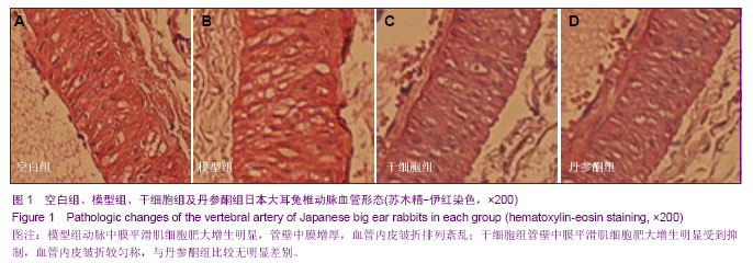
2.1 实验动物数量分析 将40只日本大耳兔随机分为4组:空白组、模型组、丹参酮组、干细胞组,全部进入结果分析,无脱失。 2.2 病理检查观察椎动脉血管形态 与空白组相比,模型组动脉中膜平滑肌细胞肥大增生明显,管壁中膜增厚,血管内皮皱折排列紊乱(图1A,B);与模型组相比,干细胞组管壁中膜平滑肌细胞肥大增生明显受到抑制,血管内皮皱折较匀称(图1C);丹参酮与干细胞组组比较无明显差别(图1D)。"

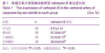
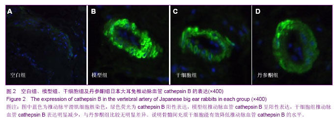
2.3 免疫荧光染色检测椎动脉血管cathepsin B表达 空白组椎动脉血管cathepsin B表达极少,图中蓝色为椎动脉平滑肌细胞核染色(图2A);与空白组相比,模型组椎动脉血管cathepsin B呈阳性表达,差异有显著性意义(P < 0.05),图中蓝色为椎动脉平滑肌细胞核染色,绿色荧光为阳性表达(图2B);与模型组相比,干细胞组椎动脉血管cathepsin B表达明显减少,差异有显著性意义(P < 0.05)(图2C);干细胞组与丹参酮组比较差异无显著性意义(P > 0.05)(图2D,表1)。"
"

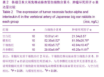
2.4 ELISA检测椎动脉血管白细胞介素6、肿瘤坏死因子α的表达 空白组椎动脉白细胞介素6、肿瘤坏死因子α表达极少;与空白组相比,模型组椎动脉血管白细胞介素6、肿瘤坏死因子α表达明显增加,差异有显著性意义(P < 0.05);与模型组相比,干细胞组椎动脉血管白细胞介素6、肿瘤坏死因子α明显减少,差异有显著性意义(P < 0.05);干细胞组与丹参酮组比较差异无显著性意义(P > 0.05)(表2)。"
| [1] Yoon SH, Kim KJ, Chung SK,et al. Inflammatory myofibroblastic tumor in the intradural extramedullary space of the lumbar spine with spondylolisthesis: case report and review of the literature.Eur Spine J. 2010;19 Suppl 2:S153-157.[2] Nagashima H, Morio Y, Yamane K,et al.Tumor necrosis factor-alpha, interleukin-1beta, and interleukin-6 in the cerebrospinal fluid of patients with cervical myelopathy and lumbar radiculopathy.Eur Spine J. 2009;18(12):1946-1950. [3] Rodríguez-Mañas L, El-Assar M, Vallejo S,et al. Endothelial dysfunction in aged humans is related with oxidative stress and vascular inflammation. Aging Cell. 2009;8(3):226-238. [4] Tatenkulova SN, Mareev VIu, Zykov KA,et al.The role of inflammatory factors in pathogenesis of ischemic heart disease. Kardiologiia. 2009;49(1):4-8.[5] Lin XQ, Zhao HY, Sun Q. Acupuncture and massage therapy for 23 cases of pharyngitis caused by cervical vertebra disease。Zhongguo Zhen Jiu. 2012;32(2):166.[6] Schmid RA, Schwenzer K, Weiss M,et al. Monostotic Paget's Disease of a cervical vertebra: differential diagnosis with F-18 FDG positron emission tomography using a coincidence technique and with Tc-99m dicarboxypropane diphosphonate. Clin Nucl Med. 2002;27(7):537-538.[7] Marumo F, Nomura T, Nishikawa H.Transverse fractures of the spinous process of the 7th cervical vertebra in RDT patients: an Al related disease. Int J Artif Organs. 1987;10(2): 93-96.[8] Nowak C, Solecki J. Absence of the body of the cervical vertebra in Bechterew's disease.Wiad Lek. 1981;34(4): 325-329.[9] Dawn B, Bolli R. Adult bone marrow-derived cells: regenerative potential, plasticity, and tissue commitment. Basic Res Cardiol. 2005;100(6):494-503.[10] Grove JE, Bruscia E, Krause DS. Plasticity of bone marrow-derived stem cells.Stem Cells. 2004;22(4):487-500.[11] Caplan AI.Why are MSCs therapeutic? New data: new insight. J Pathol. 2009;217(2):318-324.[12] Maul TM, Chew DW, Nieponice A,et al. Mechanical stimuli differentially control stem cell behavior: morphology, proliferation, and differentiation.Biomech Model Mechanobiol. 2011;10(6):939-953.[13] Patel SA, Sherman L, Munoz J,et al. Immunological properties of mesenchymal stem cells and clinical implications.Arch Immunol Ther Exp (Warsz). 2008;56(1):1-8. [14] Parekkadan B, Milwid JM. Mesenchymal stem cells as therapeutics. Annu Rev Biomed Eng. 2010;12:87-117.[15] Yi T, Song SU. Immunomodulatory properties of mesenchymal stem cells and their therapeutic applications. Arch Pharm Res. 2012;35(2):213-221.[16] Urbán VS, Kiss J, Kovács J,et al. Mesenchymal stem cells cooperate with bone marrow cells in therapy of diabetes.Stem Cells. 2008;26(1):244-253. [17] van Laar JM, Tyndall A. Adult stem cells in the treatment of autoimmune diseases.Rheumatology (Oxford). 2006;45(10): 1187-1193.[18] Wu Y, Wang J, Scott PG,et al. Bone marrow-derived stem cells in wound healing: a review.Wound Repair Regen. 2007; 15 Suppl 1:S18-26.[19] Ren G, Chen X, Dong F,et al. Concise review: mesenchymal stem cells and translational medicine: emerging issues.Stem Cells Transl Med. 2012;1(1):51-58.[20] Mahmood A, Lu D, Lu M,et al.Treatment of traumatic brain injury in adult rats with intravenous administration of human bone marrow stromal cells. Neurosurgery. 2003;53(3): 697-702.[21] Shou Z, Shen L, Xiong P. Assessing the validity of a novel model of vertebral artery type of cervical syndrome induced by injecting sclerosing agent next to transverse process of cervical vertebra.J Huazhong Univ Sci Technolog Med Sci. 2010;30(1):85-88.[22] Denis DJ, Shedid D, Shehadeh M,et al. Cervical spondylosis: a rare and curable cause of vertebrobasilar insufficiency. Eur Spine J. 2013. [Epub ahead of print][23] Rahimizadeh A, Soufiani H.Intramedullary arachnoid cyst in association with cervical spondylosis: case report.Spine J. 2013. pii: S1529-9430(13)00523-8. [24] Fujimori T, Le H, Ziewacz JE,et al. Is there a difference in range of motion, neck pain, and outcomes in patients with ossification of posterior longitudinal ligament versus those with cervical spondylosis, treated with plated laminoplasty. Neurosurg Focus. 2013;35(1):E9.[25] Ding Q, Yan M, Zhou J,et al. Clinical effects of innovative tuina manipulations on treating cervical spondylosis of vertebral artery type and changes in cerebral blood flow.J Tradit Chin Med. 2012;32(3):388-392.[26] Guo K, Li L, Zhan HS,et al. Systematic review of clinical randomized controlled trials on manipulation treatment for vertebral artery type of cervical spondylosis.Zhongguo Gu Shang. 2012;25(1):9-13.[27] Liao YY, Sun DM, Zhong CP,et al. Evaluation of the immediate effect of acupuncture on cervical spondylosis of vertebral artery type based on orthogonal design.Zhongguo Zhen Jiu. 2011;31(6):499-502.[28] Gao H, Ye YJ. Manipulative treatment of vertebral artery type of cervical spondylosis. Zhongguo Gu Shang. 2010;23(4): 257-260.[29] Chen D, Zhong J, Hong YB,et al. Influence of needle-pricking bleeding combined with pulling-rotating manipulation on blood rheology in patients with vertebral artery type cervical spondylosis.Zhen Ci Yan Jiu. 2009;34(5):344-348.[30] Hong ES, Deng MY, Cheng LH,et al. Effect of vertebral manipulation therapy on vertebro-basilar artery blood flow in cervical spondylosis of vertebral artery type. Zhongguo Zhong Xi Yi Jie He Za Zhi. 2005;25(8):742-744.[31] Lewis AC. Interleukin-6 in the pathogenesis of posterior capsule opacification and the potential role for interleukin-6 inhibition in the future of cataract surgery. Med Hypotheses. 2013;80(4):466-474. [32] Ichikawa T, Sugiura H, Koarai A,et al. 25-hydroxycholesterol promotes fibroblast-mediated tissue remodeling through NF-κB dependent pathway.Exp Cell Res. 2013;319(8): 1176-1186.[33] Liu A, Gao X, Zhang Q,et al. Cathepsin B inhibition attenuates cardiac dysfunction and remodeling following myocardial infarction by inhibiting the NLRP3 pathway.Mol Med Rep. 2013;8(2):361-366.[34] Dai C, Zhang B, Liu X,et al. Pyrimethamine sensitizes pituitary adenomas cells to temozolomide through cathepsin B-dependent and caspase-dependent apoptotic pathways.Int J Cancer. 2013;133(8):1982-1993. [35] Lin SA, Patel M, Suresch D,et al. Quantitative Longitudinal Imaging of Vascular Inflammation and Treatment by Ezetimibe in apoE Mice by FMT Using New Optical Imaging Biomarkers of Cathepsin Activity and α(v)β(3) Integrin.Int J Mol Imaging. 2012;2012:189254.[36] Zhang T, Yang ZY, Bo XP,et al. Cathepsin B promoted the apoptosis of vascular smooth muscle cells as induced by tumor necrosis factor-α. Zhonghua Yi Xue Za Zhi. 2011; 91(12):845-849.[37] Screen M, Dean W, Cross JC,et al. Cathepsin proteases have distinct roles in trophoblast function and vascular remodelling. Development. 2008;135(19):3311-3320. [38] González-Vela MC, Garijo MF, Fernández F,et al. Cathepsin D in host stromal cells is associated with more highly vascular and aggressive invasive breast carcinoma.Histopathology. 1999;34(1):35-42.[39] Yamaguchi T, Naruishi K, Arai H,et al. IL-6/sIL-6R enhances cathepsin B and L production via caveolin-1-mediated JNK-AP-1 pathway in human gingival fibroblasts.J Cell Physiol. 2008;217(2):423-432.[40] Sendler M, Dummer A, Weiss FU,et al.Tumour necrosis factor α secretion induces protease activation and acinar cell necrosis in acute experimental pancreatitis in mice.Gut. 2013; 62(3):430-439. [41] Boyd AS, Fairchild PJ. Approaches for immunological tolerance induction to stem cell-derived cell replacement therapies. Expert Rev Clin Immunol. 2010;6(3):435-448.[42] Sakaguchi S, Miyara M, Costantino CM,et al. FOXP3+ regulatory T cells in the human immune system.Nat Rev Immunol. 2010;10(7):490-500.[43] Tang Q, Boden EK, Henriksen KJ,et al. Distinct roles of CTLA-4 and TGF-beta in CD4+CD25+ regulatory T cell function.Eur J Immunol. 2004;34(11):2996-3005.[44] Kong YC, Morris GP, Brown NK,et al. Autoimmune thyroiditis: a model uniquely suited to probe regulatory T cell function.J Autoimmun. 2009;33(3-4):239-246. [45] Morris GP, Kong YC. Interference with CD4+CD25+ T-cell-mediated tolerance to experimental autoimmune thyroiditis by glucocorticoid-induced tumor necrosis factor receptor monoclonal antibody.J Autoimmun. 2006;26(1): 24-31. [46] Maloy KJ, Salaun L, Cahill R,et al. CD4+CD25+ T(R) cells suppress innate immune pathology through cytokine-dependent mechanisms.J Exp Med. 2003;197(1): 111-119.[47] Tsai YG, Chiou YL, Chien JW,et al. Induction of IL-10+ CD4+ CD25+ regulatory T cells with decreased NF-κB expression during immunotherapy.Pediatr Allergy Immunol. 2010;21(1 Pt 2):e166-73.[48] Liu Y, Wang L, Predina J,et al. Inhibition of p300 impairs Foxp3(+) T regulatory cell function and promotes antitumor immunity.Nat Med. 2013;19(9):1173-1177.[49] Yin X, Yin Y, Cao FL,et al. Tanshinone IIA attenuates the inflammatory response and apoptosis after traumatic injury of the spinal cord in adult rats.PLoS One. 2012;7(6):e38381.[50] Wu X, Liu L, Xie H,et al. Tanshinone IIA prevents uric acid nephropathy in rats through NF-κB inhibition.Planta Med. 2012;78(9):866-873. |
| [1] | Pu Rui, Chen Ziyang, Yuan Lingyan. Characteristics and effects of exosomes from different cell sources in cardioprotection [J]. Chinese Journal of Tissue Engineering Research, 2021, 25(在线): 1-. |
| [2] | Lin Qingfan, Xie Yixin, Chen Wanqing, Ye Zhenzhong, Chen Youfang. Human placenta-derived mesenchymal stem cell conditioned medium can upregulate BeWo cell viability and zonula occludens expression under hypoxia [J]. Chinese Journal of Tissue Engineering Research, 2021, 25(在线): 4970-4975. |
| [3] | Wang Xianyao, Guan Yalin, Liu Zhongshan. Strategies for improving the therapeutic efficacy of mesenchymal stem cells in the treatment of nonhealing wounds [J]. Chinese Journal of Tissue Engineering Research, 2021, 25(7): 1081-1087. |
| [4] | Wang Shiqi, Zhang Jinsheng. Effects of Chinese medicine on proliferation, differentiation and aging of bone marrow mesenchymal stem cells regulating ischemia-hypoxia microenvironment [J]. Chinese Journal of Tissue Engineering Research, 2021, 25(7): 1129-1134. |
| [5] | Kong Desheng, He Jingjing, Feng Baofeng, Guo Ruiyun, Asiamah Ernest Amponsah, Lü Fei, Zhang Shuhan, Zhang Xiaolin, Ma Jun, Cui Huixian. Efficacy of mesenchymal stem cells in the spinal cord injury of large animal models: a meta-analysis [J]. Chinese Journal of Tissue Engineering Research, 2021, 25(7): 1142-1148. |
| [6] | Hou Jingying, Yu Menglei, Guo Tianzhu, Long Huibao, Wu Hao. Hypoxia preconditioning promotes bone marrow mesenchymal stem cells survival and vascularization through the activation of HIF-1α/MALAT1/VEGFA pathway [J]. Chinese Journal of Tissue Engineering Research, 2021, 25(7): 985-990. |
| [7] | Shi Yangyang, Qin Yingfei, Wu Fuling, He Xiao, Zhang Xuejing. Pretreatment of placental mesenchymal stem cells to prevent bronchiolitis in mice [J]. Chinese Journal of Tissue Engineering Research, 2021, 25(7): 991-995. |
| [8] | Liang Xueqi, Guo Lijiao, Chen Hejie, Wu Jie, Sun Yaqi, Xing Zhikun, Zou Hailiang, Chen Xueling, Wu Xiangwei. Alveolar echinococcosis protoscolices inhibits the differentiation of bone marrow mesenchymal stem cells into fibroblasts [J]. Chinese Journal of Tissue Engineering Research, 2021, 25(7): 996-1001. |
| [9] | Fan Quanbao, Luo Huina, Wang Bingyun, Chen Shengfeng, Cui Lianxu, Jiang Wenkang, Zhao Mingming, Wang Jingjing, Luo Dongzhang, Chen Zhisheng, Bai Yinshan, Liu Canying, Zhang Hui. Biological characteristics of canine adipose-derived mesenchymal stem cells cultured in hypoxia [J]. Chinese Journal of Tissue Engineering Research, 2021, 25(7): 1002-1007. |
| [10] | Geng Yao, Yin Zhiliang, Li Xingping, Xiao Dongqin, Hou Weiguang. Role of hsa-miRNA-223-3p in regulating osteogenic differentiation of human bone marrow mesenchymal stem cells [J]. Chinese Journal of Tissue Engineering Research, 2021, 25(7): 1008-1013. |
| [11] | Lun Zhigang, Jin Jing, Wang Tianyan, Li Aimin. Effect of peroxiredoxin 6 on proliferation and differentiation of bone marrow mesenchymal stem cells into neural lineage in vitro [J]. Chinese Journal of Tissue Engineering Research, 2021, 25(7): 1014-1018. |
| [12] | Zhu Xuefen, Huang Cheng, Ding Jian, Dai Yongping, Liu Yuanbing, Le Lixiang, Wang Liangliang, Yang Jiandong. Mechanism of bone marrow mesenchymal stem cells differentiation into functional neurons induced by glial cell line derived neurotrophic factor [J]. Chinese Journal of Tissue Engineering Research, 2021, 25(7): 1019-1025. |
| [13] | Duan Liyun, Cao Xiaocang. Human placenta mesenchymal stem cells-derived extracellular vesicles regulate collagen deposition in intestinal mucosa of mice with colitis [J]. Chinese Journal of Tissue Engineering Research, 2021, 25(7): 1026-1031. |
| [14] | Pei Lili, Sun Guicai, Wang Di. Salvianolic acid B inhibits oxidative damage of bone marrow mesenchymal stem cells and promotes differentiation into cardiomyocytes [J]. Chinese Journal of Tissue Engineering Research, 2021, 25(7): 1032-1036. |
| [15] | Chen Junyi, Wang Ning, Peng Chengfei, Zhu Lunjing, Duan Jiangtao, Wang Ye, Bei Chaoyong. Decalcified bone matrix and lentivirus-mediated silencing of P75 neurotrophin receptor transfected bone marrow mesenchymal stem cells to construct tissue-engineered bone [J]. Chinese Journal of Tissue Engineering Research, 2021, 25(4): 510-515. |
| Viewed | ||||||
|
Full text |
|
|||||
|
Abstract |
|
|||||