Chinese Journal of Tissue Engineering Research ›› 2020, Vol. 24 ›› Issue (27): 4265-4271.doi: 10.3969/j.issn.2095-4344.2795
Bone cement augmented proximal femoral nail antirotation for type A3.3 intertrochanteric femoral fractures in elderly: a finite element analysis
Chen Xinmin1, Luo Sijia2, Xia Zhuowei2, Lin Ziling3, Zheng Liqin1, Li Musheng1
1First Clinical School of Guangzhou University of Chinese Medicine, Guangzhou 510405, Guangdong Province, China; 2Department of Orthopedics, Fengcheng Hospital of Traditional Chinese Medicine, Fengcheng 331100, Jiangxi Province, China; 3Department of Orthopedic Trauma, the First Affiliated Hospital of Guangzhou University of Chinese Medicine, Guangzhou 510405, Guangdong Province, China
-
Received:2019-12-23Revised:2019-12-28Accepted:2020-02-26Online:2020-09-28Published:2020-09-05 -
Contact:Lin Ziling, MD, Chief physician, Professor, Master’s supervisor, Department of Orthopedic Trauma, the First Affiliated Hospital of Guangzhou University of Chinese Medicine, Guangzhou 510405, Guangdong Province, China -
About author:Chen Xinmin, Master, First Clinical School of Guangzhou University of Chinese Medicine, Guangzhou 510405, Guangdong Province, China -
Supported by:the National Natural Science Foundation of China, No. 81673996
CLC Number:
Cite this article
Chen Xinmin, Luo Sijia, Xia Zhuowei, Lin Ziling, Zheng Liqin, Li Musheng.
Bone cement augmented proximal femoral nail antirotation for type A3.3 intertrochanteric femoral fractures in elderly: a finite element analysis [J]. Chinese Journal of Tissue Engineering Research, 2020, 24(27): 4265-4271.
share this article
1.4 材料 ①计算机:联想Thinkpad P53移动图形工作站;②实验软件:详见表1。 "
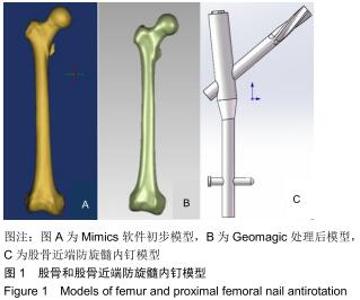
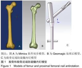
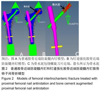
1.5.2 建立PFNA模型 参照第2代AO PFNA的具体参数,在Solidworks 2017软件中新建零件,通过拉伸、旋转、螺旋线、组合、放样等步骤得到PFNA内固定三维模型,PFNA内固定模型基本参数为:主钉长170 mm,螺旋刀片直径 10 mm,长度105 mm,颈干角130°,5°外翻角,见图1。 "

1.5.4 材料属性、边界条件与分析 参照郑利钦等[19]关于高龄股骨近端松质骨、皮质骨和PFNA内固定的密度、弹性模量、泊松比、屈服应力等参数设置2个模型[20-32]。B模型骨水泥材料参数设置为:表观密度为1.18 g/cm3,弹性模量为220 000 MPa,泊松比0.2,屈服应力100 MPa[33],边界条件均设置为股骨远端完全固定,根据志愿者体质量,在与股骨干长轴呈10°,垂直于股骨头球面,线性加载大小为630 N的载荷。各部件相互作用设置骨与骨之间摩擦系数为0.46[34],骨与内固定的摩擦系数为0.3,螺旋刀片与主钉设置为0.23,设置完成后均以K文件保存,见图2。 "
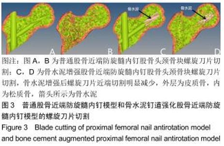
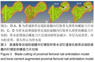
2.1 螺旋刀片切割 A模型首先在螺旋刀片近端周围产生切割,随着股骨头颈骨块的内翻和旋转,与主钉接触的松质骨单元受力后开始变形、消失,与螺旋刀片接触的外侧壁皮质骨随着应力增大出现变形、消失;B模型螺旋刀片骨水泥周围和股骨头颈骨块与主钉接触的部分松质骨单元消失,但消失的松质骨单元减少,切割程度较A模型明显减轻,见图3。 "
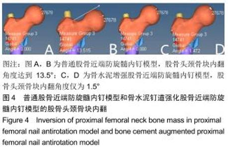
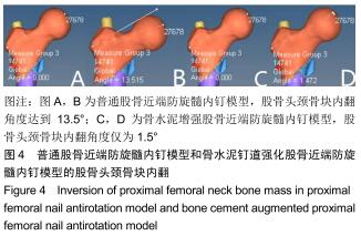
2.2 股骨颈内翻 A模型股骨头颈骨块受力后下沉,与小转子骨块相抵,随着载荷增大,螺旋刀片切割,小转子相接触的皮质骨变形、消失,股骨头颈骨块内翻,内翻角度达13.5°;B模型股骨头颈骨块受力后,骨水泥钉道强化螺旋刀片,其周围松质骨仅部分消失,切割程度较轻,内固定为股骨头颈骨块提供有效支撑,内翻角度仅1.5°,见图4。 "
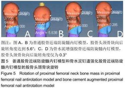
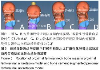
2.3 股骨颈旋转 A模型股骨头颈骨块随着载荷增大,出现螺旋刀片切割和内翻,由于相抵触的小转子骨块无法提供有效支撑,股骨头颈骨块向后旋转,但骨块受主钉阻挡,向后旋转角度为5.6°;B模型股骨头颈骨块螺旋刀片切割程度轻,内翻角度小,骨块相对稳定,向后旋转角度仅0.3°,见图5。 "
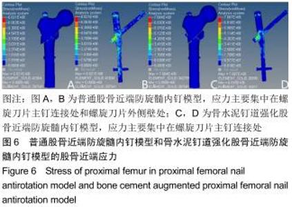
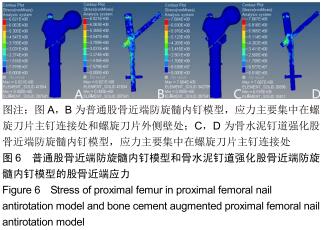
2.4 股骨近端应力 A模型股骨头颈骨块内翻严重,应力主要集中于螺旋刀片和主钉连接处和螺旋刀片外侧壁处;B模型骨水泥钉道强化后,股骨头颈骨块主要靠内固定支撑,应力主要分布在螺旋刀片和主钉连接处。A模型最大应力达682.1 MPa,B模型最大应力达768.4 MPa,见图6。 "
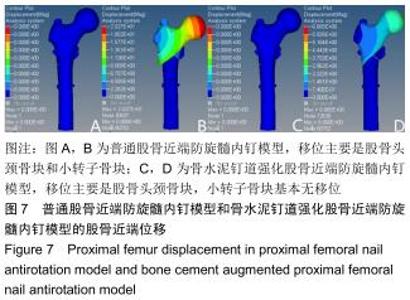
2.5 股骨近端位移 A模型股骨头颈骨块受力下沉内翻,与之相抵的小转子骨块亦下沉,移位主要是股骨头颈骨块和小转子骨块两个部分,最大位移达20.3 mm;B模型移位主要是股骨头颈骨块,小转子骨块基本无移位,最大位移仅6.7 mm。A、B模型最大位移均是股骨头,见图7。 "

|
[1] WANG J, WANG Y, LIU WD, et al. Hip fractures in Hefei, China: the Hefei osteoporosis project. J Bone Miner Metab. 2014; 32(2): 206-214. [2] XIA WB, HE SL, XU L, et al. Rapidly increasing rates of hip fracture in Beijing, China. J Bone Miner Res. 2012;27(1): 125-129.
[3] HERNLUND E, SVEDBOM A, IVERGÅRD M, et al. Osteoporosis in the European Union: medical management, epidemiology and economic burden. A report prepared in collaboration with the International Osteoporosis Foundation (IOF) and the European Federation of Pharmaceutical Industry Associations (EFPIA). Arch Osteoporos. 2013; 8: 136.
[4] ADIB HAJBAGHERY M, ABBASINIA M. Quality of life of the elderly after hip fracture surgery: a case-control study. J Caring Sci. 2013;2(1): 53-59.
[5] CLOSE JD, SWARTZ K, DEU R. Hip fracture in older patients: tips and tools to speed recovery. J Fam Pract. 2013; 62(9): 484-492.
[6] WANG J, MA JX, LU B, et al. Comparative finite element analysis of three implants fixing stable and unstable subtrochanteric femoral fractures: proximal femoral nail antirotation (pfna), proximal femoral locking plate (pflp), and reverse less invasive stabilization system (liss). Orthop Traumatol Surg Res. 2019.
[7] GUO Y, YANG HP, DOU QJ, et al. Efficacy of femoral nail anti-rotation of helical blade in unstable intertrochanteric fracture. Eur Rev Med Pharmacol Sci. 2017;21(3 Suppl): 6-11.
[8] VÉLEZ M, PALACIOS-BARAHONA U, ARANGO-POSADA MM, et al. Functional results and complications of the use of the proximal femoral nail in the treatment of intertrochanteric hip fractures. Acta Ortop Mex. 2018;32(3): 126-130.
[9] XU R, RU J, JI F, et al. Comparison of efficacy, complications and TGF-β2 expression between DHS and PFNA in elderly patients with osteoporotic femoral intertrochanteric fracture. Exp Ther Med. 2018; 16(1): 394-399.
[10] FOGAGNOLO F, KFURI M JR, PACCOLA CA. Intramedullary fixation of pertrochanteric hip fractures with the short AO-ASIF proximal femoral nail. Arch Orthop Trauma Surg. 2004; 124(1): 31-37.
[11] 武政,刘向栋,常宝生. PFNA治疗高龄不稳定性股骨转子间骨折内固定失败的危险因素分析[J]. 局解手术学杂志,2017,26(8): 616-619.
[12] 段志斌. 股骨近端防旋髓内钉术后螺旋刀片穿出股骨头的影响因素分析[D]. 南昌:南昌大学,2018.
[13] 王茂林,易志坚,卢明刚,等.防旋股骨近端髓内钉治疗股骨粗隆间骨折术后失效原因分析[J]. 中国骨与关节杂志,2019,8(7): 504-507.
[14] KIM Y, BAHK WJ, YOON YC, et al. Radiologic healing of lateral femoral wall fragments after intramedullary nail fixation for A3.3 intertrochanteric fractures. Arch Orthop Trauma Surg. 2015;135(10): 1349-1356.
[15] GUPTA RK, SANGWAN K, KAMBOJ P, et al. Unstable trochanteric fractures: the role of lateral wall reconstruction. Int Orthop. 2010;34(1): 125-129.
[16] 曾秋涛,赵洪斌,林勇,等.带孔螺旋刀片骨水泥增强型PFNA治疗转子间骨折的疗效分析[J]. 江西医药, 2017,52(12): 1330-1331. [17] 王兆稳,袁治国,白登彦,等. PFNA联合骨水泥注入治疗老年骨质疏松性股骨粗隆间骨折[J].甘肃医药,2017,36(12):1048-1050.
[18] 何祥鑫,林梓凌,李鹏飞,等. 基于Hypermesh 14.0和LS-DYNA的老年转子间骨折有限元建模分析[J].中国组织工程研究,2018, 22(11):1725-1730.
[19] 郑利钦,林梓凌,何祥鑫,等.动态载荷下股骨转子间区域皮质骨厚度对骨折类型影响的有限元分析[J].医学研究生学报,2018, 31(10):1043-1046.
[20] MORGAN EF, KEAVENY TM. Dependence of yield strain of human trabecular bone on anatomic site. J Biomech, 2001, 34(5): 569-577.
[21] KAWABATA Y, MATSUO K, NEZU Y, et al. The risk assessment of pathological fracture in the proximal femur using a CT-based finite element method. J Orthop Sci, 2017, 22(5): 931-937.
[22] MA Z, CHEN J, LAN F. Biomechanical response and injury of occupant's pelvis in side impacts: effects of the femoral head and loading conditions. J Mech Med Biol. 2014;14(6): 1440001.
[23] YENI YN, BROWN CU, NORMAN TL. Influence of bone composition and apparent density on fracture toughness of the human femur and tibia. Bone, 1998, 22(1): 79-84.
[24] MCCALDEN RW, MCGEOUGH JA, BARKER MB, et al. Age-related changes in the tensile properties of cortical bone. The relative importance of changes in porosity, mineralization, and microstructure. J Bone Joint Surg Am.1993;75(8): 1193-1205.
[25] DONG XN, GUO XE. The dependence of transversely isotropic elasticity of human femoral cortical bone on porosity. J Biomech.2004;37(8):1281-1287.
[26] BAYRAKTAR HH, MORGAN EF, NIEBUR GL, et al. Comparison of the elastic and yield properties of human femoral trabecular and cortical bone tissue. J Biomech. 2004; 37(1): 27-35.
[27] SHIBO G, XUANHUI Q, XINBO H, et al. Powder injection molding of Ti-6Al-4V alloy. J Mater Proc Technol. 2006;173(3): 310-314.
[28] HINÜBER C, KLEEMANN C, FRIEDERICHS RJ, et al. Biocompatibility and mechanical properties of diamond-like coatings on cobalt-chromium-molybdenum steel and titanium-aluminum-vanadium biomedical alloys. J Biomed Mater Res A. 2010;95(2): 388-400.
[29] GOFFIN JM, PANKAJ P, SIMPSON AH, et al. Does bone compaction around the helical blade of a proximal femoral nail anti-rotation (PFNA) decrease the risk of cut-out?: A subject-specific computational study. Bone Joint Res. 2013; 2(5): 79-83.
[30] CARNEY KS, PEREIRA JM, REVILOCK DM, et al. Jet engine fan blade containment using an alternate geometry. Int J Impact Eng. 2009; 36(5):720-728.
[31] HELGASON B, VICECONTI M, RÚNARSSON TP, et al. On the mechanical stability of porous coated press fit titanium implants: a finite element study of a pushout test. J Biomech. 2008;41(8): 1675-1681. [32] BOBBILI R, RAMAKRISHNA B, MADHU V. Dynamic compressive behavior and fracture modeling of Titanium alloy IMI 834. J Alloys Comp. 2017;714:225-231.
[33] DHANOPIA A, BHARGAVA M. Finite element analysis of human fractured femur bone implantation with PMMA thermoplastic prosthetic plate. Proc Eng. 2016;173: 1658-1665.
[34] EBERLE S, GERBER C, VON OLDENBURG G, et al. A biomechanical evaluation of orthopaedic implants for hip fractures by finite element analysis and in-vitro tests. Proc Inst Mech Eng H. 2010;224(10): 1141-1152.
[35] 武建超,师政伟,李吉鹏,等.股骨粗隆间骨折外侧壁损伤的研究进展[J].中国修复重建外科杂志, 2018,21(12):1605-1610.
[36] HAN L, LIU JJ, HU YG, et al. Controlled study on Gamma nail and proximal femoral locking plate for unstable intertrochanteric femoral fractures with broken lateral wall. Sci Rep. 2018;8(1): 11114.
[37] HU SJ, ZHANG SM, YU GR. Treatment of femoral subtrochanteric fractures with proximal lateral femur locking plates. Acta Ortop Bras.2012;20(6): 329-333.
[38] 陈鹏,傅德皓.股骨近端防旋髓内钉治疗老年股骨转子间骨折内固定失败原因分析[J]. 中国修复重建外科杂志, 2019,33(10): 1270-1274.
[39] 张世民,马卓,杜守超.股骨近端外侧壁的解剖学研究及其对转子间骨折内固定的意义[J]. 中国临床解剖学杂志, 2016, 34(1): 39-42.
[40] STOCKS GW. Treatment of reverse obliquity fractures of the intertrochanteric region of the femur. J Bone Joint Surg Am. 2002;84(5): 869-870.
[41] 王嘉嘉,徐美涛,张帅,等.经皮加压钢板治疗老年患者股骨逆粗隆间骨折17例临床观察[J]. 海军医学杂志, 2017,38(4):346-349.
[42] 王炎,汪海滨,史法见,等.PFNA和DHS固定治疗股骨粗隆间骨折的比较[J].中国矫形外科杂志,2019,27(24):2223-2227.
[43] 张景涛,雷卫军,周广伟.PFNA和LPFP在老年股骨粗隆间骨折治疗中的疗效比较[J].中国老年学杂志,2019,39(24):6001-6003.
[44] NIE B,CHEN X,LI J,et al. The medial femoral wall can play a more important role in unstable intertrochanteric fractures compared with lateral femoral wall: a biomechanical study. J Orthop Surg Res. 2017;12(1):197.
[45] 官煜超,李开南.股骨转子间骨折内侧壁损伤的研究现状[J]. 中国矫形外科杂志,2018,26(12):1116-1119.
[46] GAO Z,LV Y,ZHOU F,et al. Risk factors for implant failure after fixation of proximal femoral fractures with fracture of the lateral femoral wall. Injury. 2018;49(2):315-322.
[47] 李尧,胡传真,茅凌洲,等.股骨近端防旋髓内钉联合小钢板重建外侧壁治疗AO/OTA 31-A3型股骨转子间骨折[J].中国修复重建外科杂志, 2019,33(10):1223-1227.
[48] 姜昊,曹烈虎,潘思华,等.有限切开钛缆捆扎辅助PFNA与单纯微创辅助PFNA治疗老年骨质疏松性31-A3型股骨粗隆间骨折的疗效对比[J]. 中国骨与关节杂志,2019, 8(3):191-195.
[49] 王勇,潘骏.标准骨水泥强化型PFNA治疗老年股骨粗隆间不稳定骨折[J].中国矫形外科杂志, 2018, 26(18):1653-1658.
[50] 张磊,唐晓菊,黄有荣,等. 聚甲基丙烯酸甲酯骨水泥及磷酸钙骨水泥的材料性能及改性的研究进展[J].广西医学,2019,41(16): 2114-2118+2122. |
| [1] | Xu Feng, Kang Hui, Wei Tanjun, Xi Jintao. Biomechanical analysis of different fixation methods of pedicle screws for thoracolumbar fracture [J]. Chinese Journal of Tissue Engineering Research, 2021, 25(9): 1313-1317. |
| [2] | Zhang Tongtong, Wang Zhonghua, Wen Jie, Song Yuxin, Liu Lin. Application of three-dimensional printing model in surgical resection and reconstruction of cervical tumor [J]. Chinese Journal of Tissue Engineering Research, 2021, 25(9): 1335-1339. |
| [3] | Chen Xinmin, Li Wenbiao, Xiong Kaikai, Xiong Xiaoyan, Zheng Liqin, Li Musheng, Zheng Yongze, Lin Ziling. Type A3.3 femoral intertrochanteric fracture with augmented proximal femoral nail anti-rotation in the elderly: finite element analysis of the optimal amount of bone cement [J]. Chinese Journal of Tissue Engineering Research, 2021, 25(9): 1404-1409. |
| [4] | Du Xiupeng, Yang Zhaohui. Effect of degree of initial deformity of impacted femoral neck fractures under 65 years of age on femoral neck shortening [J]. Chinese Journal of Tissue Engineering Research, 2021, 25(9): 1410-1416. |
| [5] | Zhang Shangpu, Ju Xiaodong, Song Hengyi, Dong Zhi, Wang Chen, Sun Guodong. Arthroscopic suture bridge technique with suture anchor in the treatment of acromioclavicular dislocation [J]. Chinese Journal of Tissue Engineering Research, 2021, 25(9): 1417-1422. |
| [6] | Zhou Jihui, Li Xinzhi, Zhou You, Huang Wei, Chen Wenyao. Multiple problems in the selection of implants for patellar fracture [J]. Chinese Journal of Tissue Engineering Research, 2021, 25(9): 1440-1445. |
| [7] | Chen Junming, Yue Chen, He Peilin, Zhang Juntao, Sun Moyuan, Liu Youwen. Hip arthroplasty versus proximal femoral nail antirotation for intertrochanteric fractures in older adults: a meta-analysis [J]. Chinese Journal of Tissue Engineering Research, 2021, 25(9): 1452-1457. |
| [8] | Hu Kai, Qiao Xiaohong, Zhang Yonghong, Wang Dong, Qin Sihe. Treatment of displaced intra-articular calcaneal fractures with cannulated screws and plates: a meta-analysis of 15 randomized controlled trials [J]. Chinese Journal of Tissue Engineering Research, 2021, 25(9): 1465-1470. |
| [9] | Tang Hui, Yao Zhihao, Luo Daowen, Peng Shuanglin, Yang Shuanglin, Wang Lang, Xiao Jingang. High fat and high sugar diet combined with streptozotocin to establish a rat model of type 2 diabetic osteoporosis [J]. Chinese Journal of Tissue Engineering Research, 2021, 25(8): 1207-1211. |
| [10] | Li Zhongfeng, Chen Minghai, Fan Yinuo, Wei Qiushi, He Wei, Chen Zhenqiu. Mechanism of Yougui Yin for steroid-induced femoral head necrosis based on network pharmacology [J]. Chinese Journal of Tissue Engineering Research, 2021, 25(8): 1256-1263. |
| [11] | Xu Yulin, Shen Shi, Zhuo Naiqiang, Yang Huilin, Yang Chao, Li Yang, Zhao Heng, Zhao Lu. Biomechanical comparison of three different plate fixation methods for acetabular posterior column fractures in standing and sitting positions [J]. Chinese Journal of Tissue Engineering Research, 2021, 25(6): 826-830. |
| [12] | Cai Qunbin, Zou Xia, Hu Jiantao, Chen Xinmin, Zheng Liqin, Huang Peizhen, Lin Ziling, Jiang Ziwei. Relationship between tip-apex distance and stability of intertrochanteric femoral fractures with proximal femoral anti-rotation nail: a finite element analysis [J]. Chinese Journal of Tissue Engineering Research, 2021, 25(6): 831-836. |
| [13] | Hou Guangyuan, Zhang Jixue, Zhang Zhijun, Meng Xianghui, Duan Wen, Gao Weilu. Bone cement pedicle screw fixation and fusion in the treatment of degenerative spinal disease with osteoporosis: one-year follow-up [J]. Chinese Journal of Tissue Engineering Research, 2021, 25(6): 878-883. |
| [14] | He Li, Tian Wei, Xu Song, Zhao Xiaoyu, Miao Jun, Jia Jian. Factors influencing the efficacy of lumbopelvic internal fixation in the treatment of traumatic spinopelvic dissociation [J]. Chinese Journal of Tissue Engineering Research, 2021, 25(6): 884-889. |
| [15] | Yang Weiqiang, Ding Tong, Yang Weike, Jiang Zhengang. Combined variable stress plate internal fixation affects changes of bone histiocyte function and bone mineral density at the fractured end of goat femur [J]. Chinese Journal of Tissue Engineering Research, 2021, 25(6): 890-894. |
| Viewed | ||||||
|
Full text |
|
|||||
|
Abstract |
|
|||||