Chinese Journal of Tissue Engineering Research ›› 2020, Vol. 24 ›› Issue (22): 3467-3473.doi: 10.3969/j.issn.2095-4344.2266
Previous Articles Next Articles
Combination of magnesium ion with mineralized collagen intervenes osteogenic differentiation of mouse preosteoblasts
Sun Xirao1, Sun Baozhai1, Zhang Zhenbao2
- 12nd Affiliated Hospital of Jinzhou Medical University, Jinzhou 121000, Liaoning Province, China; 2Graduate School of Jinzhou Medical University, Jinzhou 121000, Liaoning Province, China
-
Received:2019-09-29Revised:2019-10-08Accepted:2019-11-07Online:2020-08-08Published:2020-04-26 -
About author:Sun Xirao, Master, Attending physician, 2nd Affiliated Hospital of Jinzhou Medical University, Jinzhou 121000, Liaoning Province, China -
Supported by:the Natural Science Foundation of Liaoning Province (Key Project), No. 20180530071; the Natural Science Foundation of Liaoning Province, No. 2019-MS-141
CLC Number:
Cite this article
Sun Xirao, Sun Baozhai, Zhang Zhenbao. Combination of magnesium ion with mineralized collagen intervenes osteogenic differentiation of mouse preosteoblasts[J]. Chinese Journal of Tissue Engineering Research, 2020, 24(22): 3467-3473.
share this article
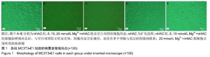
2.1 倒置显微镜观察细胞状态变化 从图1可以看到,含0,5,10 mmol/L镁离子nHAC浸提液培养的小鼠前成骨细胞贴壁情况良好,与空白对照组无明显差异,细胞形态呈长梭形,表面有多个突触与邻近的细胞相联系;含5,10 mmol/L镁离子nHAC浸提液培养的细胞密度较空白对照组和单纯nHAC浸提液实验组高,而含有20 mmol/L镁离子nHAC浸提液培养的细胞呈现明显的核浓缩。 "
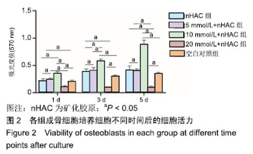
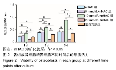
2.2 MTT法检测细胞活力结果 培养1,3,5 d时,nHAC组、5 mmol/L Mg2++nHAC组细胞活力与对照组比较差异无显著性意义(P > 0.05),10 mmol/L Mg2++nHAC组、 20 mmol/L Mg2++nHAC组细胞活力与对照组比较差异有显著性意义(P < 0.05);nHAC组细胞活力与5 mmol/L Mg2++nHAC组比较差异无显著性意义(P > 0.05),与 10 mmol/L Mg2++nHAC组、20 mmol/L Mg2++nHAC组细胞活力比较差异有显著性意义(P < 0.05);5,10, 20 mmol/L Mg2++nHAC组细胞活力两两比较差异均有显著性意义(P < 0.05),见图2。 "
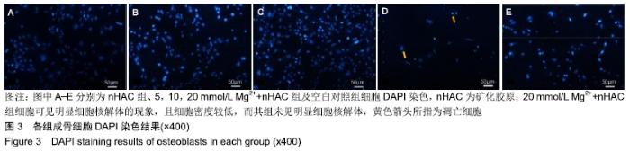
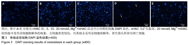
2.3 DAPI染色检测细胞凋亡结果 DAPI荧光染色结果见图3所示,当细胞发生凋亡时其细胞核呈现致密核染或呈碎块致密核染,20 mmol/L Mg2++nHAC组细胞可见明显细胞核解体的现象,且细胞密度较低,而其组未见明显细胞核解体。 "
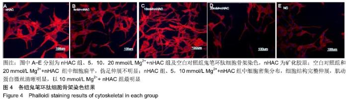
2.4 鬼笔环肽细胞骨架染色观察细胞微丝肌动蛋白 空白对照组和20 mmol/L Mg2++nHAC 组中细胞扁平,伪足伸展不明显;nHAC组、5,10 mmol/L Mg2++nHAC组中细胞密集分布,细胞结构完整伸展,肌动蛋白微丝清晰明显,以10 mmol/L Mg2++nHAC组最明显,见图4。 "

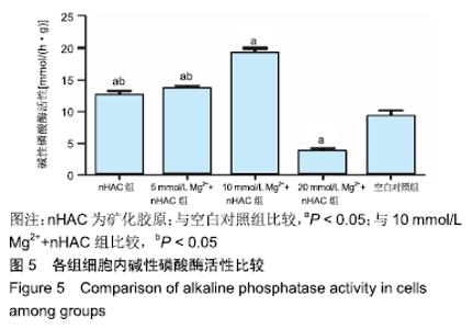
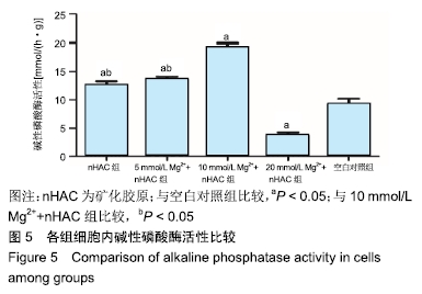
2.5 细胞内碱性磷酸酶活性检测结果 nHAC组、5, 10 mmol/L Mg2++nHAC组细胞内碱性磷酸酶活性高于空白对照组(P < 0.05),10 mmol/L Mg2++nHAC组高于nHAC组、5 mmol/L Mg2++nHAC组(P < 0.05),nHAC组、 5 mmol/L Mg2++nHAC组间比较差异无显著性意义(P > 0.05),20 mmol/L Mg2++nHAC组低于空白对照组(P < 0.05),见图5。 "

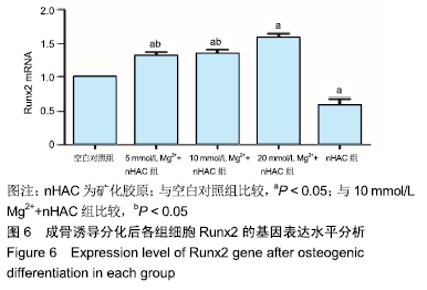
2.6 实时定量PCR检测结果 相对于空白对照组, nHAC组、5,10 mmol/L Mg2++nHAC组可以明显促进Runx2基因表达(P < 0.05),20 mmol/L Mg2++nHAC组可抑制Runx2基因表达(P < 0.05);nHAC组和5 mmol/L Mg2++nHAC组Runx2基因表达水平比较差异无显著性意义(P > 0.05),但均低于10 mmol/L Mg2++nHAC组(P < 0.05),见图6。 "
| [1] 王敏,冯希平,翁雨来.牙周病致牙槽骨缺损的治疗进展[J].上海口腔医学,2002,11(3):261-264. [2] 马冲,于寰,马勇强,等.牙周再生术治疗慢性牙周炎不同程度骨缺损的疗效评价[J].牙体牙髓牙周病学杂志,2018,28(8):455-460. [3] HOWAIT M, ALBASSAM A, YAMADA C, et al. Elevated Expression of Macrophage Migration Inhibitory Factor Promotes Inflammatory Bone Resorption Induced in a Mouse Model of Periradicular Periodontitis.J Immunol. 2019;202(7): 2035-2043. [4] HAN J, MA B, LIU H, et al. Hydroxyapatite nanowires modified polylactic acid membrane plays barrier/osteoinduction dual roles and promotes bone regeneration in a rat mandible defect model.Biomed Mater Res A.2018;106(12):3099-3110. [5] 郝柯屹,田杰华,吕鸣樾,等.GBR植骨相较于自体骨移植远期稳定性的优势[J].口腔医学,2019,39(1):67-69. [6] LAVERTY DP, KELLY R, ADDISON O. Survival of dental implants placed in autogenous bone grafts and bone flaps in head and neck oncology patients: a systematic review.Int J Implant Dent.2018;4(1):19. [7] KOU JM, FU TY, JIA XJ, et al. Clinical Observations on Repair of Non-Infected Bone Nonunion by Using Mineralized Collagen Graft.J Biomater Tissue Eng. 2014;4(12): 1107-1112(6). [8] 闫永发,王春兰,范月静.纳米羟基磷灰石复合胶原植入术治疗牙周病骨缺损的疗效观察[J].中华临床医师杂志, 2011,5(1): 176-177. [9] FENG QL, CUI FZ, ZHANG W. Nano-hydroxyapatite/collagen composite for bone repair.Zhongguo Yi Xue Ke Xue Yuan Xue Bao.2002;24(2):124-128. [10] RAHMAN MS, RANA MM, SPITZHORN LS, et al. Fabrication of biocompatible porous scaffolds based on hydroxyapatite/ collagen/chitosan composite for restoration of defected maxillofacial mandible bone.Prog Biomater. 2019;8(3): 137-154. [11] ZHU J, ZHANG K, LUO K, et al. Mineralized Collagen Modified Polymethyl Methacrylate Bone Cement for Osteoporotic Compression Vertebral Fracture at One-Year Follow-up. Spine(Phila Pa 1976).2018;44(12):827-838. [12] 王程越,姚玉胜,王树峰,等.AZ31镁合金在垂直牵张犬下颌骨成骨的初步研究[J].口腔医学,2013,33(7):456-460 [13] 王树峰,李春荣,王程越,等.微弧氧化AZ31镁合金的生物相容性[J].中国组织工程研究,2012,16(38):7101-7106. [14] 于晴,王程越,杨静馨,等.矿化胶原与镁钙合金的复合材料用于犬拔牙位点保存的实验研究[J].中国临床解剖学杂志, 2017,35(5): 532-536,542. [15] YU Q, WANG C, YANG J, et al. Mineralized collagen/Mg-Ca alloy combined scaffolds with improved biocompatibility for enhanced bone response following tooth extraction.Biomed Mater.2018;13(6):065008. [16] GUO CW, YU Q, WANG CY, et al. Evaluation of alveolar bone repair with mineralized collagen block reinforced with Mg-Ca alloy rods.J Biomater Tissue Eng.2018;8:1-10. [17] WANG CY, SUN Y, CHEN Z, et al. Application of the magnesium rod and porous mineralized collagen plug in site preservation in dogs.J Biomater Tissue Eng.2016;6(3): 171-179. [18] STAIGER MP, PIETAK AM, HUADMAI J, et al. Magnesium and its Alloys as Orthopedic Biomaterials.Biomaterials. 2006; 27(9):1728-1734. [19] SONG G. Control of biodegradation of biocompatable magnesium alloys. Corros Sci.2007;49(4):1696-1701. [20] MIAO H, ZHANG D, CHEN C, et al. Research on Biodegradable Mg–Zn–Gd Alloys for Potential Orthopedic Implants: In Vitro and in Vivo Evaluations. ACS Biomater Sci Eng. 2019;5(3):1623-1634. [21] ZHU D, COCKERILL I, SU Y, et al. Mechanical Strength, Biodegradation, and in Vitro and in Vivo Biocompatibility of Zn Biomaterials.ACS Appl Mater Interfaces. 2019;11(7): 6809-6819. [22] 陈宏,王成成,康亚斌,等.镁合金微弧氧化的研究现状[J].表面技术,2019,48(7):49-60. [23] JI RN, PENG GC, ZHANG SG, et al. The fabrication of a CeO 2 coating via cathode plasma electrolytic deposition for the corrosion resistance of AZ31 magnesium alloy.Ceram Int. 2018;44(16). [24] SWETHA CV, DUMPALA R, ANAND KS, et al. Influence of heat treatment on the machinability and corrosion behavior of AZ91 Mg alloy.J Magnes Alloy.2018;6(1):52-58. [25] LIU Y, CURIONI M, ZHU L. Correlation between electrochemical impedance measurements and corrosion rates of Mg-1Ca alloy in simulated body fluid. Electrochimica Acta.2018;264:101-108. [26] MUKAEVA VR, KULYASOVA OB, FARRAKHOV RG, et al. Mechanical properties and corrosion behavior of Mg-1Zn- 0.2Ca alloy with various grain size. IOP Conf Ser: Mater Sci Eng.2019;479(1):012075. [27] 荣欢,朱明,朱青,等.AZ91D镁合金表面无铬转化膜/杂化复合涂层的耐蚀性能[J].材料保护,2018,51(4):1-6. [28] QIAN BY, MIAO W, QIU M, et al. Influence of Voltage on the Corrosion and Wear Resistance of MicroArc Oxidation Coating on Mg-8Li-2Ca Alloy.Acta Metallurgica Sinica(English Letters).2019;32(2):54-64. [29] DONG TS, LI XB, FU BG, et al. Influence of Ca on microstructure and corrosion resistance of Mg-14Li alloy. China Foundry.2018;5(2):132-138. [30] QI H, HEISE S, ZHOU J, et al. Electrophoretic Deposition of Bioadaptive Drug Delivery Coatings on Magnesium Alloy for Bone Repair.ACS Appl Mater Interfaces. 2019;11(8): 8625-8634. [31] 冯庆玲,崔福斋,张伟.纳米羟基磷灰石/胶原骨修复材料[J].中国医学科学院学报,2002,24(2):124-128. [32] 黄永辉,沈铁城,徐晓峰,等.纳米羟基磷灰石/胶原骨修复骨缺损的效果评估[J].中国组织工程研究,2006,10(37):51-53. [33] 纳米羟基磷灰石/胶原复合材料修复猪下颌骨缺损后血管内皮生长因子的变化[J].中国组织工程研究,2019,23(26): 4148-4153. [34] WANG J, MA XY, FENG YF, et al.Magnesium Ions Promote the Biological Behaviour of Rat Calvarial Osteoblasts by Activating the PI3K/Akt Signalling Pathway.Biol Trace Elem Res.2017;179(2):284-293. [35] 王健,马翔宇,冯亚非,等.镁离子对成骨细胞活力和分化的促进作用及其机制研究[J].现代生物医学进展,2015,15(15):46-49. [36] DAYAGHI E, BAKHSHESHI-RAD HR, HAMZAH E, et al. Magnesium-zinc scaffold loaded with tetracycline for tissue engineering application: In vitro cell biology and antibacterial activity assessment.Mater Sci Eng C Mater Biol Appl. 2019; 102:53-65. [37] YOSHIZAWA S, BROWN A, BARCHOWSKY A, et al. Magnesium ion stimulation of bone marrow stromal cells enhances osteogenic activity, simulating the effect of magnesium alloy degradation.Acta Biomater. 2014;10(6): 2834-2842. [38] 吴聪颖.微丝的基本性质与细胞核肌动蛋白[J].中国细胞生物学学报,2019,41(3):67-72. [39] 常新,侯志明,柴田恭明,等.碱性磷酸酶和骨钙素在成骨过程中表达的定量研究[J].华西口腔医学杂志,2005,23(5):424-426. [40] HORITA M, NISHIDA K, HASEI J, et al. Involvement of ADAM12 in Chondrocyte Differentiation by Regulation of TGF-β1–Induced IGF-1 and RUNX-2 Expressions.Calcif Tissue Int.2019;105(1):97-106. [41] WU X, ZHANG Y, XING Y, et al. High-fat and high-glucose microenvironment decreases Runx2 and TAZ expression and inhibits bone regeneration in the mouse.J Orthop Surg Res. 2019;14(1):55. [42] ZHANG X, ZU H, ZHAO D, et al. Ion channel functional protein kinase TRPM7 regulates Mg ions to promote the osteoinduction of human osteoblast via PI3K pathway:, In vitro, simulation of the bone-repairing effect of Mg-based alloy implant.Acta Biomater.2017;63:369-382. [43] HIRATSUKA T, UEZONO M, TAKAKUDA K, et al. Enhanced bone formation onto the bone surface using a hydroxyapatite/ collagen bone‐like nanocomposite.J Biomed Mater Res B Appl Biomater.2019. doi: 10.1002/jbm.b.34397. [Epub ahead of print] [44] JIANG X, ZHONG Y, ZHENG L ,et al. Nano-hydroxyapatite/collagen film as a favorable substrate to maintain the phenotype and promote the growth of chondrocytes culturedin vitro.Int J Mol Med. 2018;41(4): 2150-2158. |
| [1] | Gao Yujin, Peng Shuanglin, Ma Zhichao, Lu Shi, Cao Huayue, Wang Lang, Xiao Jingang. Osteogenic ability of adipose stem cells in diabetic osteoporosis mice [J]. Chinese Journal of Tissue Engineering Research, 2022, 26(7): 999-1004. |
| [2] | Liang Xuezhen, Yang Xi, Li Jiacheng, Luo Di, Xu Bo, Li Gang. Bushen Huoxue capsule regulates osteogenic and adipogenic differentiation of rat bone marrow mesenchymal stem cells via Hedgehog signaling pathway [J]. Chinese Journal of Tissue Engineering Research, 2022, 26(7): 1020-1026. |
| [3] | Le Guoping, Zhang Ming, Xi Licheng, Luo Hanwen. Preparation and in vitro evaluation of vancomycin hydrochloride@polylactic acid-glycolic acid copolymer-chitosan-hyaluronic acid composite sustained-release microspheres [J]. Chinese Journal of Tissue Engineering Research, 2022, 26(4): 528-534. |
| [4] | He Guanyu, Xu Baoshan, Du Lilong, Zhang Tongxing, Huo Zhenxin, Shen Li. Biomimetic orientated microchannel annulus fibrosus scaffold constructed by silk fibroin [J]. Chinese Journal of Tissue Engineering Research, 2022, 26(4): 560-566. |
| [5] | Wang Jing, Yang Jiuju, Wang Ningning, Liu Chao. Effect of modified three-dimensional-printed titanium scaffold on osteogenic differentiation of adipose-derived mesenchymal stem cells [J]. Chinese Journal of Tissue Engineering Research, 2022, 26(27): 4265-4271. |
| [6] | Sun Kai, Li Ruixin, Gao Lilan, Fan Meng, Zhang Xizheng, Li Hui. Biocompatibility of silk fibroin/type II collagen/hydroxyapatite three-phase composite scaffold [J]. Chinese Journal of Tissue Engineering Research, 2022, 26(27): 4283-4287. |
| [7] | Chen Yue, Luo Feng, Su Dan, Yang Diyuan, Pu Qi, Zhang Min, Gui Wenjuan, Sun Xiaorong. Hemostatic efficacy, degradation in vivo and biological evaluation of fish scale collagen [J]. Chinese Journal of Tissue Engineering Research, 2022, 26(27): 4353-4359. |
| [8] | Gao Zhao, Zhao Yuhao, He Yixiang, Zhao Haiyan, Wang Wenji. DNA hydrogel based on drug delivery and bone tissue engineering [J]. Chinese Journal of Tissue Engineering Research, 2022, 26(27): 4379-4385. |
| [9] | Huang Tao, Jia Zhiqiang, Zhao Xiaoguang, Wang Lei, Fang Liping, Zhai Wenjing, Zhai Shafei, Zhou Yongxin. LncRNA-HOTAIR can regulate differentiation of adipose derived mesenchymal stem cells into osteoblasts [J]. Chinese Journal of Tissue Engineering Research, 2022, 26(25): 3951-3955. |
| [10] | Yu Chunbo, Li Dayu, Fan Fang, Li Changfu. Transcription factor-miRNA-mRNA network analysis of osteogenic differentiation of adipose-derived stem cells [J]. Chinese Journal of Tissue Engineering Research, 2022, 26(24): 3908-3913. |
| [11] | Chen Chichi, Zhang Yu, He Jiachen, Shi Qin. Osteogenic differentiation of bone marrow mesenchymal stem cells in obese mice [J]. Chinese Journal of Tissue Engineering Research, 2022, 26(24): 3846-3851. |
| [12] | You Wulin, Huang Guicheng, Wang Jianwei. Osteogenic differentiation potential of microencapsulated transgenic bone marrow mesenchymal stem cells cocultured with osteoblasts [J]. Chinese Journal of Tissue Engineering Research, 2022, 26(24): 3852-3857. |
| [13] | Cui Yinpeng, Guo Ai, Ma Lifeng, Liu Zhenjiang. Ouabain induces in vitro differentiation of human bone marrow mesenchymal stem cells into osteoblasts [J]. Chinese Journal of Tissue Engineering Research, 2022, 26(24): 3796-3801. |
| [14] | Jin Ke, Wu Xiaoling, Luo Xiaoling, Xu Pengfei, Xu Xiaomei. Osteogenic differentiation of human periodontal ligament stem cells under estrogen and crosstalk between the two pathways [J]. Chinese Journal of Tissue Engineering Research, 2022, 26(24): 3886-3891. |
| [15] | Han Zhi, Wang Zhimiao, Gaxi Sijia, Lu Qingling, Guo Tao. Tissue engineered cartilage constructed by polyurethane composite chondrocytes [J]. Chinese Journal of Tissue Engineering Research, 2022, 26(22): 3455-3459. |
| Viewed | ||||||
|
Full text |
|
|||||
|
Abstract |
|
|||||