[1] MACHT SD, KRIZEK TJ.Sutures and suturing-Current concepts. J Oral Surg.1978;36:710-712.
[2] KULKARNI S, DODWAD V, CHAVA V. Healing of periodontal flaps when closed with silk sutures and N-butyl cyanoacrylate: a clinical and histological study.Indian J Dent Res. 2007;18(2): 72-77.
[3] KAJBAFZADEH AM, A BOLGHASEM IH, ESHGHI P, et al. Single -donor fibrin sealant fo rrepair of urethrocutaneous fistulae following multiple hypospadias and epispadias repairs. J Pediatr Uro1.2011;7(4):422-427.
[4] PADHYE A, POL DG.Clinical evaluation of the efficacy of N-butyl cyanoacrylate as bioadhesive material in comparison to conventional silk sutures in modified widman flap surgery in anterior region.JIDA.2011;5:899‑904.
[5] 张飞,赵君,孟兴凯,李剑锋.合成α-氰基丙烯酸正丁酯医用胶用于肝脏止血[J].中国组织工程研究,2016,20(47):7112-7118.
[6] 王蕊,吴楠,吴燕,翁传煌,等.医用组织黏合剂黏合兔眼巩膜穿通伤口与传统缝合的对比实验研究[J].创伤外科杂志,2008,10(5): 443-446.
[7] BHASKAR SN, CUTRIGHT DE.Healing of skin wounds with butyl cyanoacrylate.J Dent Res. 1969;48:294-297.
[8] MEHTA MJ, SHAH KH, BHATT RG.Osteosynthesis of mandibular fractures with N-butyl cyanoacrylate: a pilot study.J Oral Maxillofac Surg.1987;45:393-396.
[9] ULLER W, EL-SOBKY S, ALOMARI AI, et al.Preoperative Embolization of Venous Malformations Using n-Butyl Cyanoacrylate.Vasc Endovascular Surg.2018;52(4):269-274.
[10] 王艳红,顾汉卿.医用黏合剂的发展及临床应用进展[J].透析与人工器官,2008,19(3):23-32.
[11] 刘芝兰,张健民,陈立贵.医用粘合剂的研究及临床应用进展[J].粘接,2002,23(1):28-31.
[12] 李现令,鲁守彦,刘婷婷.生物止血材料医用胶黏合儿童额面部开放性伤口创面的可行性[J].中国组织工程研究,2015,19(21): 3350-3354.
[13] 卢迪,叶桓.医用胶在处理小儿头面部皮肤伤口中的应用[J].蚌埠医学院学报,2014,39(1):87.
[14] 李学正.“504”(α-氰基丙烯酸正丁酯)粘合剂应用于人工镫骨术的初步观察[J].湖南医学院学报,1980,5(2):146-149.
[15] 鲁守东.内镜下注射福爱乐医用胶治疗食道静脉曲张破裂出血32例[J].中国医药指南,2013,11(7):552-553.
[16] MIZRAH IS, BICKEL A, BEN LAYISH EB.Use of tissue adhesives in there pair of lacer-ations in children.J Pediatr Surg.1988;23:312-313.
[17] ABOU NEEL EA, SALIH V, REVELL PA, et al.Viscoelastic and biological performance of low modulus,reactive calcium phosphate-filled,degradable,poly mericbone ad hesives.Acta Biomater.2012;8(1):313-320.
[18] CHEN WL, LIN CT, HSIEH CY, et al.Comparison of the bacteriostatic effects, corneal cytotoxicity, and the ability to seal corneal incisions among three different tissue adhesives. Cornea.2007;26(10):1228-1234.
[19] CHOW A, MARSHALL H, ZACHARAKIS E, et al.Use of tissue glue for surgical incision closure: a systematic review and meta-analysis of randomized controlled trials.J Am Coll Surg. 2010;211(1):114-125.
[20] 栗雪艳.α-氰基丙烯酸酯类医用胶的合成与性能研究[D].广州:华南理工大学,2013.
[21] COOVER HW, JOYNER FB, SHEARER NH, et al. Chemistry and performance of cyanoacrylate adhesives. Soc Plast Eng. 1959;12(2):413-417.
[22] SHAPIRO AJ, DINSMORE RC, NORTH JH.Tensile strength of wound closure with cyanoacrylate glue.Am Surg. 2001; 67(11):1113-1115.
[23] 徐梅,赵敏,李鸿.α-氰基丙烯酸正辛酯在小梁切除术中应用的初步报告[J].眼外伤职业眼病杂志,2007,29(8):581-583.
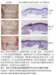
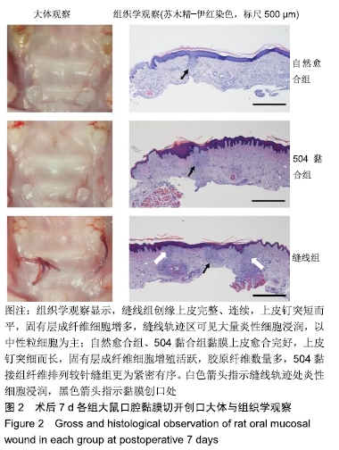
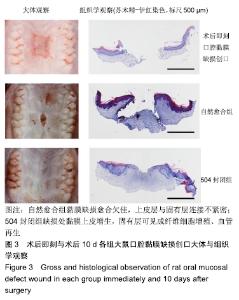
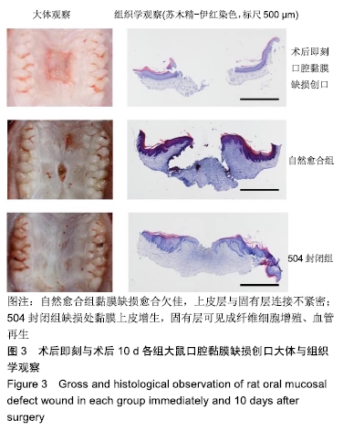
|