Chinese Journal of Tissue Engineering Research ›› 2020, Vol. 24 ›› Issue (13): 2061-2067.doi: 10.3969/j.issn.2095-4344.2060
Previous Articles Next Articles
Acellular amniotic membrane scaffold combined with human amniotic mesenchymal stem cells transfected with Scleraxis lentivirus can promote tendon-bone healing in rabbits
Zhang Jun, Yang Jibin, Jin Ying, Zou Gang, Tang Jingfeng, Ge Zhen, Yang Qifan, Liu Yi
- First Department of Orthopedics, Affiliated Hospital of Zunyi Medical University, Zunyi 563000, Guizhou Province, China
-
Received:2019-09-24Revised:2019-09-30Accepted:2019-11-15Online:2020-05-08Published:2020-03-10 -
Contact:Liu Yi, Professor, Master’s supervisor, First Department of Orthopedics, Affiliated Hospital of Zunyi Medical University, Zunyi 563000, Guizhou Province, China -
About author:Zhang Jun, Master candidate, First Department of Orthopedics, Affiliated Hospital of Zunyi Medical University, Zunyi 563000, Guizhou Province, China -
Supported by:the Science and Technology Project of Guizhou Province, No. LH[2017]7015
CLC Number:
Cite this article
Zhang Jun, Yang Jibin, Jin Ying, Zou Gang, Tang Jingfeng, Ge Zhen, Yang Qifan, Liu Yi. Acellular amniotic membrane scaffold combined with human amniotic mesenchymal stem cells transfected with Scleraxis lentivirus can promote tendon-bone healing in rabbits[J]. Chinese Journal of Tissue Engineering Research, 2020, 24(13): 2061-2067.
share this article
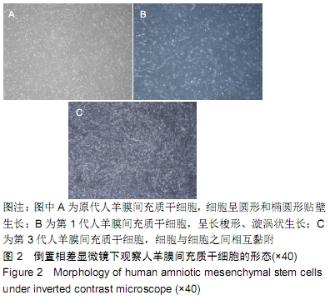
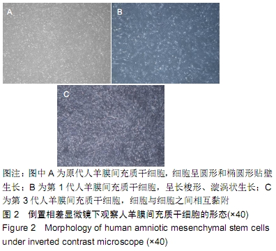
2.1 人羊膜间充质干细胞的形态 原代人羊膜间充质干细胞呈圆形或椭圆形生长,见图2A;经过传代培养后细胞逐渐呈现长梭形涡旋样贴壁生长,见图2B;第3代人羊膜间充质干细胞形态最为典型,细胞与细胞之间相互黏附,见图2C。 "
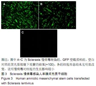
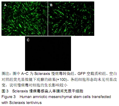
2.2 Scleraxis慢病毒感染人羊膜间充质干细胞 以最适感染复数转染第3代人羊膜间充质干细胞,96 h后在荧光显微镜下观察可见:Scleraxis慢病毒转染组和GFP空载质粒组可见明显的绿色荧光表达,空白对照组未见绿色荧光,见图3A-C。在普通显微镜下观察各组细胞形态均未见明显改变,说明慢病毒对细胞的生长影响较小。 "
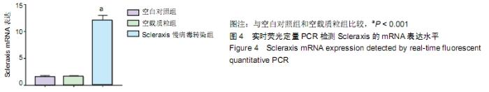
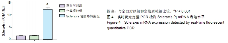
2.3 实时荧光定量PCR检测Scleraxis慢病毒感染人羊膜间充质干细胞后Scleraxis的mRNA表达水平 Scleraxis慢病毒转染组的Scleraxis mRNA表达水平明显高于其他两组,差异有显著性意义(P < 0.05),而GFP空载质粒组和空白对照组的Scleraxis mRNA表达水平未见明显差异(P > 0.05),见图4。 "
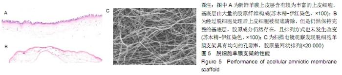
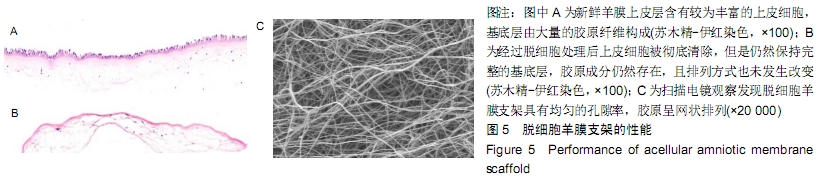
2.4 脱细胞羊膜支架的性能 通过苏木精-伊红染色发现新鲜羊膜上皮层含有较为丰富的上皮细胞,基底层由大量的胶原纤维构成,见图5A;经过脱细胞处理后其上皮细胞被彻底清除,但是仍然保持完整的基底层,胶原成分仍然存在,且排列方式也未发生改变,见图5B;通过扫描电镜观察发现脱细胞羊膜支架具有均匀的孔隙率,胶原呈网状排列,见图5C。 "
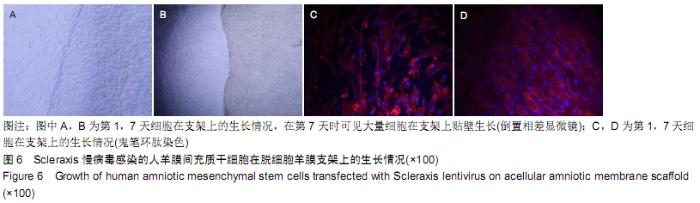
2.5 经Scleraxis慢病毒感染的人羊膜间充质干细胞在脱细胞羊膜支架上的生长情况 将Scleraxis慢病毒转染的第3代人羊膜间充质干细胞种植到脱细胞羊膜支架上并连续观察细胞在支架上的生长情况。第1天在普通倒置相差显微镜下观察可见有少量的细胞黏附在支架上生长,见图6A;连续培养后,细胞逐渐增多,在第7天时见大量细胞在支架上贴壁生长,见图6B。通过鬼笔环肽染色进一步确定细胞在支架上的生长情况,第1天有少量的细胞在支架上生长,见图6C;第7天可见密集的细胞分布在支架表面,且细胞形态较好,见图6D。 "

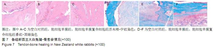
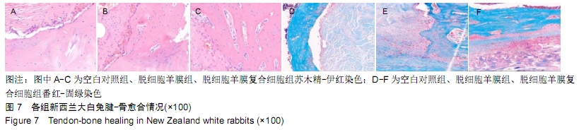
2.6 各组新西兰大白兔腱-骨愈合情况 术后3个月苏木精-伊红染色发现空白对照组腱-骨交界面有大量的炎性细胞存在,并且腱-骨之间的间隙较大,说明腱-骨愈合情况较差;相比较而言,脱细胞羊膜组腱-骨交界面的间隙逐渐缩小,并且炎性细胞也逐渐减少;脱细胞羊膜复合细胞组腱-骨交界面之间几乎看不到明显的间隙,也很少有炎性细胞的存在,有大量排列规整的胶原纤维,说明其愈合较好。通过番红-固绿染色发现空白对照组腱-骨之间间隙较大,有大量的炎性细胞存在;脱细胞羊膜组虽然间隙逐渐缩窄,且炎性细胞逐渐减少,但是未能观察到纤维软骨的生成;脱细胞羊膜复合细胞组可见纤维软骨的生成,说明其愈合较好,见图7。 "
| [1] YANG G, ROTHRAUFF BB, TUAN RS. Tendon and ligament regeneration and repair: clinical relevance and developmental paradigm. Birth Defects Res C Embryo Today. 2013;99(3): 203-222. [2] SHERMAN SL, CHALMERS PN, YANKE AB, et al. Graft tensioning during knee ligament reconstruction: principles and practice. J Am Acad Orthop Surg. 2012;20(10):633-645. [3] LU HH, THOMOPOULOS S. Functional attachment of soft tissues to bone: development, healing, and tissue engineering. Annu Rev Biomed Eng. 2013;15:201-226. [4] LIU YX, THOMOPOULOS S, BIRMAN V, et al. Bi-material attachment through a compliant interfacial system at the tendon-to-bone insertion site. Mech Mater. 2012;49(1): 83-92. [5] KATZEL EB, WOLENSKI M, LOISELLE AE, et al. Impact of Smad3 loss of function on scarring and adhesion formation during tendon healing. J Orthop Res. 2011;29(5):684-693. [6] ATESOK K, FU FH, WOLF MR, et al. Augmentation of tendon-to-bone healing. J Bone Joint Surg Am. 2014;96(6): 513-521. [7] VAN KAMPEN C, ARNOCZKY S, PARKS P, et al. Tissue-engineered augmentation of a rotator cuff tendon using a reconstituted collagen scaffold: a histological evaluation in sheep. Muscles Ligaments Tendons J. 2013; 3(3):229-235. [8] KAWAKAMI Y, TAKAYAMA K, MATSUMOTO T, et al. Anterior Cruciate Ligament-Derived Stem Cells Transduced With BMP2 Accelerate Graft-Bone Integration After ACL Reconstruction. Am J Sports Med. 2017;45(3):584-597. [9] SUN L, QU L, ZHU R, et al. Effects of Mechanical Stretch on Cell Proliferation and Matrix Formation of Mesenchymal Stem Cell and Anterior Cruciate Ligament Fibroblast. Stem Cells Int. 2016;2016:9842075. [10] LU J, CHAMBERLAIN CS, JI ML, et al. Tendon-to-Bone Healing in a Rat Extra-articular Bone Tunnel Model: A Comparison of Fresh Autologous Bone Marrow and Bone Marrow-Derived Mesenchymal Stem Cells. Am J Sports Med. 2019;47(11):2729-2736. [11] RBIA N, BULSTRA LF, LEWALLEN EA, et al. Seeding decellularized nerve allografts with adipose-derived mesenchymal stromal cells: An in vitro analysis of the gene expression and growth factors produced. J Plast Reconstr Aesthet Surg. 2019;72(8):1316-1325. [12] KUSUMA GD, MENICANIN D, GRONTHOS S, et al. Ectopic Bone Formation by Mesenchymal Stem Cells Derived from Human Term Placenta and the Decidua. PLoS One. 2015; 10(10):e0141246. [13] YANG M, LIU H, WANG Y, et al. Hypoxia reduces the osteogenic differentiation of peripheral blood mesenchymal stem cells by upregulating Notch-1 expression. Connect Tissue Res. 2019;60(6):583-596. [14] HASHIMOTO R, KATOH Y, MIYAMOTO Y, et al. Increased extracellular and intracellular Ca²⁺ lead to adipocyte accumulation in bone marrow stromal cells by different mechanisms. Biochem Biophys Res Commun. 2015;457(4): 647-652. [15] CHEN X, LIU Y, DING W, et al. Mechanical stretch-induced osteogenic differentiation of human jaw bone marrow mesenchymal stem cells (hJBMMSCs) via inhibition of the NF-κB pathway. Cell Death Dis. 2018;9(2):207. [16] BORNES TD, JOMHA NM, MULET-SIERRA A, et al. Porous Scaffold Seeding and Chondrogenic Differentiation of BMSC-seeded Scaffolds. Bio Protoc. 2015;5(24): e1693. [17] QIANG Y, LIANG G, YU L. Human amniotic mesenchymal stem cells alleviate lung injury induced by ischemia and reperfusion after cardiopulmonary bypass in dogs. Lab Invest. 2016;96(5):537-546. [18] EVANS MA, BROUGHTON BRS, DRUMMOND GR, et al. Amnion epithelial cells - a novel therapy for ischemic stroke?Neural Regen Res. 2018;13(8):1346-1349. [19] CARLBERG AL, TUAN RS, HALL DJ. Regulation of scleraxis function by interaction with the bHLH protein E47. Mol Cell Biol Res Commun. 2000;3(2):82-86. [20] HU JS, OLSON EN, KINGSTON RE. HEB, a helix-loop-helix protein related to E2A and ITF2 that can modulate the DNA-binding ability of myogenic regulatory factors. Mol Cell Biol. 1992;12(3):1031-1042. [21] LIU Y, WATANABE H, NIFUJI A, et al. Overexpression of a single helix-loop-helix-type transcription factor, scleraxis, enhances aggrecan gene expression in osteoblastic osteosarcoma ROS17/2.8 cells. J Biol Chem. 1997;272(47): 29880-29885. [22] ALBERTON P, POPOV C, PRÄGERT M, et al. Conversion of human bone marrow-derived mesenchymal stem cells into tendon progenitor cells by ectopic expression of scleraxis. Stem Cells Dev. 2012;21(6):846-858. [23] MADRY H, KOHN D, CUCCHIARINI M. Direct FGF-2 gene transfer via recombinant adeno-associated virus vectors stimulates cell proliferation, collagen production, and the repair of experimental lesions in the human ACL. Am J Sports Med. 2013;41(1):194-202. [24] WANG CJ, WENG LH, HSU SL, et al. pCMV-BMP-2- transfected cell-mediated gene therapy in anterior cruciate ligament reconstruction in rabbits. Arthroscopy. 2010;26(7): 968-976. [25] CHEN B, LI B, QI YJ, et al. Enhancement of tendon-to-bone healing after anterior cruciate ligament reconstruction using bone marrow-derived mesenchymal stem cells genetically modified with bFGF/BMP2. Sci Rep. 2016;6:25940. [26] KAWAKAMI Y, TAKAYAMA K, MATSUMOTO T, et al. Anterior Cruciate Ligament-Derived Stem Cells Transduced With BMP2 Accelerate Graft-Bone Integration After ACL Reconstruction. Am J Sports Med. 2017;45(3):584-597. [27] PENG W, ZHANG J, ZHANG H, et al. Effects of lentiviral transfection containing bFGF gene on the biological characteristics of rabbit BMSCs. J Cell Biochem. 2018; 119(10): 8389-8397. [28] ILHAN A, YOLCU U, GUNDOGAN FC. Amniotic membrane: new concepts for an old dressing. Wound Repair Regen. 2015;23(1):145. [29] KARALASHVILI L, KAKABADZE A, VYSHNEVSKA G, et al. Acellular human amniotic membrane as a three-dimensional scaffold for the treatment of mucogingival defects. Georgian Med News. 2015;(244-245):84-89. [30] ZELEN CM, SNYDER RJ, SERENA TE, et al. The use of human amnion/chorion membrane in the clinical setting for lower extremity repair: a review. Clin Podiatr Med Surg. 2015; 32(1):135-146. [31] 朱喜忠,刘子铭,吴术红,等.Scleraxis慢病毒基因感染人羊膜间充质干细胞向肌腱细胞的定向分化[J].中国组织工程研究, 2017, 21(33):5382-5387. [32] LIU PF, GUO L, ZHAO DW, et al. Study of human acellular amniotic membrane loading bone marrow mesenchymal stem cells in repair of articular cartilage defect in rabbits. Genet Mol Res. 2014;13(3):7992-8001. [33] ZHANG Y, YU J, ZHANG J, et al. Simvastatin With PRP Promotes Chondrogenesis of Bone Marrow Stem Cells In Vitro and Wounded Rat Achilles Tendon-Bone Interface Healing In Vivo. Am J Sports Med. 2019;47(3):729-739. [34] ZHANG J, YUAN T, ZHENG N, et al. The combined use of kartogenin and platelet-rich plasma promotes fibrocartilage formation in the wounded rat Achilles tendon entheses. Bone Joint Res. 2017;6(4):231-244. |
| [1] | Zhang Tongtong, Wang Zhonghua, Wen Jie, Song Yuxin, Liu Lin. Application of three-dimensional printing model in surgical resection and reconstruction of cervical tumor [J]. Chinese Journal of Tissue Engineering Research, 2021, 25(9): 1335-1339. |
| [2] | Zeng Yanhua, Hao Yanlei. In vitro culture and purification of Schwann cells: a systematic review [J]. Chinese Journal of Tissue Engineering Research, 2021, 25(7): 1135-1141. |
| [3] | Xu Dongzi, Zhang Ting, Ouyang Zhaolian. The global competitive situation of cardiac tissue engineering based on patent analysis [J]. Chinese Journal of Tissue Engineering Research, 2021, 25(5): 807-812. |
| [4] | Wu Zijian, Hu Zhaoduan, Xie Youqiong, Wang Feng, Li Jia, Li Bocun, Cai Guowei, Peng Rui. Three-dimensional printing technology and bone tissue engineering research: literature metrology and visual analysis of research hotspots [J]. Chinese Journal of Tissue Engineering Research, 2021, 25(4): 564-569. |
| [5] | Ma Ziyue, Ju Xiaochen, Zhang Lei, Sun Rongxin. Tendon-bone healing in anterior cruciate ligament reconstruction with and without remnant preservation [J]. Chinese Journal of Tissue Engineering Research, 2021, 25(4): 582-587. |
| [6] | Chang Wenliao, Zhao Jie, Sun Xiaoliang, Wang Kun, Wu Guofeng, Zhou Jian, Li Shuxiang, Sun Han. Material selection, theoretical design and biomimetic function of artificial periosteum [J]. Chinese Journal of Tissue Engineering Research, 2021, 25(4): 600-606. |
| [7] | Liu Fei, Cui Yutao, Liu He. Advantages and problems of local antibiotic delivery system in the treatment of osteomyelitis [J]. Chinese Journal of Tissue Engineering Research, 2021, 25(4): 614-620. |
| [8] | Li Xiaozhuang, Duan Hao, Wang Weizhou, Tang Zhihong, Wang Yanghao, He Fei. Application of bone tissue engineering materials in the treatment of bone defect diseases in vivo [J]. Chinese Journal of Tissue Engineering Research, 2021, 25(4): 626-631. |
| [9] | Zhang Zhenkun, Li Zhe, Li Ya, Wang Yingying, Wang Yaping, Zhou Xinkui, Ma Shanshan, Guan Fangxia. Application of alginate based hydrogels/dressings in wound healing: sustained, dynamic and sequential release [J]. Chinese Journal of Tissue Engineering Research, 2021, 25(4): 638-643. |
| [10] | Chen Jiana, Qiu Yanling, Nie Minhai, Liu Xuqian. Tissue engineering scaffolds in repairing oral and maxillofacial soft tissue defects [J]. Chinese Journal of Tissue Engineering Research, 2021, 25(4): 644-650. |
| [11] | Xing Hao, Zhang Yonghong, Wang Dong. Advantages and disadvantages of repairing large-segment bone defect [J]. Chinese Journal of Tissue Engineering Research, 2021, 25(3): 426-430. |
| [12] | Chen Siqi, Xian Debin, Xu Rongsheng, Qin Zhongjie, Zhang Lei, Xia Delin. Effects of bone marrow mesenchymal stem cells and human umbilical vein endothelial cells combined with hydroxyapatite-tricalcium phosphate scaffolds on early angiogenesis in skull defect repair in rats [J]. Chinese Journal of Tissue Engineering Research, 2021, 25(22): 3458-3465. |
| [13] | Wang Hao, Chen Mingxue, Li Junkang, Luo Xujiang, Peng Liqing, Li Huo, Huang Bo, Tian Guangzhao, Liu Shuyun, Sui Xiang, Huang Jingxiang, Guo Quanyi, Lu Xiaobo. Decellularized porcine skin matrix for tissue-engineered meniscus scaffold [J]. Chinese Journal of Tissue Engineering Research, 2021, 25(22): 3473-3478. |
| [14] | Mo Jianling, He Shaoru, Feng Bowen, Jian Minqiao, Zhang Xiaohui, Liu Caisheng, Liang Yijing, Liu Yumei, Chen Liang, Zhou Haiyu, Liu Yanhui. Forming prevascularized cell sheets and the expression of angiogenesis-related factors [J]. Chinese Journal of Tissue Engineering Research, 2021, 25(22): 3479-3486. |
| [15] | Liu Chang, Li Datong, Liu Yuan, Kong Lingbo, Guo Rui, Yang Lixue, Hao Dingjun, He Baorong. Poor efficacy after vertebral augmentation surgery of acute symptomatic thoracolumbar osteoporotic compression fracture: relationship with bone cement, bone mineral density, and adjacent fractures [J]. Chinese Journal of Tissue Engineering Research, 2021, 25(22): 3510-3516. |
| Viewed | ||||||
|
Full text |
|
|||||
|
Abstract |
|
|||||