Chinese Journal of Tissue Engineering Research ›› 2017, Vol. 21 ›› Issue (29): 4629-4634.doi: 10.3969/j.issn.2095-4344.2017.29.007
Previous Articles Next Articles
5-Azacytidine combined with angiotensin II induces differentiation of adipose-derived mesenchymal stem cells into cardiomyocytes
Wang Jing1, Han Jiang-hong1, Li Qiong2
- 1Sanquan College of Xinxiang Medical University, Xinxiang 453000, Henan Province, China; 2Xinxiang Medical University, Xinxiang 453000, Henan Province, Chin
-
Revised:2017-05-02Online:2017-10-18Published:2017-11-08 -
Contact:Li Qiong, M.D., Associate professor, Xinxiang Medical University, Xinxiang 453000, Henan Province, China -
About author:Wang Jing, Master, Lecturer, Sanquan College of Xinxiang Medical University, Xinxiang 453000, Henan Province, China -
Supported by:the Scientific Development Program of Henan Province, No. 2015HNJ0031
CLC Number:
Cite this article
Wang Jing, Han Jiang-hong, Li Qiong. 5-Azacytidine combined with angiotensin II induces differentiation of adipose-derived mesenchymal stem cells into cardiomyocytes[J]. Chinese Journal of Tissue Engineering Research, 2017, 21(29): 4629-4634.
share this article
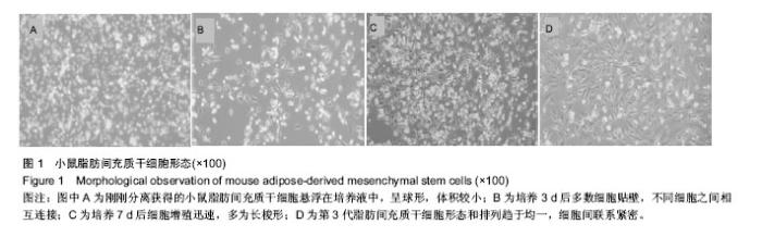
2.1 实验动物数量分析 20只4周龄ICR小鼠全部进入结果分析,中途无脱落。 2.2 小鼠脂肪间充质干细胞形态 倒置显微镜下观察,刚刚分离获得的小鼠脂肪间充质干细胞悬浮在培养液中,呈球形,体积较小;培养3 d后多数细胞贴壁,呈梭形、星形,不同细胞之间相互连接;培养7 d后细胞增殖迅速,多为长梭形,放射状;传代培养后细胞形态和排列趋于均一,细胞间联系紧密,见图1。"

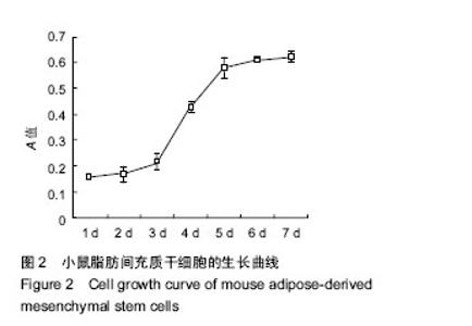
2.3 小鼠脂肪间充质干细胞生长曲线 第3代小鼠脂肪间充质干细胞的生长曲线呈现“S”形,第1-3天为潜伏期,第4天进入对数生长期,第5-7天细胞生长达到峰值,进入平稳期,见图2。"

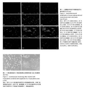
2.4 小鼠脂肪间充质干细胞鉴定结果 采用免疫荧光技术对第4代脂肪间充质干细胞进行鉴定,结果显示:第4代脂肪间充质干细胞表面CD90、CD105及CD73分别表现为绿色、绿色及红色荧光,CD45无荧光表达,说明第4代脂肪间充质干细胞具备间充质干细胞表面标记,见 图3。 2.5 诱导分化后心肌细胞形态 5-氮杂胞苷组诱导后细胞增殖速度下降,超过50%细胞出现脱壁、降解、死亡。光镜下观察细胞明显增大、体积不等、胞质粗糙,细胞的数量较同期5-氮杂胞苷联合血管紧张素Ⅱ组的细胞数量明显减少。5-氮杂胞苷联合血管紧张素Ⅱ组诱导后1周细胞开始脱壁凋亡,增殖速度降低,第10天细胞增殖速度开始趋向加快,第4周后,出现内皮细胞的形态特征,见图4。"


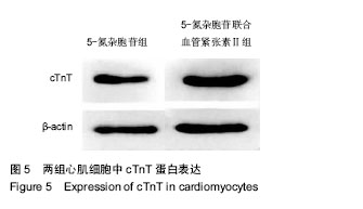
2.6 两组心肌细胞中cTnT蛋白表达 诱导7 d时,5-氮杂胞苷联合血管紧张素Ⅱ组心肌细胞中cTnT蛋白表达量显著高于5-氮杂胞苷,见图5。"
| [1] Ishihara K, Nakayama K, Akieda S, et al. Simultaneous regeneration of full-thickness cartilage and subchondral bone defects in vivo using a three-dimensional scaffold-free autologous construct derived from high-density bone marrow-derived mesenchymal stem cells. J Orthop Surg Res. 2014;9:98.[2] Heywood HK, Nalesso G, Lee DA, et al. Culture expansion in low-glucose conditions preserves chondrocyte differentiation and enhances their subsequent capacity to form cartilage tissue in three-dimensional culture. Biores Open Access. 2014;3(1):9-18.[3] Najar M, Raicevic G, Fayyad-Kazan H, et al. Impact of different mesenchymal stromal cell types on T-cell activation, proliferation and migration. Int Immunopharmacol. 2013; 15(4):693-702.[4] 庞荣清,何洁,李福兵,等.一种简单的人脐带间充质干细胞分离培养方法[J].中华细胞与干细胞杂志:电子版,2011,1(2):30-33.[5] 陈运贤,欧瑞明,钟雪云,等.自体骨髓干细胞原位移植治疗急性心肌梗死的临床研究[J].中国病理生理杂志,2003,19(4):452-454.[6] 牛丽丽,曹丰,郑敏,等.同种异体移植骨髓间充质干细胞治疗大鼠心肌梗死[J].中华内科杂志, 2004,43(3):186-190.[7] 王宝珠,马依彤,王春兰,等.大鼠自体脂肪干细胞移植治疗心肌梗死动物模型的建立[J].中华实验外科杂志,2010,27(9):1187.[8] 石金鑫,刘剑锋,王海滨,等.棕色脂肪干细胞与白色脂肪干细胞移植对心肌梗死大鼠心功能的影响[J].中国医药导报,2013,10(15): 18-21.[9] Melief SM, Zwaginga JJ, Fibbe WE, et al. Adipose tissue-derived multipotent stromal cells have a higher immunomodulatory capacity than their bone marrow-derived counterparts. Stem Cells Transl Med. 2013;2(6):455-463.[10] Strioga M, Viswanathan S, Darinskas A, et al. Same or not the same? Comparison of adipose tissue-derived versus bone marrow-derived mesenchymal stem and stromal cells. Stem Cells Dev. 2012;21(14):2724-2752.[11] Liu C, Fan Y, Zhou L, et al. Pretreatment of mesenchymal stem cells with angiotensin II enhances paracrine effects, angiogenesis, gap junction formation and therapeutic efficacy for myocardial infarction. Int J Cardiol. 2015;188:22-32. [12] Qin Q, Wang J, Yan Y, et al. Angiotensin Ⅱ induces the differentiation of mouse epicardial progenitor cells into vascular smooth muscle-like cells. Biochem Biophys Res Commun. 2016;480(4):696-701. [13] Hou J, Yan P, Guo T, et al. Cardiac stem cells transplantation enhances the expression of connexin 43 via the ANG II/AT1R/TGF-beta1 signaling pathway in a rat model of myocardial infarction. Exp Mol Pathol. 2015;99(3):693-701. [14] Fan Y, Wang L, Liu C, et al. Local renin-angiotensin system regulates hypoxia-induced vascular endothelial growth factor synthesis in mesenchymal stem cells. Int J Clin Exp Pathol. 2015;8(3):2505-2514. [15] Xue C, Zhang J, Lv Z, et al. Angiotensin II promotes differentiation of mouse c-kit-positive cardiac stem cells into pacemaker-like cells. Mol Med Rep. 2015;11(5):3249-3258.[16] Castelo-Branco MT, Soares ID, Lopes DV, et al. Intraperitoneal but not intravenous cryopreserved mesenchymal stromal cells home to the inflamed colon and ameliorate experimental colitis. PLoS One. 2012;7(3):e33360.[17] Nagyova M, Slovinska L, Blasko J, et al. A comparative study of PKH67, DiI, and BrdU labeling techniques for tracing rat mesenchymal stem cells. In Vitro Cell Dev Biol Anim. 2014; 50(7):656-663.[18] 国强华,宋维鹏,王庆胜,等.组织块贴壁法培养人肠系膜动脉平滑肌细胞的研究[J].武警医学,2012,23(12):1033-1035.[19] Park JR, Kim E, Yang J, et al. Isolation of human dermis derived mesenchymal stem cells using explants culture method: expansion and phenotypical characterization. Cell Tissue Bank. 2015;16(2):209-218.[20] Mori Y, Ohshimo J, Shimazu T, et al. Improved explant method to isolate umbilical cord-derived mesenchymal stem cells and their immunosuppressive properties. Tissue Eng Part C Methods. 2015;21(4):367-372.[21] Gittel C, Brehm W, Burk J, et al. Isolation of equine multipotent mesenchymal stromal cells by enzymatic tissue digestion or explant technique: comparison of cellular properties. BMC Vet Res. 2013;9:221.[22] Boyette LB, Creasey OA, Guzik L, et al. Human bone marrow-derived mesenchymal stem cells display enhanced clonogenicity but impaired differentiation with hypoxic preconditioning. Stem Cells Transl Med. 2014;3(2):241-254.[23] Brini AT, Amodeo G, Ferreira LM, et al. Therapeutic effect of human adipose-derived stem cells and their secretome in experimental diabetic pain. Sci Rep. 2017;7(1):9904.[24] Liew LJ, Ong HT, Dilley RJ.Isolation and Culture of Adipose-Derived Stromal Cells from Subcutaneous Fat. Methods Mol Biol. 2017;1627:193-203.[25] Muñoz-Criado I, Meseguer-Ripolles J, Mellado-López M, et al. Human Suprapatellar Fat Pad-Derived Mesenchymal Stem Cells Induce Chondrogenesis and Cartilage Repair in a Model of Severe Osteoarthritis. Stem Cells Int. 2017;2017:4758930.[26] Balducci L, Alessandri G.Isolation, et al. Expansion, and Immortalization of Human Adipose-Derived Mesenchymal Stromal Cells from Biopsies and Liposuction Specimens. Methods Mol Biol. 2016;1416:259-274.[27] 刘红,俞小芳,滕杰,等.低氧预处理对小鼠骨髓间充质干细胞迁移能力的影响[J].中华医学杂志,2012,92(10):709-713.[28] Lönne M, Lavrentieva A, Walter JG, et al. Analysis of oxygen-dependent cytokine expression in human mesenchymal stem cells derived from umbilical cord. Cell Tissue Res. 2013;353(1):117-122.[29] 刘林奇,鲁峰,高建华.血管内皮生长因子参与血管形成的机制研究进展[J].中华实验外科杂志,2012,29(4):770-772.[30] Lee Y, Jung J, Cho KJ, et al. Increased SCF/c-kit by hypoxia promotes autophagy of human placental chorionic plate-derived mesenchymal stem cells via regulating the phosphorylation of mTOR. J Cell Biochem. 2013;114(1): 79-88.[31] 刘林奇,高建华,袁艺,等.低氧预处理对人脂肪来源干细胞的活性及表面标志的影响[J].中国美容整形外科杂志,2013,24(3): 180-183.[32] 刘毅,王有虎,哈小琴.转染HGF基因的MSCs对移植颗粒脂肪存活率的影响[J].中华医学美学美容杂志,2011,17(1):12-16.[33] 戴晓俊,王飓,陶凯,等.影响自体游离脂肪移植成活率相关因素的研究进展[J].中国美容整形外科杂志,2013,24(4):232-234.[34] 王克勇,张福业,王永刚,等.人脐带间充质干细胞辅助的一种脂肪移植的实验研究[J].东南大学学报,2013,32(6):733-737. |
| [1] | Yao Xiaoling, Peng Jiancheng, Xu Yuerong, Yang Zhidong, Zhang Shuncong. Variable-angle zero-notch anterior interbody fusion system in the treatment of cervical spondylotic myelopathy: 30-month follow-up [J]. Chinese Journal of Tissue Engineering Research, 2022, 26(9): 1377-1382. |
| [2] | Wang Jing, Xiong Shan, Cao Jin, Feng Linwei, Wang Xin. Role and mechanism of interleukin-3 in bone metabolism [J]. Chinese Journal of Tissue Engineering Research, 2022, 26(8): 1260-1265. |
| [3] | Xiao Hao, Liu Jing, Zhou Jun. Research progress of pulsed electromagnetic field in the treatment of postmenopausal osteoporosis [J]. Chinese Journal of Tissue Engineering Research, 2022, 26(8): 1266-1271. |
| [4] | An Weizheng, He Xiao, Ren Shuai, Liu Jianyu. Potential of muscle-derived stem cells in peripheral nerve regeneration [J]. Chinese Journal of Tissue Engineering Research, 2022, 26(7): 1130-1136. |
| [5] | Tian Chuan, Zhu Xiangqing, Yang Zailing, Yan Donghai, Li Ye, Wang Yanying, Yang Yukun, He Jie, Lü Guanke, Cai Xuemin, Shu Liping, He Zhixu, Pan Xinghua. Bone marrow mesenchymal stem cells regulate ovarian aging in macaques [J]. Chinese Journal of Tissue Engineering Research, 2022, 26(7): 985-991. |
| [6] | Wen Dandan, Li Qiang, Shen Caiqi, Ji Zhe, Jin Peisheng. Nocardia rubra cell wall skeleton for extemal use improves the viability of adipogenic mesenchymal stem cells and promotes diabetes wound repair [J]. Chinese Journal of Tissue Engineering Research, 2022, 26(7): 1038-1044. |
| [7] | Zhu Bingbing, Deng Jianghua, Chen Jingjing, Mu Xiaoling. Interleukin-8 receptor enhances the migration and adhesion of umbilical cord mesenchymal stem cells to injured endothelium [J]. Chinese Journal of Tissue Engineering Research, 2022, 26(7): 1045-1050. |
| [8] | Fang Xiaolei, Leng Jun, Zhang Chen, Liu Huimin, Guo Wen. Systematic evaluation of different therapeutic effects of mesenchymal stem cell transplantation in the treatment of ischemic stroke [J]. Chinese Journal of Tissue Engineering Research, 2022, 26(7): 1085-1092. |
| [9] | Guo Jia, Ding Qionghua, Liu Ze, Lü Siyi, Zhou Quancheng, Gao Yuhua, Bai Chunyu. Biological characteristics and immunoregulation of exosomes derived from mesenchymal stem cells [J]. Chinese Journal of Tissue Engineering Research, 2022, 26(7): 1093-1101. |
| [10] | Zhang Jinglin, Leng Min, Zhu Boheng, Wang Hong. Mechanism and application of stem cell-derived exosomes in promoting diabetic wound healing [J]. Chinese Journal of Tissue Engineering Research, 2022, 26(7): 1113-1118. |
| [11] | Hou Jingying, Guo Tianzhu, Yu Menglei, Long Huibao, Wu Hao. Hypoxia preconditioning targets and downregulates miR-195 and promotes bone marrow mesenchymal stem cell survival and pro-angiogenic potential by activating MALAT1 [J]. Chinese Journal of Tissue Engineering Research, 2022, 26(7): 1005-1011. |
| [12] | Liang Xuezhen, Yang Xi, Li Jiacheng, Luo Di, Xu Bo, Li Gang. Bushen Huoxue capsule regulates osteogenic and adipogenic differentiation of rat bone marrow mesenchymal stem cells via Hedgehog signaling pathway [J]. Chinese Journal of Tissue Engineering Research, 2022, 26(7): 1020-1026. |
| [13] | Huang Chuanjun, Zou Yu, Zhou Xiaoting, Zhu Yangqing, Qian Wei, Zhang Wei, Liu Xing. Transplantation of umbilical cord mesenchymal stem cells encapsulated in RADA16-BDNF hydrogel promotes neurological recovery in an intracerebral hemorrhage rat model [J]. Chinese Journal of Tissue Engineering Research, 2022, 26(4): 510-515. |
| [14] | He Yunying, Li Lingjie, Zhang Shuqi, Li Yuzhou, Yang Sheng, Ji Ping. Method of constructing cell spheroids based on agarose and polyacrylic molds [J]. Chinese Journal of Tissue Engineering Research, 2022, 26(4): 553-559. |
| [15] | He Guanyu, Xu Baoshan, Du Lilong, Zhang Tongxing, Huo Zhenxin, Shen Li. Biomimetic orientated microchannel annulus fibrosus scaffold constructed by silk fibroin [J]. Chinese Journal of Tissue Engineering Research, 2022, 26(4): 560-566. |
| Viewed | ||||||
|
Full text |
|
|||||
|
Abstract |
|
|||||