Chinese Journal of Tissue Engineering Research ›› 2017, Vol. 21 ›› Issue (25): 4020-4025.doi: 10.3969/j.issn.2095-4344.2017.25.014
Previous Articles Next Articles
Combined use of low-dose 17-beta estradiol and bone marrow mesenchymal stem cell transplantation for spinal cord repair
Kang Cong1, Meng Xian-yong2, Yang Xin-ming2, Cheng Yao-yu1, Zhang Zhen-liang1
- 1Hebei North University, Zhangjiakou 075000, Hebei Province, China; 2Department of Orthopedics, the First Affiliated Hospital of Hebei North University, Zhangjiakou 075000, Hebei Province, China
-
Revised:2017-03-20Online:2017-09-08Published:2017-10-09 -
Contact:Meng Xian-yong, Associate chief physician, Department of Orthopedics, the First Affiliated Hospital of Hebei North University, Zhangjiakou 075000, Hebei Province, China -
About author:Kang Cong, Studying for master’s degree, Hebei North University, Zhangjiakou 075000, Hebei Province, China -
Supported by:the Medical Research Project of Hebei Provincial Health Department in 2011, No. 20110176
CLC Number:
Cite this article
Kang Cong, Meng Xian-yong, Yang Xin-ming, Cheng Yao-yu, Zhang Zhen-liang. Combined use of low-dose 17-beta estradiol and bone marrow mesenchymal stem cell transplantation for spinal cord repair[J]. Chinese Journal of Tissue Engineering Research, 2017, 21(25): 4020-4025.
share this article
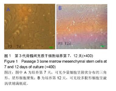
2.1 骨髓间充质干细胞的分离培养 第3代骨髓间充质干细胞培养第7天,可见少量细胞呈簇状分布的三角形、星形细胞聚集,第12天时可见较多梭形细胞呈漩涡状铺满瓶底(图1)。"

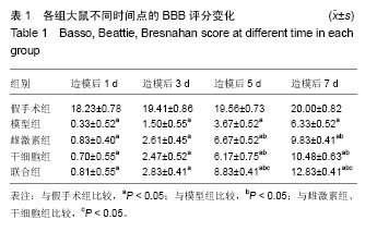
2.2 大鼠双后肢运动功能的BBB评分 造模前,各组大鼠的BBB评分均为21分;脊髓损伤当天,大鼠双后肢运动功能完全丧失呈迟缓性瘫痪,造模后第1,3天,模型组、雌激素组、干细胞组、联合组BBB评分比较差异无显著性意义(P > 0.05);随着时间的推移,各组大鼠后肢功能逐渐恢复,BBB评分逐渐升高,这是大鼠自然恢复的结果;造模第3天,雌激素组、干细胞组、联合组大鼠后肢功能开始逐渐恢复,雌激素组、干细胞组造模后第5,7天的BBB评分明显高于模型组(P < 0.05),联合组造模后第5,7天的BBB评分且明显高于雌激素组、干细胞组(P < 0.05),见表1。"
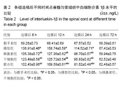
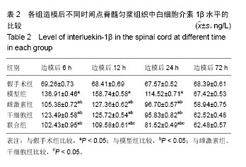
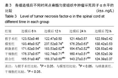
2.3 脊髓组织白细胞介素1β、肿瘤坏死因子α水平测定结果 与假手术组相比,其余4组脊髓损伤后至12 h白细胞介素1β呈持续上升趋势,可见由白细胞介素1β介导的炎症反应在损伤后12 h到达顶峰;损伤后72 h,受损节段脊髓组织白细胞介素1β水平基本恢复正常;与模型组相比,从造模12 h开始,雌激素组、干细胞组、联合组大鼠白细胞介素1β、肿瘤坏死因子α逐渐降低,联合组造模后12,24 h的白细胞介素1β、肿瘤坏死因子α水平低于雌激素组、干细胞组(P < 0.05),见表2,3,结果表明联合治疗抗炎作用优于单独使用雌激素或干细胞移植。"
"
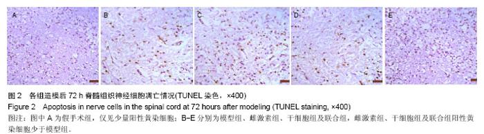
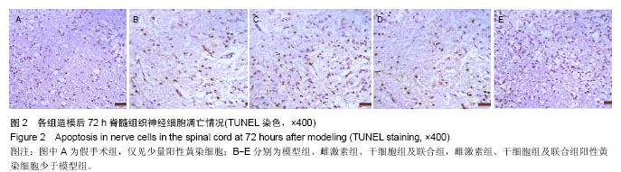
2.4 TUNEL染色结果 TUNEL染色显示,凋亡阳性细胞的胞核内存在棕黄色颗粒。假手术组组切片组织中仅见少量散在的TUNEL阳性细胞,神经元细胞结构完整,核膜清晰;其余各组造模后不同时间点均可检测到较多TUNEL阳性细胞,呈现典型的凋亡特征性,凋亡的细胞结构模糊,细胞核固缩,胞浆浓染。 模型组、雌激素组、干细胞组、联合组造模后6 h阳性细胞较假手术组增多,主要位于灰质;12 h时,损伤部位阳性细胞明显增多,达到损伤高峰,白质中也开始出现阳性细胞;24 h时,白质中阳性细胞数量减少;72 h时,阳性细胞较前减少且阳性细胞主要位于白质脱髓鞘区域,见图2。 造模后72 h,假手术组、模型组、雌激素组、干细胞组、联合组的TUNEL阳性细胞率分别为(2.74±0.51)%、(36.42±2.46)%、(15.56±2.85)%、(18.92±2.73)%、(6.97± 2.08)%;模型组TUNEL阳性细胞率高于假手术组(P < 0.05),雌激素组、干细胞组、联合组TUNEL阳性细胞率低于模型组(P < 0.05),联合组低于雌激素组、干细胞组(P < 0.05)。"
| [1] Mosquera L,Jennifer M,Colón,José M,et al.Tamoxifen and estradiol improved locomotor function and increased spared tissue in rats after spinal cord injury: their antioxidant effect and role of estrogen receptor alpha.Brain Res.2014;1561(2):成11-22.[2] Lin CW,Chen B,Huang KL,et al.Inhibition of autophagy by estradiol promotes locomotor recovery after spinal cord injury in rats.Neurosci Bull.2016;32(2):137-144.[3] Letaif OB,Cristante AF,Barros Filho TE,et al.Effects of estrogen on functional and neurological recovery after spinal cord injury: An experimental study with rats.Clinics(Sao Paulo).2015; 70(10):700-705.[4] Okuda A,Horii-Hayashi N,Sasagawa T,et al.Bone marrow stomal cell sheets may promote axonal regeneration and functional recovery with suppression of glial scar formation after spinal cord transection injury in rats.J Neurosurg Spine. 2016;25:1-8.[5] Han S,Wang B,Li X,et al.Bone marrow-derived mesenchymal stem cells in three-dimensional culture promote neuronal regeneration by neurotrophic protection and immunomodulation. J Biomed Mater Res A.2016;104(7):1759-1769.[6] Baydin A,Cokluk C,Aydin K.A new minimally invasive experimental spinal cord injury model in rabbits.Minim Invasive Neurosurg.2007;50(3):170-172.[7] Basso DM,Beattie MS,Bresnahan JC.A sensitive and reliable locomotor rating seale for open field testing in rats.J Neurotrauma.1995;12(1):1-21.[8] 王继猛,梁卓文,胡文昱,等.激光照射对脊髓损伤大鼠TNF-α、IL-6和IL-10表达的影响[J].中国激光,2014,2(2):0204003,1-5.[9] Yin X,Yin Y,Cao FL,et al.Tanshinone IIA attenuates the inflammatory response and apoptosis after traumatic injury of the spinal cord in adult rats.PLoS One.2012;7(6):e38381.[10] Gokce EC,Kahveci R,Gokce A,et al.Neuroprotective effects of thymoquinone against spinal cord ischemia-reperfusion injury by attenuation of inflammation, oxidative stress, and apoptosis.J Neurosurg Spine.2016;24(6):949-959.[11] Sullivan PG,Krishnamurthy S,Petel SP,et al.Temporal characterization of mitochondrial bioenergetics sfter spinal cord injury.J Neurotrauma.2007;24(6):991-999.[12] Pan W,Kastin AJ.Spinal cord injury changes cytokine transport.Neurol Disord Drug Targets.2016;15(9):1139-1150.[13] Han D,Wu C,Xiong Q,et al.Anti-inflammatory mechanism of bone marrow mesenchymal stem cell transplantation in rat model of spinal cord injury.Cell Biochem Biophys. 2015; 71(3):1341-1347.[14] Yan P,Li Q,Kim JM,et al.Cellular localization of tumornecrosis factor-α following acute spinal cord injury in adult rats.J Neurotrauma.2001;18(5):563-568.[15] Ankeny DP,Mctigue DM,Jakeman LB.Bone marrow transplants provide tissue protection and directional guidance for axons after contusive spinal cord injury in rats.Exp Neurol. 2004;190(1):17-31.[16] Patel SP,Sullivan PG,Pandya JD,et al.Differential effects of the mitochondrial uncoupling agent,2,4-dinitrophenol,or the nitroxide antioxidant,Tempol,on synaptic or nonsynaptic mitochondria after spinal cord injury.J Neurosci Res.2009; 87(1):130-140. [17] Roozbehi A,Joghataie MT,Mehdizadeh M,et al.The effects of cyclosporin-A on functional outcome and axonal regrowth following spinal cord injury in adult rats.Acta Med Iran.2012; 50(4):226-232.[18] Ritfeld GJ,Patel A,Chou A,et al.The role of brain-derived neurotrophic factor in bone marrow stromal cell-mediated spinal cordrepair.Cell Transplant.2015;24(11):2209-20. [19] 任义行,杨新明,孟宪勇,等.白细胞介素6受体单克隆抗体与骨髓间充质干细胞可减少急性脊髓损伤神经元的凋亡[J].中国组织工程研究,2016,20(14):1981-1988.[20] 李洪秋,王哲,阿良,等.骨髓间充质干细胞移植对大鼠脊髓损伤后氧化应激的影响[J].脊柱外科杂志,2010,8(3):157-161.[21] 边立功,刘承杏,李兴国,等.17-β雌二醇提高巯醇抗氧化物(酶)治疗脊髓损伤的作用[J].解剖学报,2010,41(2):185-190.[22] Ray SK,Samntaray S,Banik NL.Future directions for using estrogen receptor agonists in the treatment of acute and chronic spinal cord injury.Neural Regen Res. 2016;11(9): 1418-1419. [23] Li XM,Yang Q,Li XB,et al.Estrogen-like neuroprotection of isopsoralen against spinal cord injury through estrogen receptor ERα.Metab Brain Dis.2017;32(1):259-265. [24] Cheng Q,Meng J,Wang XS,et al.G-1 exerts neuroprotective effects through G protein-coupled estrogen receptor 1 following spinal cord injury in mice.Biosci Rep.2016;36(4). pii:e00373. [25] Amini Pishva A,Akbari M,Farahabadi A,et al.Effect of Estrogen Therapy on TNF-α and iNOS Gene Expression in Spinal Cord Injury Model.Acta Med Iran.2016;54(5):296-301.[26] Brotfain E,Gruenbaum SE,Boyko M,et al.Neuroprotection by Estrogen and Progesterone in Traumatic Brain Injury and Spinal Cord Injury.Curr Neuropharmacol.2016;14(6):641-653.[27] Letaif OB,Cristante AF,Barros Filho TE,et al.Effects of estrogen on functional and neurological recovery after spinal cord injury: An experimental study with rats.Clinics(Sao Paulo). 2015;70(10):700-705. [28] Chakrabarti M,Das A,Samantaray S,et al.Molecular mechanisms of estrogen for neuroprotection in spinal cord injury and traumatic brain injury.Rev Neurosci. 2016;27(3): 271-281. [29] Chen J,Hu R,Ge H,et al.G-protein-coupled receptor 30-mediated antiapoptotic effect of estrogen on spinal motor neurons following injury and its underlying mechanisms.Mol Med Rep.2015;12(2):1733-1740. [30] Lee JY,Choi HY,Na WH,et al.17β-estradiol inhibits MMP-9 and SUR1/TrpM4 expression and activation and thereby attenuates BSCB disruption/hemorrhage after spinal cord injury in male rats.Endocrinology.2015;156(5):1838-1850. [31] Hu JZ,Long H,Wu TD,et al.The effect of estrogen-related receptor α on the regulation of angiogenesis after spinal cord injury.Neuroscience.2015;290:570-580. [32] Mosquera L,Colón JM,Santiago JM,et al.Tamoxifen and estradiol improved locomotor function and increased spared tissue in rats after spinal cord injury: their antioxidant effect and role of estrogen receptor alpha.Brain Res. 2014; 1561: 11-22. [33] Prossnitz ER.G protein-coupled estrogen receptor: a new therapeutic target in stroke and traumatic brain/spinal cord injury?Crit Care Med.2012;40(12):3323-3325. [34] Samantaray S,Das A,Matzelle DC,et al.Administration of low dose estrogen attenuates persistent inflammation, promotes angiogenesis, and improves locomotor function following chroni spinal cord injury in rats. J Neurochem.2016; 137(4): 604-617. [35] Cox A,Varma A,Barry J,et al.Nanoparticle estrogen in rat spinal cord injury elicits rapid anti-inflammatory effects in plasma,cerebrospinal fluid,and tissue.J Neurotrauma. 2015; 32(18):1413-1421.[36] 程姝丽,丁寅.雌激素对小鼠骨髓间充质干细胞凋亡的抑制作用及其机制研究[J].细胞与分子免疫学杂志,2012,28(5):514-516. |
| [1] | Yao Xiaoling, Peng Jiancheng, Xu Yuerong, Yang Zhidong, Zhang Shuncong. Variable-angle zero-notch anterior interbody fusion system in the treatment of cervical spondylotic myelopathy: 30-month follow-up [J]. Chinese Journal of Tissue Engineering Research, 2022, 26(9): 1377-1382. |
| [2] | Wang Jing, Xiong Shan, Cao Jin, Feng Linwei, Wang Xin. Role and mechanism of interleukin-3 in bone metabolism [J]. Chinese Journal of Tissue Engineering Research, 2022, 26(8): 1260-1265. |
| [3] | Xiao Hao, Liu Jing, Zhou Jun. Research progress of pulsed electromagnetic field in the treatment of postmenopausal osteoporosis [J]. Chinese Journal of Tissue Engineering Research, 2022, 26(8): 1266-1271. |
| [4] | Tian Chuan, Zhu Xiangqing, Yang Zailing, Yan Donghai, Li Ye, Wang Yanying, Yang Yukun, He Jie, Lü Guanke, Cai Xuemin, Shu Liping, He Zhixu, Pan Xinghua. Bone marrow mesenchymal stem cells regulate ovarian aging in macaques [J]. Chinese Journal of Tissue Engineering Research, 2022, 26(7): 985-991. |
| [5] | Hou Jingying, Guo Tianzhu, Yu Menglei, Long Huibao, Wu Hao. Hypoxia preconditioning targets and downregulates miR-195 and promotes bone marrow mesenchymal stem cell survival and pro-angiogenic potential by activating MALAT1 [J]. Chinese Journal of Tissue Engineering Research, 2022, 26(7): 1005-1011. |
| [6] | Liang Xuezhen, Yang Xi, Li Jiacheng, Luo Di, Xu Bo, Li Gang. Bushen Huoxue capsule regulates osteogenic and adipogenic differentiation of rat bone marrow mesenchymal stem cells via Hedgehog signaling pathway [J]. Chinese Journal of Tissue Engineering Research, 2022, 26(7): 1020-1026. |
| [7] | Wen Dandan, Li Qiang, Shen Caiqi, Ji Zhe, Jin Peisheng. Nocardia rubra cell wall skeleton for extemal use improves the viability of adipogenic mesenchymal stem cells and promotes diabetes wound repair [J]. Chinese Journal of Tissue Engineering Research, 2022, 26(7): 1038-1044. |
| [8] | Zhu Bingbing, Deng Jianghua, Chen Jingjing, Mu Xiaoling. Interleukin-8 receptor enhances the migration and adhesion of umbilical cord mesenchymal stem cells to injured endothelium [J]. Chinese Journal of Tissue Engineering Research, 2022, 26(7): 1045-1050. |
| [9] | Fang Xiaolei, Leng Jun, Zhang Chen, Liu Huimin, Guo Wen. Systematic evaluation of different therapeutic effects of mesenchymal stem cell transplantation in the treatment of ischemic stroke [J]. Chinese Journal of Tissue Engineering Research, 2022, 26(7): 1085-1092. |
| [10] | Guo Jia, Ding Qionghua, Liu Ze, Lü Siyi, Zhou Quancheng, Gao Yuhua, Bai Chunyu. Biological characteristics and immunoregulation of exosomes derived from mesenchymal stem cells [J]. Chinese Journal of Tissue Engineering Research, 2022, 26(7): 1093-1101. |
| [11] | Zhang Jinglin, Leng Min, Zhu Boheng, Wang Hong. Mechanism and application of stem cell-derived exosomes in promoting diabetic wound healing [J]. Chinese Journal of Tissue Engineering Research, 2022, 26(7): 1113-1118. |
| [12] | An Weizheng, He Xiao, Ren Shuai, Liu Jianyu. Potential of muscle-derived stem cells in peripheral nerve regeneration [J]. Chinese Journal of Tissue Engineering Research, 2022, 26(7): 1130-1136. |
| [13] | Huang Chuanjun, Zou Yu, Zhou Xiaoting, Zhu Yangqing, Qian Wei, Zhang Wei, Liu Xing. Transplantation of umbilical cord mesenchymal stem cells encapsulated in RADA16-BDNF hydrogel promotes neurological recovery in an intracerebral hemorrhage rat model [J]. Chinese Journal of Tissue Engineering Research, 2022, 26(4): 510-515. |
| [14] | He Yunying, Li Lingjie, Zhang Shuqi, Li Yuzhou, Yang Sheng, Ji Ping. Method of constructing cell spheroids based on agarose and polyacrylic molds [J]. Chinese Journal of Tissue Engineering Research, 2022, 26(4): 553-559. |
| [15] | He Guanyu, Xu Baoshan, Du Lilong, Zhang Tongxing, Huo Zhenxin, Shen Li. Biomimetic orientated microchannel annulus fibrosus scaffold constructed by silk fibroin [J]. Chinese Journal of Tissue Engineering Research, 2022, 26(4): 560-566. |
| Viewed | ||||||
|
Full text |
|
|||||
|
Abstract |
|
|||||