Chinese Journal of Tissue Engineering Research ›› 2017, Vol. 21 ›› Issue (18): 2858-2863.doi: 10.3969/j.issn.2095-4344.2017.18.012
Previous Articles Next Articles
Three-dimensional printing of strontium-containing mesoporous bioactive glass scaffolds with varied macropore morphologies: an in vitro cytological experiment
- Department of Orthopedics, Shanghai Pudong Hospital, Fudan University, Shanghai 200120, China
-
Received:2017-02-11Online:2017-06-28Published:2017-07-07 -
Contact:Yu Bao-qing, M.D., Chief physician, Department of Orthopedics, Shanghai Pudong Hospital, Fudan University, Shanghai 200120, China -
About author:Zhang Xu, Studying for doctorate, Department of Orthopedics, Shanghai Pudong Hospital, Fudan University, Shanghai 200120, China -
Supported by:The Shanghai Science and Technology Commission Foundation for Basic Research, No. 13JC1407302; Disciplines Group Construction Project of Pudong Health Bureau of Shanghai, No. PWZxq2014-03; Major Scientific Research Projects of Shanghai Municipal Health Bureau, No. 20134039
CLC Number:
Cite this article
Zhang Xu, Wu Liang-hao, Li De-jian, Ao Rong-guang, Chen Fan-cheng, Yu Bin, Yu Bao-qing.
share this article
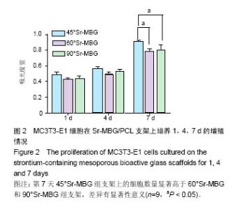
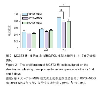
2.1 MC3T3-E1细胞在Sr-MBG/PCL复合支架上的增殖能力 图2为MC3T3-E1细胞在45°Sr-MBG、60°Sr-MBG和90°Sr-MBG复合支架上培养1,4,7 d后的增殖情况。CCK-8方法检测结果表明,3组支架材料都能够支持细胞增殖,第1天和第4天45°Sr-MBG组支架上的细胞数量略高于60°Sr-MBG和90°Sr-MBG组支架,但是差异无显著性意义 (P > 0.05);第7天45°Sr-MBG组支架上的细胞数量显著高于60°Sr-MBG和90°Sr-MBG组支架,而且差异有显著性意义(P < 0.05)。"
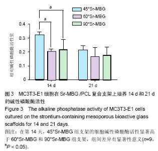
2.2 MC3T3-E1细胞在Sr-MBG/PCL复合支架上的碱性磷酸酶活性 图3为MC3T3-E1细胞在45°Sr-MBG、60°Sr-MBG和90°Sr-MBG组支架上培养14 d和21 d的碱性磷酸酶活性。从图中可以看出,在第14天,45°Sr-MBG组支架的细胞碱性磷酸酶活性显著高于60°Sr-MBG和90°Sr-MBG组支架,组间差异有显著性意义(P < 0.05)。在第21天,3组支架的碱性磷酸酶活性差异无显著性意义(P > 0.05)。"

| [1]Zhou Y, Wu C, Xiao Y. The stimulation of proliferation and differentiation of periodontal ligament cells by the ionic products from Ca7Si2P2O16 bioceramics. Acta Biomater. 2012;8(6):2307-2316.[2]Zhang Y, Fan W, Ma Z, et al. The effects of pore architecture in silk fibroin scaffolds on the growth and differentiation of mesenchymal stem cells expressing BMP7. Acta Biomater. 2010;6(8):3021-3028.[3]Cho SY, Park HH, Jin HJ. Controlling pore size of electrospun silk fibroin scaffold for tissue engineering. Polym Korea. 2012; 36(5):651-655.[4]Hench LL, Wilson J. Surface-active biomaterials. Science. 1984;226(4675):630-636.[5]Zhu M, Shi JL, He QJ, et al. An emulsification-solvent evaporation route to mesoporous bioactive glass microspheres for bisphosphonate drug delivery. J Mater Sci. 2012; 47(5):2256-2263.[6]Wang D, Lin H, Jiang J, et al. One-pot synthesis of magnetic, macro/mesoporous bioactive glasses for bone tissue engineering. Sci Technol Adv Mater. 2013;14(2):025004.[7]Baino F, Fiorilli S, Mortera R, et al. Mesoporous bioactive glass as a multifunctional system for bone regeneration and controlled drug release. J Appl Biomater Funct Mater. 2012;10(1):12-21.[8]Wu C, Chang J. Mesoporous bioactive glasses: structure characteristics, drug/growth factor delivery and bone regeneration application. Interface Focus. 2012;2(3):292-306.[9]Wang X, Chen W, Liu Q, et al. Function and mechanism of mesoporous bioactive glass adsorbed epidermal growth factor for accelerating bone tissue regeneration. Biomed Mater. 2017;12(2):025020.[10]Shoaib M, Saeed A, Akhtar J, et al. Potassium-doped mesoporous bioactive glass: Synthesis, characterization and evaluation of biomedical properties. Mater Sci Eng C Mater Biol Appl. 2017;75:836-844. [11]Qi X, Pei P, Zhu M, et al. Three dimensional printing of calcium sulfate and mesoporous bioactive glass scaffolds for improving bone regeneration in vitro and in vivo. Sci Rep. 2017;7:42556.[12]Schumacher M, Reither L, Thomas J, et al. Calcium phosphate bone cement/mesoporous bioactive glass composites for controlled growth factor delivery. Biomater Sci. 2017;5(3):578-588. [13]Zhang Q, Chen X, Geng S, et al. Nanogel-based scaffolds fabricated for bone regeneration with mesoporous bioactive glass and strontium: In vitro and in vivo characterization. J Biomed Mater Res A. 2017;105(4):1175-1183. [14]Wang X, Li W. Biodegradable mesoporous bioactive glass nanospheres for drug delivery and bone tissue regeneration. Nanotechnology. 2016;27(22):225102. [15]Wu C, Xia L, Han P, et al. Europium-Containing Mesoporous Bioactive Glass Scaffolds for Stimulating in Vitro and in Vivo Osteogenesis. ACS Appl Mater Interfaces. 2016;8(18): 11342-11354.[16]Marie PJ, Ammann P, Boivin G, et al. Mechanisms of action and therapeutic potential of strontium in bone. Calcif Tissue Int. 2001;69(3):121-129.[17]Gentleman E, Fredholm YC, Jell G, et al. The effects of strontium-substituted bioactive glasses on osteoblasts and osteoclasts in vitro. Biomaterials. 2010;31(14):3949-3956.[18]Zreiqat H, Ramaswamy Y, Wu C, et al. The incorporation of strontium and zinc into a calcium-silicon ceramic for bone tissue engineering. Biomaterials. 2010;31(12):3175-3184.[19]Tsang VL, Bhatia SN. Three-dimensional tissue fabrication. Adv Drug Deliv Rev. 2004;56(11):1635-1647.[20]Rao RB, Krafcik KL, Morales AM, et al. Microfabricated Deposition Nozzles for Direct-Write Assembly of Three-Dimensional Periodic Structures. Advanced Materials. 2005; 17(3):289-293.[21]Franco J, Hunger P, Launey ME, et al. Direct write assembly of calcium phosphate scaffolds using a water-based hydrogel. Acta Biomater. 2010;6(1):218-228.[22]Khan MN, Islam JM, Khan MA. Fabrication and characterization of gelatin-based biocompatible porous composite scaffold for bone tissue engineering. J Biomed Mater Res A. 2012;100(11):3020-3028.[23]Wang Z, Sakakibara T, Sudo A, et al. Porosity of β-tricalcium phosphate affects the results of lumbar posterolateral fusion. J Spinal Disord Tech. 2013;26(2):E40-45.[24]Yan X, Huang X, Yu C, et al. The in-vitro bioactivity of mesoporous bioactive glasses. Biomaterials. 2006;27(18):3396-3403.[25]Takeuchi A, Mishina Y, Miyaishi O, et al. Heterozygosity with respect to Zfp148 causes complete loss of fetal germ cells during mouse embryogenesis. Nat Genet. 2003;33(2): 172-176.[26]Melchels FP, Barradas AM, van Blitterswijk CA, et al. Effects of the architecture of tissue engineering scaffolds on cell seeding and culturing. Acta Biomater. 2010;6(11):4208-4217.[27]Yun HS, Park JW, Kim SH, et al. Effect of the pore structure of bioactive glass balls on biocompatibility in vitro and in vivo. Acta Biomater. 2011;7(6):2651-2660.[28]Wang C, Zhao Q, Wang M, et al. Cryogenic 3D printing for producing hierarchical porous and rhBMP-2-loaded Ca-P/PLLA nanocomposite scaffolds for bone tissue engineering. Biofabrication. 2017;9(2):025031. [29]Sears N, Dhavalikar P, Whitely M,et al. Fabrication of biomimetic bone grafts with multi-material 3D printing. Biofabrication. 2017;9(2):025020.[30]Wang Y, Wang K, Li X, et al. 3D fabrication and characterization of phosphoric acid scaffold with a HA/β-TCP weight ratio of 60:40 for bone tissue engineering applications. PLoS One. 2017;12(4):e0174870.[31]Shao H, Ke X, Liu A,et al. Bone regeneration in 3D printing bioactive ceramic scaffolds with improved tissue/material interface pore architecture in thin-wall bone defect. Biofabrication. 2017;9(2):025003.[32]Qi X, Pei P, Zhu M, et al.Three dimensional printing of calcium sulfate and mesoporous bioactive glass scaffolds for improving bone regeneration in vitro and in vivo. Sci Rep. 2017;7:42556. [33]Bendtsen ST, Quinnell SP, Wei M. Development of a novel alginate-polyvinyl alcohol-hydroxyapatite hydrogel for 3D bioprinting bone tissue engineered scaffolds. J Biomed Mater Res A. 2017;105(5):1457-1468.[34]Egorov AA, Fedotov AY, Mironov AV, et al. 3D printing of mineral-polymer bone substitutes based on sodium alginate and calcium phosphate. Beilstein J Nanotechnol. 2016;7: 1794-1799. [35]Li C, Jiang C, Deng Y, et al. RhBMP-2 loaded 3D-printed mesoporous silica/calcium phosphate cement porous scaffolds with enhanced vascularization and osteogenesis properties. Sci Rep. 2017;7:41331.[36]Causa F, Battista E, Della Moglie R, et al. Surface investigation on biomimetic materials to control cell adhesion: the case of RGD conjugation on PCL. Langmuir. 2010;26(12): 9875-9884.[37]Huang ZH, Song B, Chen YF, et al. Effect of polycaprolactone-ascobic acid scaffold in repairing articular cartilage defects in rabbits. Nan Fang Yi Ke Da Xue Xue Bao. 2017;37(5):607-613.[38]Esmaeilzadeh J, Hesaraki S, Hadavi SM, et al. Poly (d/l) lactide/polycaprolactone/bioactive glasss nanocomposites materials for anterior cruciate ligament reconstruction screws: The effect of glass surface functionalization on mechanical properties and cell behaviors. Mater Sci Eng C Mater Biol Appl. 2017;77:978-989. [39]Kong J, Yu Y, Pei X, et al.Polycaprolactone nanocomposite reinforced by bioresource starch-based nanoparticles. Int J Biol Macromol. 2017;102:1304-1311.[40]Gomes S, Rodrigues G, Martins G, et al. Evaluation of nanofibrous scaffolds obtained from blends of chitosan, gelatin and polycaprolactone for skin tissue engineering. Int J Biol Macromol. 2017;102:1174-1185. [41]Shim JH, Won JY, Park JH, et al. Effects of 3D-Printed Polycaprolactone/β-Tricalcium Phosphate Membranes on Guided Bone Regeneration. Int J Mol Sci. 2017;18(5): E899.[42]Wu C, Ramaswamy Y, Kwik D, et al. The effect of strontium incorporation into CaSiO3 ceramics on their physical and biological properties. Biomaterials. 2007;28(21):3171-3181.[43]Zhang J, Zhao S, Zhu Y, et al. Three-dimensional printing of strontium-containing mesoporous bioactive glass scaffolds for bone regeneration. Acta Biomater. 2014;10(5):2269-2281.[44]Valerio P, Pereira MM, Goes AM, et al. The effect of ionic products from bioactive glass dissolution on osteoblast proliferation and collagen production. Biomaterials. 2004; 25(15):2941-2948. |
| [1] | Yao Xiaoling, Peng Jiancheng, Xu Yuerong, Yang Zhidong, Zhang Shuncong. Variable-angle zero-notch anterior interbody fusion system in the treatment of cervical spondylotic myelopathy: 30-month follow-up [J]. Chinese Journal of Tissue Engineering Research, 2022, 26(9): 1377-1382. |
| [2] | Wang Jing, Xiong Shan, Cao Jin, Feng Linwei, Wang Xin. Role and mechanism of interleukin-3 in bone metabolism [J]. Chinese Journal of Tissue Engineering Research, 2022, 26(8): 1260-1265. |
| [3] | Xiao Hao, Liu Jing, Zhou Jun. Research progress of pulsed electromagnetic field in the treatment of postmenopausal osteoporosis [J]. Chinese Journal of Tissue Engineering Research, 2022, 26(8): 1266-1271. |
| [4] | An Weizheng, He Xiao, Ren Shuai, Liu Jianyu. Potential of muscle-derived stem cells in peripheral nerve regeneration [J]. Chinese Journal of Tissue Engineering Research, 2022, 26(7): 1130-1136. |
| [5] | Zhang Jinglin, Leng Min, Zhu Boheng, Wang Hong. Mechanism and application of stem cell-derived exosomes in promoting diabetic wound healing [J]. Chinese Journal of Tissue Engineering Research, 2022, 26(7): 1113-1118. |
| [6] | Chen Xiaoxu, Luo Yaxin, Bi Haoran, Yang Kun. Preparation and application of acellular scaffold in tissue engineering and regenerative medicine [J]. Chinese Journal of Tissue Engineering Research, 2022, 26(4): 591-596. |
| [7] | Kang Kunlong, Wang Xintao. Research hotspot of biological scaffold materials promoting osteogenic differentiation of bone marrow mesenchymal stem cells [J]. Chinese Journal of Tissue Engineering Research, 2022, 26(4): 597-603. |
| [8] | Shen Jiahua, Fu Yong. Application of graphene-based nanomaterials in stem cells [J]. Chinese Journal of Tissue Engineering Research, 2022, 26(4): 604-609. |
| [9] | Zhang Tong, Cai Jinchi, Yuan Zhifa, Zhao Haiyan, Han Xingwen, Wang Wenji. Hyaluronic acid-based composite hydrogel in cartilage injury caused by osteoarthritis: application and mechanism [J]. Chinese Journal of Tissue Engineering Research, 2022, 26(4): 617-625. |
| [10] | Li Hui, Chen Lianglong. Application and characteristics of bone graft materials in the treatment of spinal tuberculosis [J]. Chinese Journal of Tissue Engineering Research, 2022, 26(4): 626-630. |
| [11] | Gao Cangjian, Yang Zhen, Liu Shuyun, Li Hao, Fu Liwei, Zhao Tianyuan, Chen Wei, Liao Zhiyao, Li Pinxue, Sui Xiang, Guo Quanyi. Electrospinning for rotator cuff repair [J]. Chinese Journal of Tissue Engineering Research, 2022, 26(4): 637-642. |
| [12] | Yang Sidi, Wang Qian, Xu Nuo, Wang Ronghan, Jin Chuanqi, Lu Ying, Dong Ming. Biodentine enhances the proliferation and differentiation of osteoblasts through upregulating bone morphogenetic protein-2 [J]. Chinese Journal of Tissue Engineering Research, 2022, 26(4): 516-520. |
| [13] | He Yunying, Li Lingjie, Zhang Shuqi, Li Yuzhou, Yang Sheng, Ji Ping. Method of constructing cell spheroids based on agarose and polyacrylic molds [J]. Chinese Journal of Tissue Engineering Research, 2022, 26(4): 553-559. |
| [14] | He Guanyu, Xu Baoshan, Du Lilong, Zhang Tongxing, Huo Zhenxin, Shen Li. Biomimetic orientated microchannel annulus fibrosus scaffold constructed by silk fibroin [J]. Chinese Journal of Tissue Engineering Research, 2022, 26(4): 560-566. |
| [15] | Guan Jian, Jia Yanfei, Zhang Baoxin , Zhao Guozhong. Application of 4D bioprinting in tissue engineering [J]. Chinese Journal of Tissue Engineering Research, 2022, 26(3): 446-455. |
| Viewed | ||||||
|
Full text |
|
|||||
|
Abstract |
|
|||||