Chinese Journal of Tissue Engineering Research ›› 2017, Vol. 21 ›› Issue (13): 2074-2080.doi: 10.3969/j.issn.2095-4344.2017.13.018
Previous Articles Next Articles
The use of placental mesenchymal stem cells to repair the damaged endometrium
Niu Ting, Li Ai-bin, Chen Li
- Reproductive Medical Center, Renmin Hospital of Wuhan University, Wuhan 430060, Hubei Province, China
-
Revised:2016-12-09Online:2017-05-08Published:2017-06-09 -
Contact:Li Ai-bin, M.D., Chief physician, Reproductive Medical Center, Renmin Hospital of Wuhan University, Wuhan 430060, Hubei Province, China -
About author:Niu Ting, Studying for master’s degree, Reproductive Medical Center, Renmin Hospital of Wuhan University, Wuhan 430060, Hubei Province, China -
Supported by:the Natural Science Foundation of Hubei Province, No. 2014CFB206
CLC Number:
Cite this article
Niu Ting, Li Ai-bin, Chen Li. The use of placental mesenchymal stem cells to repair the damaged endometrium[J]. Chinese Journal of Tissue Engineering Research, 2017, 21(13): 2074-2080.
share this article
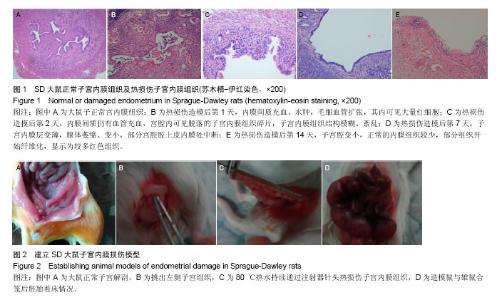
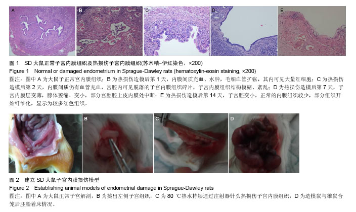
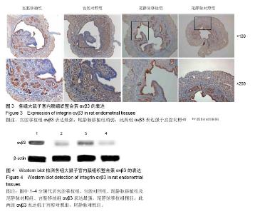
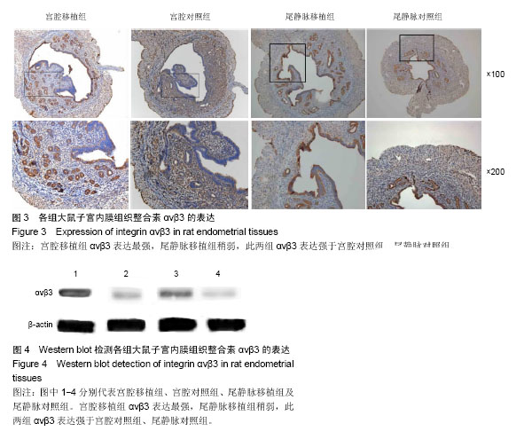
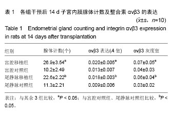
2.1 大鼠子宫内膜损伤造模情况 2.1.1 大鼠正常子宫大体解剖及组织病理切片苏木精-伊红染色 显微镜下观察大鼠正常子宫内膜组织,可见宫腔形态不规则,分为内膜层、肌层和外膜层,子宫内膜呈单层柱状上皮,覆盖子宫腔表面和腺腔,子宫内膜腺体主要位于黏膜下层和基底层,呈椭圆形或圆形(图1A)。 2.1.2 热损伤处理后大鼠子宫内膜组织 建模后1 d,大体可见子宫充血、肿大,苏木精-伊红染色显微镜下观察可见内膜间质充血、水肿,毛细血管扩张,其内可见大量红细胞;处理后2 d,大体见子宫水肿减轻,苏木精-伊红染色镜下可见内膜间质仍有血管充血,宫腔内可见脱落的子宫内膜组织碎片,子宫内膜组织结构模糊、紊乱;处理后7 d,大体可见受损子宫与周围组织粘连,苏木精-伊红染色镜下可见子宫内膜层变薄,腺体萎缩、变小,部分宫腔腔上皮内膜处中断;处理后14 d,大体见子宫形态缩小、苍白,苏木精-伊红染色见子宫腔变小,正常的内膜组织较少, 部分组织开始纤维化,苏木精-伊红染色显示为较多红色组织(图1B-E)。 2.1.3 生育能力检测 造模后第15天,将造模大鼠与雄性鼠合笼,观察到大鼠阴栓,记录为孕0.5 d,雌鼠受孕12 d 后处死,可见处理侧子宫萎缩变小,外观显示为白色条索状,子宫胚鼠数目为一两个,而对侧正常子宫侧可见胚鼠6-8只(图2);统计学分析结果显示:内膜损伤侧子宫与正常子宫侧胚泡数目比较差异有显著性意义(1.00±0.82,6.00±0.97,P < 0.05)。 2.2 细胞移植后修复效果检测 2.2.1 细胞移植后大鼠子宫内膜腺体计数 细胞移植后,通过阴道脱落细胞涂片法确定大鼠动情周期,在第3个动情周期的动情期断颈处死大鼠,对各组横切面子宫内膜腺体计数,结果见表1,其中宫腔移植组子宫内膜腺体计数最多,尾静脉移植组腺体个数稍少,单因素方差分析检验各组子宫内膜腺体计数,各组间差异有显著性意义(P < 0.05);SNK检验显示,宫腔移植组腺体个数多于宫腔对照组、尾静脉移植组、尾静脉对照组(P < 0.05),尾静脉移植组多于宫腔对照组、尾静脉对照组(P < 0.05),宫腔对照组与尾静脉对照组间无差异。 2.2.2 免疫组织化学检测子宫内膜整合素ανβ3表达 见图3。 ανβ3表达于子宫内膜腺上皮和腔上皮,是子宫内膜容受性的标记之一,表达于大鼠围着床期,可用于监测子宫内膜损伤修复的指标之一,用IPP软件来计算各组间ανβ3的吸光度值,结果见表1,宫腔移植组ανβ3的表达最强,尾静脉移植组稍弱,单因素方差分析检验各组间差异有显著性意义(P < 0.05);SNK检验显示,宫腔移植组整合素ανβ3表达强于宫腔对照组、尾静脉移植组、尾静脉对照组(P < 0.05),尾静脉移植组强于宫腔对照组、尾静脉对照组(P < 0.05),宫腔对照组与尾静脉对照组间无差异。 2.2.3 Western blot检测子宫内膜整合素ανβ3的表达 见图4。 宫腔移植组ανβ3表达最强,尾静脉移植组稍弱,见表1,单因素方差分析检验各组间差异有显著性意义(P < 0.05);SNK检验显示,宫腔移植组整合素ανβ3表达强于宫腔对照组、尾静脉移植组、尾静脉对照组(P < 0.05),尾静脉移植组强于宫腔对照组、尾静脉对照组(P < 0.05),宫腔对照组与尾静脉对照组间无差异。"
"

"
| [1] Maybin JA,Critchley HO.Menstrual physiology: implications for endometrial pathology and beyond.Hum Reprod Update. 2015;21(6):748-761.[2] Gargett CE,Schwab KE,Zillwood RM,et al.Isolation and culture of epithelial progenitors and mesenchymal stem cells from human endometrium.Biol Reprod.2009;80(6):1136-1145.[3] Singh N,Mohanty S,Seth T,et al.Autologous stem cell transplantation in refractory Asherman's syndrome: A novel cell based therapy.J Hum Reprod Sci.2014;7(2):93-98.[4] Majumdar MK,Thiede MA,Haynesworth SE,et al.Human marrow-derived mesenchymal stem cells(MSCs) express hematopoietic cytokines and support long-term hematopoiesis when differentiated toward stromal and osteogenic lineages.J Hematother Stem Cells Res.2000; 9(6):841-848.[5] Dippolito G,Schiller PC,Ricordi C,et al.Age-related osteogenic potential of mesenchymal stromal stem cells from human vertebral bone marrow.J Bone Miner Res.1999;13(7): 1115-1122.[6] 赵基栋,苗宗宁,钱寒光,等.人胎盘间充质干细胞促进血管生成[J].中国组织工程研究,2013,17(23):4216-4223.[7] Zhang B,Adesanya TM,Zhang L,et al.Delivery of placenta-derived mesenchymal stem cells ameliorates ischemia induced limb injury by immunomodulation.Cell Physiol Biochem 2014;34(6):1998-2006.[8] Ding H,Zhang H,Ding H,et al.Transplantation of placenta-derived mesenchymal stem cells reduces hypoxic-ischemic brain damage in rats by ameliorating the inflammatory response. Cell Mol Immunol.2015.doi: 10.1038/cmi.2015.99. [Epub ahead of print][9] Chatterjee P,Chiasson VL,Pinzur L,et al.Human placenta-derived stromal cells decrease inflammation, placental injury and blood pressure in hypertensive pregnant mice.Clin Sci(Lond).2016;130(7):513-523.[10] 曲军英,吕一帆,林茵.姜一平:建立小鼠子宫内膜损伤模型的研究[J].福建医科大学学报,2011,45(1):34-46.[11] Yu D,Wong YM,Cheong Y,et al.Asherman syndrome-one century later.Fertil Steril.2008;89:759-779.[12] Amer MI,Abd-El-Maeboud KH.Amnion graft following hysteroscopic lysis of intrauterine adhesions.J Obstet Gynaecol Res.2006;32:559-566.[13] 赵静.骨髄间充质干细胞移植治疗薄型子宫内膜的实验研究[D].中南大学博士学位论文,2013.[14] Halbersztadt A,Pajak J,Nowicki P,et al.Implantation and antigenicity of human endometrium.Postepy Hig Med Dosw. 2006;60:71-77.[15] Lessey BA.Implantation defects in inferile women with endometrio-sis.Ann N Y Acad Sci.2002;955(3):265-280. [16] Thomas K.Endometrial integrin expression in women undergoing in vitro fertilization and the association with subsequent treatment outcome.Fertil Steril.2003;80(3): 502-507.[17] Antoniadou E,David AL.Placental stem cells.Best Pract Res Clin Obstet Gynaecol. 2016;31:13-29.[18] Yin T,He S,Su C,et al.Genetically modified human placenta-derived mesenchymal stem cells with FGF-2 and PDGF-BB enhance neovascularization in a model of hindlimb ischemia.Mol Med Rep.2015;12(4):5093-5099.[19] He S,Gleason J,Fik E,et al.Human Placenta-Derived Mesenchymal Stromal-like Cells (PDA-002) Enhance Angiogenesis via T Cell-Dependent Reprogramming of Macrophage Differentiation.StemCells.2017.doi: 10.1002/stem.2598.[Epub ahead of print][20] Zhang B,Adesanya TM,Zhang L,et al.Delivery of placenta-derived mesenchymal stem cells ameliorates ischemia induced limb injury by immunomodulation.Cell Physiol Biochem. 2014;34(6):1998-2006.[21] Jiang H,Zhang Y,Tian K,et al.Amelioration of experimental autoimmune encephalomyelitis through transplantation of placental derivedmesenchymal stem cells.Sci Rep.2017;10(7): 41837.[22] Zhang Y,Lin X,Dai Y,et al.Endometrial stem cells repair injured endometrium and induce angiogenesis via AKT and ERKpathways.Reproduction.2016;152(5):389-402.[23] Wang J,Ju B,Pan C,et al.Application of Bone Marrow-Derived Mesenchymal Stem Cells in the Treatment of Intrauterine Adhesions in Rats.Cell Physiol Biochem. 2016; 39(4): 1553-1560.[24] Hyodo S,Matsubara K,Kameda K,et al.Endometrial injury increasespopulation cells in the uterune endometrium :a decisive role of estrogen.Tohoku J Exp Med.2011; 224(1): 47-55.[25] Schüring AN,Braun J,Wüllner S,et al.mRNA-expression of ERα,ERβ, and PR in clonal stem cell cultures obtained from human endometrialbiopsies.Scientific World J.2011; 11: 1762-1769.[26] Hou X,Tan Y,Li M,et al.Wnt signaling is critical to estrogen-mediated uterine growth.Mol Endocrinol.2004; 18(12):3035-3049.[27] Tulac S, Nayak NR, Kao LC,et al. Identification, characterization,and regulation of the canonical Wnt signaling pathway in human endometrium.J Clin Endocrinol Metab. 2003; 88(8):3860-1866.[28] 牛婷,李爱斌,曹景云,等.胎盘间充质干细胞的应用研究[J].中国组织工程研究,2015,19(32):5236-5242. [29] Hur W,Lee HY,Min HS,et al.Regeneration of full-thickness skin defects by differentiated adipose-derived stem cells into fibroblast-like cells by fibroblast-conditioned medium.Stem Cell Res Ther.2017;8(1):92.[30] Redondo-Castro E,Cunningham C,Miller J,et al.Interleukin-1 primes human mesenchymal stem cells towards an anti-inflammatory and pro-trophic phenotype in vitro.Stem Cell Res Ther.2017;8(1):79.[31] Zhu J,Wang P,Yu Z,et al.Advanced glycosylation end product promotes forkhead box O1 and inhibits Wnt pathway to suppress capacities of epidermal stem cells.Am J Transl Res.2016;8(12):5569-5579.[32] Simerman AA,Phan JD,Dumesic DA,et al.Muse Cells: Nontumorigenic Pluripotent Stem Cells Present in Adult Tissues-A Paradigm Shift in Tissue Regeneration and Evolution.Stem Cells Int.2016;2016:1463258.[33] Croze RH,Thi WJ,Clegg DO.ROCK Inhibition Promotes Attachment, Proliferation, and Wound Closure in Human Embryonic Stem Cell-Derived Retinal Pigmented Epithelium.Transl Vis Sci Technol.2016;5(6):7.[34] Vasandan AB,Jahnavi S,Shashank C,et al.Human Mesenchymal stem cells program macrophage plasticity by altering their metabolic status via a PGE<sub>2</sub>-dependent mechanism.Sci Rep.2016; 6:38308.[35] Sugiura T,Hibino N,Breuer CK,et al.Tissue-engineered cardiac patch seeded with human induced pluripotent stem cell derived cardiomyocytes promoted the regeneration of host cardiomyocytes in a rat model.J Cardiothorac Surg.2016; 11(1):163.[36] Chambers DC,Enever D,Ilic N,et al.A phase 1b study of placenta-derived mesenchymal stromal cells in patients with idiopathic pulmonary fibrosis.Respirology.2014; 19(7): 1013-1018. |
| [1] | Yao Xiaoling, Peng Jiancheng, Xu Yuerong, Yang Zhidong, Zhang Shuncong. Variable-angle zero-notch anterior interbody fusion system in the treatment of cervical spondylotic myelopathy: 30-month follow-up [J]. Chinese Journal of Tissue Engineering Research, 2022, 26(9): 1377-1382. |
| [2] | An Weizheng, He Xiao, Ren Shuai, Liu Jianyu. Potential of muscle-derived stem cells in peripheral nerve regeneration [J]. Chinese Journal of Tissue Engineering Research, 2022, 26(7): 1130-1136. |
| [3] | Fang Xiaolei, Leng Jun, Zhang Chen, Liu Huimin, Guo Wen. Systematic evaluation of different therapeutic effects of mesenchymal stem cell transplantation in the treatment of ischemic stroke [J]. Chinese Journal of Tissue Engineering Research, 2022, 26(7): 1085-1092. |
| [4] | Zhang Jinglin, Leng Min, Zhu Boheng, Wang Hong. Mechanism and application of stem cell-derived exosomes in promoting diabetic wound healing [J]. Chinese Journal of Tissue Engineering Research, 2022, 26(7): 1113-1118. |
| [5] | Chen Xiaoxu, Luo Yaxin, Bi Haoran, Yang Kun. Preparation and application of acellular scaffold in tissue engineering and regenerative medicine [J]. Chinese Journal of Tissue Engineering Research, 2022, 26(4): 591-596. |
| [6] | Kang Kunlong, Wang Xintao. Research hotspot of biological scaffold materials promoting osteogenic differentiation of bone marrow mesenchymal stem cells [J]. Chinese Journal of Tissue Engineering Research, 2022, 26(4): 597-603. |
| [7] | Shen Jiahua, Fu Yong. Application of graphene-based nanomaterials in stem cells [J]. Chinese Journal of Tissue Engineering Research, 2022, 26(4): 604-609. |
| [8] | Zhang Tong, Cai Jinchi, Yuan Zhifa, Zhao Haiyan, Han Xingwen, Wang Wenji. Hyaluronic acid-based composite hydrogel in cartilage injury caused by osteoarthritis: application and mechanism [J]. Chinese Journal of Tissue Engineering Research, 2022, 26(4): 617-625. |
| [9] | Li Hui, Chen Lianglong. Application and characteristics of bone graft materials in the treatment of spinal tuberculosis [J]. Chinese Journal of Tissue Engineering Research, 2022, 26(4): 626-630. |
| [10] | Gao Cangjian, Yang Zhen, Liu Shuyun, Li Hao, Fu Liwei, Zhao Tianyuan, Chen Wei, Liao Zhiyao, Li Pinxue, Sui Xiang, Guo Quanyi. Electrospinning for rotator cuff repair [J]. Chinese Journal of Tissue Engineering Research, 2022, 26(4): 637-642. |
| [11] | He Yunying, Li Lingjie, Zhang Shuqi, Li Yuzhou, Yang Sheng, Ji Ping. Method of constructing cell spheroids based on agarose and polyacrylic molds [J]. Chinese Journal of Tissue Engineering Research, 2022, 26(4): 553-559. |
| [12] | He Guanyu, Xu Baoshan, Du Lilong, Zhang Tongxing, Huo Zhenxin, Shen Li. Biomimetic orientated microchannel annulus fibrosus scaffold constructed by silk fibroin [J]. Chinese Journal of Tissue Engineering Research, 2022, 26(4): 560-566. |
| [13] | Guan Jian, Jia Yanfei, Zhang Baoxin , Zhao Guozhong. Application of 4D bioprinting in tissue engineering [J]. Chinese Journal of Tissue Engineering Research, 2022, 26(3): 446-455. |
| [14] | Liu Jiali, Suo Hairui, Yang Han, Wang Ling, Xu Mingen. Influence of lay-down angles on mechanical properties of three-dimensional printed polycaprolactone scaffolds [J]. Chinese Journal of Tissue Engineering Research, 2022, 10(16): 2612-2617. |
| [15] | Huang Bo, Chen Mingxue, Peng Liqing, Luo Xujiang, Li Huo, Wang Hao, Tian Qinyu, Lu Xiaobo, Liu Shuyun, Guo Quanyi . Fabrication and biocompatibility of injectable gelatin-methacryloyl/cartilage-derived matrix particles composite hydrogel scaffold [J]. Chinese Journal of Tissue Engineering Research, 2022, 10(16): 2600-2606. |
| Viewed | ||||||
|
Full text |
|
|||||
|
Abstract |
|
|||||