Chinese Journal of Tissue Engineering Research ›› 2017, Vol. 21 ›› Issue (13): 1992-1997.doi: 10.3969/j.issn.2095-4344.2017.13.005
Previous Articles Next Articles
Vitamin C treatment promotes cell proliferation of human adipose-derived stem cells under high glucose conditions
Li Jiang-feng, Ding Shi-chao, Qi Ya-wei, Li Jin, Zeng Guo-fang, Lai Qiao, Liu Li, Zhang Pei-hua
- Institute of Plastic Surgery, Affiliated Hospital of Guangdong Medical University, Zhanjiang 524001, Guangdong Province, China
-
Revised:2017-02-12Online:2017-05-08Published:2017-06-09 -
Contact:Zhang Pei-hua, M.D., Chief physician, Master’s supervisor, Institute of Plastic Surgery, Affiliated Hospital of Guangdong Medical University, Zhanjiang 524001, Guangdong Province, China;Liu Li, M.D., Assistant researcher, Institute of Plastic Surgery, Affiliated Hospital of Guangdong Medical University, Zhanjiang 524001, Guangdong Province, China -
About author:Li Jiang-feng, Studying for master’s degree, Institute of Plastic Surgery, Affiliated Hospital of Guangdong Medical University, Zhanjiang 524001, Guangdong Province, China -
Supported by:the National Natural Science Foundation of China in 2015, No. 81570260; the Natural Science Foundation of Guangdong Province in 2014, No. 2014A030313535; the Science and Technology Plan of Guangdong Province in 2016, No. 2016A020214018; the Special Financial Funds for Science and Technology in Zhanjiang in 2014, No. 2014C01022; the Doctor Fund of Guangdong Medical University in 2015, No. BJ201510; the Doctoral Initial Fund of the Affiliated Hospital of Guangdong Medical University in 2015, No. BJ201505
CLC Number:
Cite this article
Li Jiang-feng, Ding Shi-chao, Qi Ya-wei, Li Jin, Zeng Guo-fang, Lai Qiao, Liu Li, Zhang Pei-hua. Vitamin C treatment promotes cell proliferation of human adipose-derived stem cells under high glucose conditions[J]. Chinese Journal of Tissue Engineering Research, 2017, 21(13): 1992-1997.
share this article

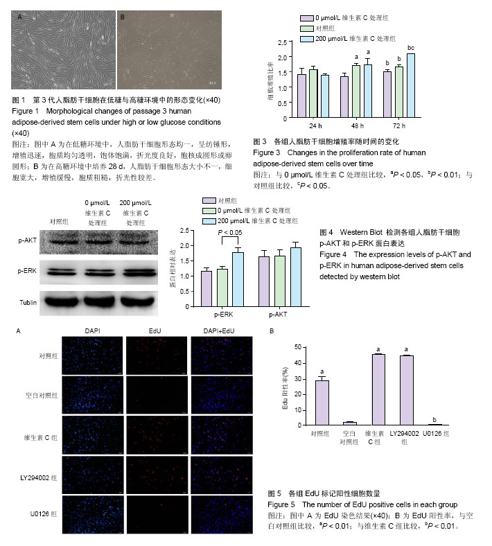
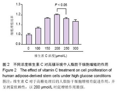
2.1 人脂肪干细胞的形态学特征 第3代人脂肪干细胞形态均一,呈纺锤形,增殖迅速,胞质均匀透明,饱体饱满,折光度良好,胞核成圆形或卵圆形(图1A);高糖环境中培养4周后,人脂肪干细胞形态大小不一,细胞宽大,增殖缓慢,胞质粗糙,折光性较差(图1B)。 2.2 CCK-8检测细胞活力确定维生素C最佳剂量 在图2中,维生素C对于高糖处理后的人脂肪干细胞增殖有促进作用,并呈剂量依赖性,以200 μmol/L时促增殖作用最强,与250 μmol/L比较差异有显著性意义(P < 0.05)。 2.3 CCK-8检测维生素C作用在时间上的变化 从图3中可见,培养24 h时,3组间人脂肪干细胞活力比较差异无显著性意义;培养48 h时,对照组与200 μmol/L维生素C处理组细胞活力比较差异无显著性意义,但两者明显高于0 μmol/L维生素C处理组(P < 0.05);培养72 h时,0 μmol/L维生素C处理组细胞有增殖趋势,但明显低于对照组和200 μmol/L维生素C处理组(P < 0.01),对照组低于200 μmol/L维生素C处理组(P < 0.05)。 2.4 Western Blot检测信号通路相关蛋白表达水平 各组间p-AKT表达量比较差异无显著性意义;200 μmol/L维生素C处理组p-ERK表达明显高于对照组、0 μmol/L维生素C处理组(P < 0.01),见图4。这提示维生素C促进高糖环境下人脂肪干细胞增殖与ERK/MAPK信号通路相关。 2.5 EdU检测细胞增殖 由图5可见,空白对照组EdU阳性率低于对照组(P < 0.01),维生素C组EdU阳性率高于空白对照组(P < 0.01);在2个通路抑制剂组中,加入ERK/MAPK通路抑制剂U0126后,EdU阳性率较维生素C组降低(P < 0.01);加入PI3K/AKT通路抑制剂LY294002后,EdU阳性率与维生素C组无差异,进一步明确了维生素C通过ERK/MAPK信号通路促进高糖环境下人脂肪干细胞的增殖。"
"

| [1] Guariguata L,Whiting DR,Hambleton I,et al.Global estimates of diabetes prevalence for 2013 and projections for 2035. Diabetes Res Clin Pract.2014;103(2):137-149.[2] Li J,Yang D,Yan J,et al.Secondary diabetic ketoacidosis and severe hypoglycaemia in patients with established type 1 diabetes mellitus in China: a multicentre registration study. Diabetes Metab Res Rev.2014;30(6):497-504.[3] Moura LI,Dias AM,Carvalho E,et al.Recent advances on the development of wound dressings for diabetic foot ulcer treatment--a review.Acta Biomater.2013; 9(7):7093-7114.[4] Suresh DH,Suryanarayan S,Sarvajnamurthy S,et al. Treatment of a non-healing Diabetic foot ulcer with platelet-rich plasma.J Cutan Aesthet Surg.2014;7(4):229-231.[5] Mulder G,Tenenhaus M,D'Souza GF.Reduction of diabetic foot ulcer healing times through use of advanced treatment modalities.Int J Low Extrem Wounds.2014; 13(4):335-346.[6] Strassburg S,Nienhueser H,Björn Stark G,et al.Co-culture of adipose-derived stem cells and endothelial cells in fibrin induces angiogenesis and vasculogenesis in a chorioallantoic membrane model.J Tissue Eng Regen Med.2016; 10(6): 496-506.[7] Strassburg S,Nienhueser H,Stark GB,et al.Human adipose-derived stem cellsenhance the angiogenic potential of endothelial progenitor cells, but not of human umbilical vein endothelial cells.Tissue Eng Part A.2013;19(1-2):166-174.[8] Liu L,Gao J,Yuan Y,et al.Hypoxia preconditioned human adipose derived mesenchymalstem cells enhance angiogenic potential via secretion of increased VEGF and bFGF. Cell Biol Int.2013;37(6):551-560.[9] Ma T,Liu H,Chen W,et al.Implanted adipose-derived stem cells attenuate small-for-size liver graft injury by secretion of VEGF in rats. Am J Transplant. 2012; 12(3):620-629.[10] Cheng NC,Hsieh TY,Lai HS,et al.High glucose-induced reactive oxygen species generation promotes stemness in human adipose-derived stem cells.Cytotherapy.2016; 18(3): 371-383.[11] Hankamolsiri W,Manochantr S,Tantrawatpan C,et al.The Effects of High Glucose on Adipogenic and Osteogenic Differentiation of Gestational Tissue-Derived MSCs.Stem Cells Int.2016;2016:1-15.[12] Wang W,Zhang X,Zheng J,et al.High glucose stimulates adipogenic and inhibits osteogenic differentiation in MG-63 cells through cAMP/protein kinase A/extracellular signal-regulated kinase pathway.Mol Cell Biochem.2010; 338(1-2):115-122.[13] Liu Z,Lei M,Jiang Y,et al.High glucose attenuates VEGF expression in rat multipotent adult progenitor cells in association with inhibition of JAK2/STAT3 signalling.J Cell Mol Med.2009;13(9b):3427-3436.[14] Cunha FF,Martins L,Martin PK,et al.A comparison of the reparative and angiogenic properties of mesenchymal stem cells derived from the bone marrow of BALB/c and C57/BL6 mice in a model of limb ischemia.Stem Cell Res Ther.2013; 4(4):86. [15] Willenborg S,Eckes B,Brinckmann J,et al.Genetic ablation of mast cells redefines the role of mast cells in skin wound healing and bleomycin-induced fibrosis.J Invest Dermatol. 2014;134(7):2005-2015. [16] Park KJ,Park E,Liu E,et al.Bone marrow-derived endothelial progenitor cells protect postischemic axons after traumatic brain injury. J Cereb Blood Flow Metab. 2014; 34(2):357-366. [17] Hu J,Cheng D,Gao X,et al.Vitamin C enhances the in vitro development of porcine pre-implantation embryos by reducing oxidative stress.Reprod Domest Anim.2012; 47:873-879.[18] Zhang P,Li J,Qi Y,et al.Vitamin C promotes the proliferation of human adipose-derived stem cells via p53-p21 pathway. Organogenesis.2016;12(3):143-151.[19] Nualart F, Salazar K, Oyarce K, et al.Typical and atypical stem cells in the brain, vitamin C effect and neuropathology. Biol Res.2012;45:243-256.[20] Zeng G,Lai K,Li J,et al.A rapid and efficient method for primary culture of human adipose-derived stem cells. Organogenesis. 2013;9:287-295.[21] Zuk PA,Zhu M,Mizuno H,et al.Multilineage cells from human adipose tissue: implications for cell-based therapies.Tissue Eng.2001;7(2):211-228.[22] Zuk PA,Zhu M,Ashjian P,et al.Human adipose tissue is a source of multipotent stem cells.Mol Biol Cell.2002;13(12): 4279-4295.[23] Bayati V,Sadeghi Y,Shokrgozar MA,et al.The evaluation of cyclic uniaxial strain on myogenic differentiation of adipose-derived stem cells.Tissue Cell.2011;43(6): 359-366.[24] Yilgor Huri P,Cook CA,Hutton DL,et al.Biophysical cues enhance myogenesis of human adipose derived stem/stromal cells.Biochem Biophys Res Commun.2013;438(1):180-185.[25] Zhang P,Moudgill N,Hager E,et al.Endothelial differentiation of adipose-derived stem cells from elderly patients with cardiovascular disease.Stem Cells Dev. 2011;20(6): 977-988.[26] Shi Z,Neoh KG,Kang ET,et al.Enhanced endothelial differentiation of adipose-derived stem cells by substrate nanotopography.J Tissue Eng Regen Med.2014; 8(1):50-58.[27] Kingham PJ,Kalbermatten DF,Mahay D,et al.Adipose-derived stem cells differentiate into a Schwann cell phenotype and promoteneurite outgrowthin vitro.Exp Neurol.2007; 207(2): 267-274.[28] Kingham PJ,Kolar MK,Novikova LN,et al.Stimulating theneurotrophicand angiogenic properties of human adipose-derivedstem cells enhances nerve repair.Stem Cells Dev. 2014;23(7):741-754.[29] Erba P,Mantovani C,Kalbermatten DF,et al.Regeneration potential and survival of transplanted undifferentiated adipose tissue-derived stem cells in peripheral nerve conduits.J Plast Reconstr Aesthet Surg.2010;63(12):e811-817.[30] Reid AJ,Sun M,Wiberg M,et al.Nerve repair with adipose-derived stem cells protects dorsal root ganglia neurons from apoptosis.Neuroscience.2011;199:515-522.[31] Hassan WU,Greiser U,Wang W.Role of adipose-derived stem cells in wound healing. Wound Repair Regen.2014; 22(3): 313-325.[32] Nie C,Zhang G,Yang D,et al.Targeted delivery of adipose-derived stem cells via acellular dermal matrix enhances wound repair in diabetic rats.J Tissue Eng Regen Med, 2015;9(3):224-235.[33] Kuo YR,Wang CT,Cheng JT,et al.Bone marrow-derived mesenchymal stem cells enhanced diabetic wound healing through recruitment of tissue regeneration in a rat model of streptozotocin-induced diabetes.Plast Reconstr Surg.2011; 128(4): 872-880. [34] 王涛.维生素C促进小鼠体细胞重编程[D].2010中国科学院研究生院,2010.[35] Lo T,Ho JH,Yang MH,et al.Glucose reduction prevents replicative senescence and increases mitochondrial respiration in human mesenchymal stem cells.Cell Transplant. 2011;20(6):813-825.[36] Chepda T,Cadau M,Girin P,et al.Monitoring of ascorbate at a constant ate in cell culture: effect on cell growth.In Vitro Cell Dev Biol Anim.2011;37:26-30.[37] Osiecki M,Ghanavi P,Atkinson K,et al.The ascorbic acid paradox.Biochem Biophys Res Commun.2010;400:466-470.[38] Huang Y,Tang X,Xie W,et al.Vitamin C enhances in vitro and in vivo development of porcine somatic cell nuclear transfer embryos.Biochem Biophys Res Commun.2011;411: 397-401.[39] Marshall CJ. Specificity of receptor tyrosine kinase signaling: transient versus sustained extracellular signal-regulated kinase activation. Cell.1985;80(2):179-185.[40] [40] Murakami J,Ishii M,Suehiro F,et al.Vascular endothelial growth factor-C induces osteogenic differentiation of human mesenchymal stem cells through the ERK and RUNX2 pathway. Biochem Biophys Res Commun.2017. pii:S0006-291X(17)30262-0. doi: 10.1016/j.bbrc.2017.02.001. [Epub ahead of print] [41] Zhu C,Yu J,Pan Q,et al.Hypoxia-inducible factor-2 alpha promotes the proliferation of human placenta-derived mesenchymal stem cells through the MAPK/ERK signaling pathway. Sci Rep.2016; 6:35489.[42] Zhang Y,Wang J,Lv Z,et al.Cox-2 promotes mesenchymal stem cells differentiation into cardiocytes by activating JNK and ERK pathway.Biochem Biophys Res Commun. 2016; 480(1):101-105. |
| [1] | Yao Xiaoling, Peng Jiancheng, Xu Yuerong, Yang Zhidong, Zhang Shuncong. Variable-angle zero-notch anterior interbody fusion system in the treatment of cervical spondylotic myelopathy: 30-month follow-up [J]. Chinese Journal of Tissue Engineering Research, 2022, 26(9): 1377-1382. |
| [2] | Wang Jing, Xiong Shan, Cao Jin, Feng Linwei, Wang Xin. Role and mechanism of interleukin-3 in bone metabolism [J]. Chinese Journal of Tissue Engineering Research, 2022, 26(8): 1260-1265. |
| [3] | Xiao Hao, Liu Jing, Zhou Jun. Research progress of pulsed electromagnetic field in the treatment of postmenopausal osteoporosis [J]. Chinese Journal of Tissue Engineering Research, 2022, 26(8): 1266-1271. |
| [4] | Tian Chuan, Zhu Xiangqing, Yang Zailing, Yan Donghai, Li Ye, Wang Yanying, Yang Yukun, He Jie, Lü Guanke, Cai Xuemin, Shu Liping, He Zhixu, Pan Xinghua. Bone marrow mesenchymal stem cells regulate ovarian aging in macaques [J]. Chinese Journal of Tissue Engineering Research, 2022, 26(7): 985-991. |
| [5] | Hou Jingying, Guo Tianzhu, Yu Menglei, Long Huibao, Wu Hao. Hypoxia preconditioning targets and downregulates miR-195 and promotes bone marrow mesenchymal stem cell survival and pro-angiogenic potential by activating MALAT1 [J]. Chinese Journal of Tissue Engineering Research, 2022, 26(7): 1005-1011. |
| [6] | Zhou Ying, Zhang Huan, Liao Song, Hu Fanqi, Yi Jing, Liu Yubin, Jin Jide. Immunomodulatory effects of deferoxamine and interferon gamma on human dental pulp stem cells [J]. Chinese Journal of Tissue Engineering Research, 2022, 26(7): 1012-1019. |
| [7] | Liang Xuezhen, Yang Xi, Li Jiacheng, Luo Di, Xu Bo, Li Gang. Bushen Huoxue capsule regulates osteogenic and adipogenic differentiation of rat bone marrow mesenchymal stem cells via Hedgehog signaling pathway [J]. Chinese Journal of Tissue Engineering Research, 2022, 26(7): 1020-1026. |
| [8] | Wang Jifang, Bao Zhen, Qiao Yahong. miR-206 regulates EVI1 gene expression and cell biological behavior in stem cells of small cell lung cancer [J]. Chinese Journal of Tissue Engineering Research, 2022, 26(7): 1027-1031. |
| [9] | Liu Feng, Peng Yuhuan, Luo Liangping, Wu Benqing. Plant-derived basic fibroblast growth factor maintains the growth and differentiation of human embryonic stem cells [J]. Chinese Journal of Tissue Engineering Research, 2022, 26(7): 1032-1037. |
| [10] | An Weizheng, He Xiao, Ren Shuai, Liu Jianyu. Potential of muscle-derived stem cells in peripheral nerve regeneration [J]. Chinese Journal of Tissue Engineering Research, 2022, 26(7): 1130-1136. |
| [11] | Fan Yiming, Liu Fangyu, Zhang Hongyu, Li Shuai, Wang Yansong. Serial questions about endogenous neural stem cell response in the ependymal zone after spinal cord injury [J]. Chinese Journal of Tissue Engineering Research, 2022, 26(7): 1137-1142. |
| [12] | Wen Dandan, Li Qiang, Shen Caiqi, Ji Zhe, Jin Peisheng. Nocardia rubra cell wall skeleton for extemal use improves the viability of adipogenic mesenchymal stem cells and promotes diabetes wound repair [J]. Chinese Journal of Tissue Engineering Research, 2022, 26(7): 1038-1044. |
| [13] | Zhu Bingbing, Deng Jianghua, Chen Jingjing, Mu Xiaoling. Interleukin-8 receptor enhances the migration and adhesion of umbilical cord mesenchymal stem cells to injured endothelium [J]. Chinese Journal of Tissue Engineering Research, 2022, 26(7): 1045-1050. |
| [14] | Luo Xiaoling, Zhang Li, Yang Maohua, Xu Jie, Xu Xiaomei. Effect of naringenin on osteogenic differentiation of human periodontal ligament stem cells [J]. Chinese Journal of Tissue Engineering Research, 2022, 26(7): 1051-1056. |
| [15] | Wang Xinmin, Liu Fei, Xu Jie, Bai Yuxi, Lü Jian. Core decompression combined with dental pulp stem cells in the treatment of steroid-associated femoral head necrosis in rabbits [J]. Chinese Journal of Tissue Engineering Research, 2022, 26(7): 1074-1079. |
| Viewed | ||||||
|
Full text |
|
|||||
|
Abstract |
|
|||||