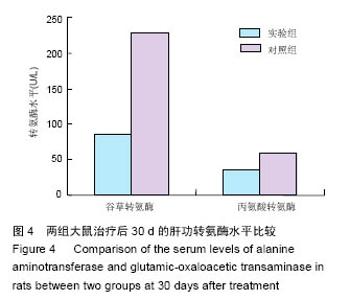
| [1]胡鹏蕴,程远,汪艳,等.去细胞化全肝生物支架循环灌注培养条件下体外细胞再植[J].中国组织工程研究, 2012, 16(18):3231-3235.[2]Shouval D, Lai CL, Chang TT,et al.Relapse of hepatitis B in HBeAg -negative chronic hepatitis B patients who discontinued successful entecavir treatment: the case for continuous antiviral therapy.J Hepatol. 2009;50(2): 289-295.[3]宋坤修,何爱咏,谢求恩.纳米羟基磷灰石/胶原复合材料结合血管内皮生长因子修复兔桡骨缺损[J].中国组织工程研究与临床康复,2009,13(24):6624-6628.[4]李福兵,徐永清,潘兴华,等.小鼠胫骨中段1/3不同缺损直径单层骨皮质缺损模型比较研究[J].中国修复重建外科杂志,2012,26(10):1221.[5]徐素彬,宋芹,彭菊花,等.护理风险管理在普外科病房的应用[J].中国医药导报,2012,9(12):154-155.[6]Fukumitsu K,Yagi H,Soto-Gutierrez A.Bioengineering in Organ Transplantation:Targeting the Liver. Transplant Proc.2011;43(6):2137-2138.[7]韦从云,周磊,赖春花,等.骨代用品实验中兔股骨远端标准骨缺损模型的建立[J].广东牙病防治,2011,18(6):306.[8]刘添皇.慢性重型肝炎病原学特征、病变基础、年龄、性别、抗病毒治疗与病死率相关性的分析[J].临床医药实践, 2010,19(1):11-12.[9]Ren H,Zhao Q,Cheng T,et al. No contribution of umbilical cord mesenchymal stromal cells to capillarization and venularization of hepatic sinusoids accompanied by hepatic differentiation in carbon tetrachloride-induced mouse liver fibrosis. Cytotherapy. 2010;12(3):371-383.[10]宋芹,陈封政,颜军,等.胶原蛋白研究进展[J].成都大学学报:自然科学版,2012,31(1):35-38.[11]Brown BN,Freund JM,Han L,et al.Comparison of three methods for the derivation of a biologic scaffold composed of adipose tissue extracellular matrix.Tissue Eng Part C.2011;17(4):411-421.[12]Kulig KM,Luo X,Finkelstein EB,et al.Biologic properties of surgical scaffold materials derived from dermal ECM.Biomaterials.2013;34(23):5776-5784.[13]Coutu DL,Mahfouz W,Loutochin O,et al.Tissue engineering of rat bladder using marrow-derived mesenchymal stem cells and bladder acellular matrix.PloS One.2014;9(12):e111966.[14]Goh SK,Bertera S,Olsen P,et al. Perfusion-decellularized pancreas as a natural 3D scaffold for pancreatic tissue and whole organ engineering.Biomaterials.2013;34(28): 6760–6772.[15]中华医学会肝病学分会,中华医学会感染病学分会.慢性乙型肝炎防治指南(2010年版)[J].临床肝胆病杂志, 2011, 27(1):Ⅰ-ⅩⅥ.[16]Wang X,Cui J,Zhang BQ,et al.Decellularized liver scaffolds effectively support the proliferation and differentiation of mouse fetal hepatic progenitors. Journal of biomedical materials research.J Biomed Mater Res A.2014;102(4):1017-1025.[17]吴阳,吴大帅,张弓,等. Pokemon自身抗体在原发性肝癌中的表达及意义[J].中华实验外科杂志, 2013,30(8):1717-1719.[18]蒋涛,任先军,阴洪,等.京尼平交联对大鼠脱细胞脊髓生物学特性的影响[J].第三军医大学学报, 2013,35(20):2164-2167.[19]张国安,宁方刚,钟京鸣,等.反复冻融配合超声振荡洗涤制备猪脱细胞真皮基质[J].中国组织工程研究与临床康复, 2007,11(41):8280-8284.[20]石海涛,莫秀梅,何创龙,等.肝素化纳米材料P(LLA-CL)的制备及其表征和抗凝血性特征[J].中国组织工程研究与临床康复,2008,12(1):10-14.[21]曾伟杰,凌友,支晓兴.心血管支架材料生物力学及生物相容性特征[J].中国组织工程研究与临床康复, 2008, 12(13):2531-2534.[22]Nottelet B,Pektok E,Mandracchia D,et al.Factorial design optimization and in vivo feasibility of poly(ε-caprolactone)- micro- and nanofiber-based small diameter vascular grafts.J Biomed Mater Res A.2009;89(4):865-875.[23]Pektok E,Nottelet B,Tille JC,et al.Degradation and healing characteristics of small-diameter poly(epsilon-caprolactone) vascular grafts in the rat systemic arterial circulation.Circulation. 2008;118(24): 2563-2570.[24]李一鹏,陈旭义,朱祥,等.脱细胞脊髓基质支架的制备及生物特性[J].中国组织工程研究,2015,19(21):3398-3402.[25]綦惠,杰永生,陈磊,等.两种交联剂制备软骨细胞移植支架的比较[J].中国医药生物技术,2013,8(6):408-413.[26]郭树章,任先军,蒋涛,等.脱细胞脊髓天然支架的制备及形态学观察[J].中国矫形外科杂志,2007,15(3):226-228.[27]Liu J,Chen J,Liu B,et al.Acellular spinal cord scaffold seeded with mesenchymal stem cells promotes long-distance axon regeneration and functional recovery in spinal cord injured rats.J Neurol Sci. 2013; 325(1):127-136.[28]牛震海,金正花,于家傲.纳米纤维支架的制备及其在组织工程皮肤中的应用[J].中华损伤与修复杂志:电子版, 2011, 6(1):118-124.[29]Sato T,Araki M,Nakajima N.Biodegradable polymer coating promotes the epithelization of tissue-engineered airway prostheses.J Thorac Cardiov Surg.2013;139(1):26-31. [30]Xu F,Kang T,Deng J,et al.Functional Nanoparticles Activate a Decellularized Liver Scaffold for Blood Detoxification.Small.2016;12(15):2067-2076. [31]Maghsoudlou P,Georgiades F,Smith H,et al. Optimization of Liver Decellularization Maintains Extracellular Matrix Micro-Architecture and Composition Predisposing to Effective Cell Seeding.PLoS One.2016;11(5):e0155324.[32]Mazza G,Simons JP,Al-Shawi R,et al.Amyloid persistence in decellularized liver: biochemical and histopathological characterization. Amyloid. 2016; 23(1):1-7. [33]Mazza G,Rombouts K,Rennie Hall A,et al. Decellularized human liver as a natural 3D-scaffold for liver bioengineering and transplantation.Sci Rep. 2015; 5:13079. [34]Hussein KH,Park KM,Teotia PK,et al.Sterilization using electrolyzed water highly retains the biological properties in tissue-engineered porcine liver scaffold. Int J Artif Organs.2013;36(11):781-792. [35]Lin YQ,Wang LR,Wang JT,et al.New advances in liver decellularization and recellularization: innovative and critical technologies.Expert Rev Gastroenterol Hepatol. 2015;9(9):1183-1191. [36]Li Q,Uygun BE,Geerts S,et al.Proteomic analysis of naturally-sourced biological scaffolds.Biomaterials. 2016;75:37-46. [37]Bao J,Sun J,Zhou Y,et al.Anticoagulant Ability and Heparinization of Decellularized Biomaterial Scaffolds.Sheng Wu Yi Xue Gong Cheng Xue Za Zhi.2015;32(3):594-598. [38]Aguado BA,Caffe JR,Nanavati D,et al.Extracellular matrix mediators of metastatic cell colonization characterized using scaffold mimics of the pre-metastatic niche.Acta Biomater.2016;33:13-24.[39]Nakanishi R,Hashimoto M,Muranaka H.Effect of cryopreservation period on rat tracheal allografts.J Heart Lung Transplant.2014;20(9):1010-1015. |


