Chinese Journal of Tissue Engineering Research ›› 2016, Vol. 20 ›› Issue (37): 5609-5616.doi: 10.3969/j.issn.2095-4344.2016.37.020
Previous Articles Next Articles
Glial scar formation and astrocyte role in spinal cord injury
Li Jian-feng, Yan Jin-yu, Xia Run-fu, Zhang Xu, Tan Xiao-hui, Guan Jian, Ye Zhen, Zhang Shu-lian
- Department of Rehabilitation, Second Affiliated Hospital of Inner Mongolia Medical University, Hohhot 010030, Inner Mongolia Autonomous Region, China
-
Online:2016-09-09Published:2016-09-09 -
Contact:Zhang Shu-lian, Associate chief nurse, Department of Rehabilitation, Second Affiliated Hospital of Inner Mongolia Medical University, Hohhot 010030, Inner Mongolia Autonomous Region, China -
About author:Li Jian-feng, M.D., Attending physician, Department of Rehabilitation, Second Affiliated Hospital of Inner Mongolia Medical University, Hohhot 010030, Inner Mongolia Autonomous Region, China -
Supported by:the National Natural Science Foundation of China, No. 81560212; the Natural Science Foundation of Inner Mongolia Autonomous Region, China, No. 2015MS0898
CLC Number:
Cite this article
Li Jian-feng, Yan Jin-yu, Xia Run-fu, Zhang Xu, Tan Xiao-hui, Guan Jian, Ye Zhen, Zhang Shu-lian. Glial scar formation and astrocyte role in spinal cord injury[J]. Chinese Journal of Tissue Engineering Research, 2016, 20(37): 5609-5616.
share this article
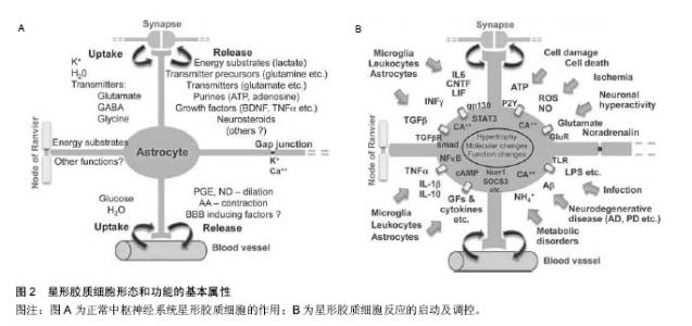
2.1 星形胶质细胞的生物学特性 星形胶质细胞、少突胶质细胞及其前体细胞和小胶质细胞等胶质细胞为中枢神经系统提供了功能和生理学的结构支持,并且在相应的病理生理状况下对损伤和疾病做出应答。少突胶质细胞在某些特定环境下,可包绕轴突形成髓鞘[4-5];小胶质细胞在脑和脊髓当中形成宿主免疫系统的吞噬细胞[6],并在中枢神经系统受到损伤后迅速发挥神经保护和修复作用[7];星形胶质细胞对神经递质调节、离子平衡、血脑屏障的维持以及基底膜和神 经周围营养因子的产生的起着至关重要的作用[8-12],而且脊髓的星形胶质细胞还可以分泌强啡肽类物质参与脊髓疼痛发生的生理过程[13]。星形胶质细胞是神经网络系统重要的组成部分,研究发现星形胶质细胞上存在K+、Na+和Ca2+等离子通道[14-15]。现在,大量证据表明Ca2+浓度增高在星形胶质细胞之间以及星形胶质细胞和神经元细胞之间的信息传递发挥着重要的作用。 星形胶质细胞Ca2+浓度变化而引发的生物效应主要包括:①由于细胞内Ca2+的释放而发生细胞内离子振荡;②在神经活动期间由谷氨酸盐和嘌呤类等递质激发形成Ca2+外流;③从星形胶质细胞中诱导释放谷氨酸盐等递质到细胞外间隙触发受体导致神经元形成电流;④将细胞信号传送至邻近星形胶质细胞[16-20]。星形胶质细胞包裹突触并且在维持其体液、离子、酸碱环境和内稳定方面发挥重要作用[21],从而保障正常的突触间信息传递。正常情况下星形胶质细胞分为静止态、活化态和增殖态,三者相互转换构成了广义上的细胞周期。在正常的中枢神经系统中,静止态和活化态的胶质细胞并存。当受到损伤时,在细胞因子作用下,静止态的细胞逐渐向活化态转化。 19世纪以来,根据星形胶质细胞的形态和解剖位置将其分为2种主要的亚型:原浆型星形胶质细胞和纤维型星形胶质细胞。原浆型星形胶质细胞存在于中枢神经系统灰质当中,纤维型星形胶质细胞主要存在 "

| [1] Carvalho ZMF, Darder JJT, Reis PAM,et al. Experiencing a traumatic spinal cord injury: analysis on the view of the theory of Watson’s transpersonal caring. J Biom Sci Eng. 2013;6(1):14-20. [2] Reis PA, Carvalho ZM, Tirado Darder JJ,et al. Cross-cultural adaptation of the Quality of Life Index Spinal Cord Injury-Version III. Rev Esc Enferm USP. 2015; 49(3):401-408. [3] Shamim MS,Ali SF,Enam SA.Non-operative management is superior to surgical stabilization in spine injury patients with complete neurological deficits: A perspective study from a developing world country, Pakistan. Surg NeurolInt.2011; 2:166. [4] Nishiyama A.Polydendrocytes: NG2 cells with many roles in development and repair of the CNS. Neuroscientist.2007;13:62-76. [5] Wilson HC, Scolding NJ, Raine CS. Co-expression of PDGF alpha receptor and NG2 by oligodendrocyte precursors in human CNS and multiple sclerosis lesions.J Neuroimmunol.2006;176:162-173. [6] Kim SU, de Vellis J. Microglia in health and disease.J Neurosci Res.2005;81:302-313. [7] Wu D,Zhang Y,Bo X, et al. Actions of neuropoietic cytokines and cyclic AMP in regenerative conditioning of rat primary sensory neurons.Exp Neurol.2007; 204:66-76. [8] Schousboe A, Westergaarde N. Transport of neuroactive amino acids in astrocytes. Neuroglia.1995; 3:246-258. [9] Walz W.Role of glial cells in the regulation of the brain ion microenvironment. Prog Neurobiol.1989;33: 309-333. [10] Wolburg H, Risau W. Formation of the blood-brain-barrier. Neuroglia.1995; 11:763-776. [11] Massey JM,Hubscher CH,Wagoner MR,et al. Chondroitinase ABC digestion of the perineuronal net promotes functional collateral sprouting in the cuneate nucleus after cervical spinal cord injury. J Neurosci. 2006;26:4406-4414. [12] Tom VJ, Doller CM,Malouf AT,et al.Astrocyte- associated fibronectin is critical for axonal regeneration in adult white matter. J Neurosci.2004;24: 9282-9290. [13] Wahlert A,Funkelstein L, Fitzsimmons B, et al. Spinal astrocytes produce and secrete dynorphin neuropeptides. Neuropeptides.2013;1:1132-1139. [14] Pice DL, Ludwig JW, Mi H, et al. Distribution of rSlo Ca2+ -activated K+ channels in rat astrocyte perivascular endfeet. Brain Res.2002;956: 183-193. [15] Krzemien DM, Schaller KL, Levinson SR, et al. Immunolocalization of Sodium Channel Isoform NaCh6 in the Nervous System. J Comp Neurol. 2000; 420(1):70-83. [16] Halassa MM,Fellin T,Haydon PG.The tripartite syn-apse: roles for gliotransmission in health and disease.Trends Mol Med.2007;13:54-63. [17] Nedergaard M, Ransom B, Goldman SA. New roles for astrocytes: redefining the functional architecture of the brain.Trends Neurosci.2003;26:523-530. [18] Perea G, Navarrete M, Araque A.Tripartite synapses: astrocytes process and control synaptic information. Trends Neurosci.2009;32:421-431. [19] Voskuhl RR, Peterson RS, Song B, et al. Reactive astrocytes form scar-like perivascular barriers to leukocytes during adaptive immune inflammation of the CNS. J Neurosci.2009;29:11511-11522. [20] Volterra A, Meldolesi J.Astrocytes, from brain glue to communication elements: the revolution continues. Nat Rev Neurosci.2005;6:626-640. [21] Brown AM, Ransom BR.Astrocyte glycogen and brain energy metabolism. Glia.2007; 55:1263-1271. [22] Pellerin L, Bouzier-Sore AK, Aubert A, et al. Activity-dependent regulation of energy metabolism by astrocytes: an update. Glia.2007;55:1251-1262. [23] Michael V, Sofroniew,Harry V,et al. Astrocytes: biology and pathology.Acta Neuropathol.2010; 119:7-35. [24] Gimenez Y, Ribotta M, Menet V, et al . The role of ast rocytes in axonal regeneration in the mammalian CNS. Prog Brain Res.2001;132 :587-610. [25] Drogemuller K, Helmuth U, Brunn A, et al. Astrocyte gp130 expression is critical for the control of Toxo-plasma encephalitis. J Immunol.2008;181: 2683-2693. [26] Koyama Y. Signaling molecules regulating phenotypic conversions of astrocytes and glial scar formation in damaged nerve tissues. Neurochem Int.2014; 78: 35-42. [27] Kozuka N,Itofusa R,Kudo Y,et al.Lipopolysaccharide and proinflammatory cytokines require different ast rocytes states to induce nitric oxide production. J Neurosci Res.2005;82(5):717-728. [28] Lebkuechner I,Möllerström E,Wilhelmsson U,et al. Heterogeneity of Notch signaling in astrocytes and the effects of GFAP and vimentin deficiency.J Neurochem. 2015;2:13202-13213. [29] Sofroniew MV. Molecular dissection of reactive as-trogliosis and glial scar formation. Trends Neurosci. 2009;32:638-647. [30] Sofroniew MV,Vinters HV.Astrocytes:biology and pathology. Acta Neuropathol. 2001;119(1):7-35. [31] Sofroniew MV. Molecular dissection of reactive astrogliosis and glial scar formation. Trends Neurosci. 2009;32(12):638-647. [32] Wilhelmsson U, Bushong EA, Price DL, et al. Redefining the concept of reactive astrocytes as cells that remain within their unique domains upon reaction to injury. Proc Natl Acad Sci USA.2006;103: 17513-17518. [33] Herrmann JE,Imura T,Song B, et al. STAT3 is a critical regulator of astrogliosis and scar formation after spinal cord injury. J Neurosci.2008;28: 7231-7243. [34] Silver J, Miller JH. Regeneration beyond the glial scar. Nature Rev Neurosci. 2004;5:146-156. [35] Sofroniew MV,Vinters HV.Astrocytes: biology and pathology. Acta Neuropathol. 2001;119(1):7-35. [36] Rolls A,Shechter R,Schwartz M.The bright side of the glial scar in CNS repair. Nat Rev Neurosci.2009; 10(3):235-241. [37] Faulkner JR,Herrmann JE,Woo MJ, et al. Reactive astrocytes protect tissue and preserve function after spinal cord injury.J Neurosci.2004;24(9):2143-2155. [38] Silver J, Miller JH. Regeneration beyond the glial scar. Nat Rev Neurosci. 2004;5(2):146-156. [39] Brambilla R, Persaud T,Hu X, et al.Transgenic inhibition of astroglial NF-kappa B improves functional outcome in experimental autoimmune encephalomyelitis by suppressing chronic central nervous system inflammation.J Immunol.2009; 182(5):2628-2640. [40] Hamby ME, Hewett JA, Hewett SJ. TGF-beta1 potentiates astrocytic nitric oxide production by expanding the population of astrocytes that express NOS-2.Glia. 2006; 54(6):566-577. [41] Takano T, Kang J, Jaiswal JK, et al. Receptor-mediated glutamate release from volume sensitive channels in astrocytes.Proc Natl Acad Sci U S A.2005; 102(45):16466-16471. [42] North HA, Pan L, McGuire TL,et al.β1-Integrin alters ependymal stem cell BMP receptor localization and attenuates astrogliosis after spinal cord injury.J Neurosci.2015;35(9):3725-33. [43] Vos PE,Jacobs B,Andriessen TM, et al. GFAP and S100B are biomarkers of traumatic brain injury: an observational cohort study. Neurology.2010;75(20): 1786-1793. [44] Fitch MT, Silve J.Astrocytes are Dynamic Participants in Central Nervous System Development and Injury Responses. Oxford University Press.2001;5:263-269. [45] Filbin MT.Myelin-associated inhibitors of axonal regeneration in the adult mammalian CNS. Nat Rev Neurosci. 2003;4:703-713. [46] Moreau-Fauvarque C, Kumanogoh A.The transmembrane semaphorin Sema4D/CD100, an inhibitor of axonal growth, is expressed on oligodendrocytes and upregulated after CNS lesion. J Neurosci.2003;23:9229-9239. [47] Morgenstern DA,Asher RA,Fawcett JW.Chondroitin sulphate pro-teoglycans in the CNS injury response. Prog Brain Res.2002;137:313-332. [48] Fitch MT, Silver J.CNS injury, glial scars, and inflammation: Inhibitory extracellular matrices and regeneration failure. Exp Neurol.2008;209:294-301. [49] Carlson SL, Parrish ME, Springer JE, et al. Acute inflammatory response in spinal cord following impact injury. Exp Neurol.1998;151:77-88. [50] Morino T, Ogata T, Horiuchi H, et al. Delayed neuronal damage related to microglia proliferation after mild spinal cord compression injury. Neurosci Res. 2003; 46: 309-318. [51] Watanabe T, Yamamoto T, Abe Y, et al. Differential activation of microglia after experimental spinal cord injury. J Neurotrauma.1999;16:255-265. [52] Tang X. Changes in distribution, cell associations, and protein expression levels of NG2, neurocan, phosphacan, brevican, versican V2, and tenascin-C during acute to chronic maturation of spinal cord scar tissue.J Neurosci Res.2003; 71:427-444. [53] Asher RA, Morgenstern DA, Moon LD, et al. Chondroitin sulphate proteoglycans: inhibitory components of the glial scar. Prog Brain Res.2001; 132:611-619. [54] Yamagata T, Saito H, Habuchi O, et al. Purification and properties of bacterial chondroitinases and chondrosulfatases. J Biol Chem.1968;243(7): 1523-1535. [55] Bradbury EJ,Carter LM.Manipulating the glial scar: Chondroitinase ABC as a therapy for spinal cord injury. Brain Res Bull.2011;84(4-5):306-316. [56] Pizzi MA,Crowe MJ.Matrix metalloproteinases and proteoglycans in axonal regeneration. Exp Neurol. 2007;204:496-511. [57] Yong VW. Metalloproteinases: mediators of pathology and regeneration in the CNS. Nat Rev Neurosci. 2005; 6:931-944. [58] Jeong SR, Kwon MJ, Lee HG, et al. Hepatocyte growth factor reduces astrocytic scar formation and promotes axonal growth beyond glial scars after spinal cord injury. Exp Neurol.2012;233(1):312-322. [59] West H, Richardson WD , Fruttiger M. Stabilization of t he retinal vascular network by reciprocal feedback between blood vessels and ast rocytes. Development. 2005;132(8):1855-1862. [60] Bradbury EJ, Moon LD, Popat RJ, et al. Chondroitinase ABC promotes functional recovery after spinal cord injury. Nature.2002;416: 636-640. [61] Tan AM, Colletti M, Rorai AT, et al. Antibodies against the NG2 proteoglycan promote the regeneration of sensory axons within the dorsal columns of the spinal cord. J Neurosci.2006;26: 4729-4739. [62] Grimpe B, Silver J.A novel DNA enzyme reduces glycosaminoglycan chains in the glial scar and allows microtransplanted dorsal root ganglia axons to regenerate beyond lesions in the spinal cord. J Neurosci. 2004;24: 1393-1397. |
| [1] | Cao Qingjun, Yang Fenghua, Wang Hua. Hippocampal astrocytes in juvenile rats with persistent epilepsy: the role of cannabinoid receptor type 2 in regulating MAPK pathway [J]. Chinese Journal of Tissue Engineering Research, 2020, 24(32): 5179-5185. |
| [2] | Gao Jianbo, Xia Bing, Li Shengyou, Yang Yujie, Ma Teng, Yu Peng, Luo Zhuojing, Huang Jinghui. Effect of nanoparticles carrying chondroitin sulfate ABC on the migration of Schwann cells in a magnetic field [J]. Chinese Journal of Tissue Engineering Research, 2020, 24(28): 4526-4532. |
| [3] | Zhou Yanxing1, Peng Xinsheng2, Hou Gan1, Li Jiangbin1, Zhang Hua1, Zhou Zhikun2, Zhou Yanfang3 . Inhibitory effect of capsaicin on fibroblast proliferation and its molecular mechanism [J]. Chinese Journal of Tissue Engineering Research, 2019, 23(7): 1018-1022. |
| [4] | Lü Cong, Sun Lin, Feng Haoyu, Ma Xun, He Yajun, Li Jisheng. Inhibition of high mobility group box 1/nuclear factor-kappa B pathway reduces apoptosis in spinal cord astrocytes after oxygen-glucose deprivation/reoxygenation [J]. Chinese Journal of Tissue Engineering Research, 2019, 23(33): 5353-5359. |
| [5] | Zhang Yong, Ma Xun, Sun Lin, Zhang Li, Guan Xiaoming, Lü Cong, Chen Xu. Exosomes derived from human umbilical cord mesenchymal stem cells attenuate edema of spinal astrocytes after oxygen-glucose deprivation/reoxygenation injury in rats [J]. Chinese Journal of Tissue Engineering Research, 2019, 23(25): 4011-4017. |
| [6] | Zhou Yan, Wang Lin, Pei Shuang, Li Yanfei, Chen Xuemei, Jia Yanjie. Bone marrow mesenchymal stem cell-derived exosomes reduce the activation of type A1 astrocytes after spinal cord injury [J]. Chinese Journal of Tissue Engineering Research, 2019, 23(21): 3294-3301. |
| [7] | Jing Ya-jun 1, Zhang Lei1, Zhang Shao-qun1, Liao Li-qing1, Yuan Shi-guo2, Li Yi-kai1. Pathological changes of the skeletal muscle after local loosening therapies [J]. Chinese Journal of Tissue Engineering Research, 2018, 22(4): 535-541. |
| [8] | Ge Wenjia, Ma Xiaorong, Ouyang Tianxiang . Aminolevulinic acid photodynamic therapy can significantly inhibit the growth of endothelial cells and induce cell apoptosis [J]. Chinese Journal of Tissue Engineering Research, 2018, 22(36): 5840-5845. |
| [9] | Lv Ying1, An Mei-wen1, Hou Chun-sheng2. Finite element analysis of skin closure stress in different directions [J]. Chinese Journal of Tissue Engineering Research, 2017, 21(4): 609-614. |
| [10] | Wu Zi-han1, Li Gao-feng2. Correlation of angiopoietin-1 with angiogenesis during scar formation [J]. Chinese Journal of Tissue Engineering Research, 2017, 21(32): 5158-5163. |
| [11] | Liu Ying-wei, Zhang Wan-li, Chi Cheng-tao, Xu Qing-yu, Lu De-zhi. Effects of hyaluronic acid on scar formation in the acellular nerve allograft [J]. Chinese Journal of Tissue Engineering Research, 2016, 20(42): 6317-6323. |
| [12] | Fan Xu-hui, Yang Bo, Hu Xiang, Guan Fang-xia. Reactive hyperplasia of glial cells induced by spinal cord injury in a rat model [J]. Chinese Journal of Tissue Engineering Research, 2016, 20(40): 6001-6006. |
| [13] | Ding Chao, Sun Qiang, Tang Cheng. Comparison of 3.0T MRI and SPECT-CT in the diagnosis of osteoporotic vertebral compression fractures [J]. Chinese Journal of Tissue Engineering Research, 2016, 20(39): 5885-5891. |
| [14] | Sun Gui-fang, Zhang Xiao-fen, Li Hong-chang, Pan Li-yun, Chen Ya-feng, Xu Ke, Feng Dian-xu. Shengjiyuhong ointment inhibits hypertrophic scar formation [J]. Chinese Journal of Tissue Engineering Research, 2016, 20(33): 4890-4898. |
| [15] | Wang Yang-yang, Li Xiao-jing, Bu Shou-shan. Biological properties of acellular dermal matrix derived from mature scar tissue versus normal skin [J]. Chinese Journal of Tissue Engineering Research, 2016, 20(30): 4496-4502. |
| Viewed | ||||||
|
Full text |
|
|||||
|
Abstract |
|
|||||