Chinese Journal of Tissue Engineering Research ›› 2019, Vol. 23 ›› Issue (33): 5353-5359.doi: 10.3969/j.issn.2095-4344.1811
Previous Articles Next Articles
Inhibition of high mobility group box 1/nuclear factor-kappa B pathway reduces apoptosis in spinal cord astrocytes after oxygen-glucose deprivation/reoxygenation
Lü Cong, Sun Lin, Feng Haoyu, Ma Xun, He Yajun, Li Jisheng
- Department of Orthopedics, Shanxi Dayi Hospital Affiliated to Shanxi Medical University, Taiyuan 030032, Shanxi Province, China
-
Revised:2019-04-06Online:2019-11-28Published:2019-11-28 -
Contact:Sun Lin, Associate chief physician, Department of Orthopedics, Shanxi Dayi Hospital Affiliated to Shanxi Medical University, Taiyuan 030032, Shanxi Province, China -
About author:Lü Cong, Master, Department of Orthopedics, Shanxi Dayi Hospital Affiliated to Shanxi Medical University, Taiyuan 030032, Shanxi Province, China -
Supported by:the National Natural Science Foundation of China, No. 81870976 (to SL)
CLC Number:
Cite this article
Lü Cong, Sun Lin, Feng Haoyu, Ma Xun, He Yajun, Li Jisheng. Inhibition of high mobility group box 1/nuclear factor-kappa B pathway reduces apoptosis in spinal cord astrocytes after oxygen-glucose deprivation/reoxygenation [J]. Chinese Journal of Tissue Engineering Research, 2019, 23(33): 5353-5359.
share this article
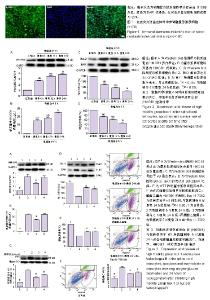
2.1 星形胶质细胞鉴定结果 取第3代细胞,用星形胶质细胞特定表达蛋白S100β对细胞进行免疫荧光染色,染色结果为星形胶质细胞纯度为95%,见图1。 2.2 氧糖剥夺/复氧后星形胶质细胞HMGB1的表达与释放及细胞活性与凋亡程度 HMGB1表达与释放:Western blot与ELISA检测显示与正常组相比,氧糖剥夺6 h/复氧6,12,24 h组HMGB1蛋白表达与释放明显增多,且氧糖剥夺6 h/复氧24 h组中HMGB1表达与释放最多的(P < 0.05),见图2A,B。 凋亡蛋白表达:与正常组相比,氧糖剥夺6 h/复氧6,12,24 h组Bcl-2与BAX表达均增多,但Bcl-2随复氧时间增长表达逐渐减少,BAX表达随复氧时间延长表达增加,因此Bcl-2/BAX比值随时间增长降低,在复氧24 h时Bcl-2/BAX比值达到最低(P < 0.05),见图2C。 细胞存活率:与正常组相比,氧糖剥夺6 h/复氧,12,24 h组星形胶质细胞活性随时间的增加减弱,复氧24 h时脊髓星形胶质细胞存活率最低(P < 0.05),见图2D。因此选择复氧24 h作为后续实验的时间点。 2.3 抑制HMGB1与核转录因子κB表达后氧糖剥夺6 h/复氧24 h星形胶质细胞的凋亡程度与成活率 HMGB1表达与释放:Western blot及ELISA结果显示与氧糖剥夺6 h/复氧24 h组比,氧糖剥夺6 h/复氧24 h+丙酮酸乙酯组星形胶质细胞内HMGB1的表达与释放减少(P < 0.05),见图3A,B。 核转录因子κB蛋白表达:Western blot结果显示与氧糖剥夺6 h/复氧24 h组比较,氧糖剥夺6 h/复氧24 h+Bay 11-7082组核转录因子κB表达明显减少(P < 0.05),见图3C。 凋亡蛋白表达:Western blot结果显示与氧糖剥夺6 h/复氧24 h组比较,氧糖剥夺6 h/复氧24 h+丙酮酸乙酯组与氧糖剥夺6 h/复氧24 h+Bay 11-7082组Bcl-2/BAX比值明显升高(P < 0.05),见图3D。 细胞存活率:MTT结果表明与氧糖剥夺6 h/复氧24 h组比较,氧糖剥夺6 h/复氧24 h+丙酮酸乙酯组与氧糖剥夺6 h/复氧24 h+Bay 11-7082组星形胶质细胞活性明显增强(P < 0.05),见图3E。 细胞凋亡:与氧糖剥夺6 h/复氧24 h组相比,氧糖剥夺6 h/复氧24 h+丙酮酸乙酯组与氧糖剥夺6 h/复氧24 h+ Bay 11-7082组星形胶质细胞凋亡率明显减少(P < 0.05),见图3F。 丙酮酸乙酯有效抑制HMGB1的表达,Bay 11-7082有效抑制了核转录因子κB的表达,两者分别降低了大鼠脊髓星形胶质细胞的凋亡程度,提高了星形胶质细胞的成活率。"

| [1]Zhang W,Fang X,Zhang C,et al.Transplantation of embryonic spinal cord neurons to the injured distal nerve promotes axonal regeneration after delayed nerve repair. Eur J Neurosci.2017;45(6):750-762.[2]Scintu F,Scorciapino L,Carta M,et al.Human astrocytes can be induced to differentiate into cells with neuronal phenotype.Exp Cell Res. 2006; 312(12):2336-2346.[3]Zhao X,Zhou KS,Li ZH,et al.Knockdown of Ski decreased the reactive astrocytes proliferation in vitro induced by oxygen-glucose deprivation/reoxygenation.J Cell Biochem.2018;119(6):4548-4558.[4]Chen J,Wang Q,Zhou W,et al.GPCR kinase 2-interacting protein-1 protects against ischemia-reperfusion injury of the spinal cord by modulating ASK1/JNK/p38 signaling.FASEB J. 2018:fj201800548.[5]Li H,Zhang X,Qi X,et al.Icariin Inhibits Endoplasmic Reticulum Stress-induced Neuronal Apoptosis after Spinal Cord Injury through Modulating the PI3K/AKT Signaling Pathway.Int J Biol Sci.2019;15(2): 277-286.[6]Jiang H,Fang J,Xing J,et al.Tilianin mediates neuroprotection against ischemic injury by attenuating CaMKII-dependent mitochondrion- mediated apoptosis and MAPK/NF-kappaB signaling.Life Sci.2019; 216:233-245.[7]Brunelle JK,Letai A.Control of mitochondrial apoptosis by the Bcl-2 family.J Cell Sci.2009;122(Pt 4):437-441.[8]Lu Z,Miao Y,Muhammad I,et al.Colistin-induced autophagy and apoptosis involves the JNK-Bcl2-Bax signaling pathway and JNK-p53-ROS positive feedback loop in PC-12 cells.Chem Biol Interact. 2017;277:62-73.[9]Haas C,Fischer I.Human astrocytes derived from glial restricted progenitors support regeneration of the injured spinal cord.J Neurotrauma.2013;30(12):1035-1052.[10]Lepore AC, O'Donnell J,Kim AS,et al.Reduction in expression of the astrocyte glutamate transporter, GLT1, worsens functional and histological outcomes following traumatic spinal cord injury. Glia. 2011;59(12):1996-2005.[11]Xu Z,Zhang K,Wang Q,et al.MicroRNA?124 improves functional recovery and suppresses Bax?dependent apoptosis in rats following spinal cord injury.Mol Med Rep.2019;19(4):2551-2560.[12]Pillai R, Scintu F, Scorciapino L,et al.Human astrocytes can be induced to differentiate into cells with neuronal phenotype.Exp Cell Res.2006;312(12):2336-2346.[13]Xu W,Gao L,Li T,et al. Mesencephalic Astrocyte-Derived Neurotrophic Factor (MANF) Protects Against Neuronal Apoptosis via Activation of Akt/MDM2/p53 Signaling Pathway in a Rat Model of Intracerebral Hemorrhage.Front Molr Neurosci.2018;11:176.[14]Tran AP,Warren PM,Silver J.The Biology of Regeneration Failure and Success After Spinal Cord Injury.Physiol Rev.2018;98(2):881-917.[15]Qiu J,Nishimura M,Wang Y,et al.Early release of HMGB-1 from neurons after the onset of brain ischemia. J Cereb Blood Flow Metab. 2008;28(5):927-938.[16]Bi Y,Zhu Y,Zhang M,et al.Effect of Shikonin on Spinal Cord Injury in Rats Via Regulation of HMGB1/TLR4/NF-kB Signaling Pathway. Cell Physiol Biochem. 2017;43(2):481-491.[17]Kang N,Hai Y,Yang J,et al.Hyperbaric oxygen intervention reduces secondary spinal cord injury in rats via regulation of HMGB1/TLR4/ NF-kappaB signaling pathway.Int J Clin Exp Pathol. 2015;8(2):1141-1153.[18]Kigerl KA,Lai W,Wallace LM,et al.High mobility group box-1 (HMGB1) is increased in injured mouse spinal cord and can elicit neurotoxic inflammation.Brain Behav Immun. 2018;72:22-33.[19]Papatheodorou A,Stein A,Bank M,et al.High-Mobility Group Box 1 (HMGB1) Is Elevated Systemically in Persons with Acute or Chronic Traumatic Spinal Cord Injury.J Neurotrauma.2017;34(3):746-754.[20]Gwak GY,Moon TG,Lee DH,et al.Glycyrrhizin attenuates HMGB1-induced hepatocyte apoptosis by inhibiting the p38-dependent mitochondrial pathway.World J Gastroenterol.2012;18(7):679-684.[21]Gong G,Xiang L,Yuan L,et al.Protective effect of glycyrrhizin, a direct HMGB1 inhibitor, on focal cerebral ischemia/reperfusion-induced inflammation, oxidative stress, and apoptosis in rats. PLoS One. 2014;9(3):e89450.[22]Yang L,Wang F,Yang L,et al.HMGB1 a-Box Reverses Brain Edema and Deterioration of Neurological Function in a Traumatic Brain Injury Mouse Model. Cell Physiol Biochem.2018;46(6):2532-2542.[23]Zhang M,Ma Y,Chai L,et al.Storax Protected Oxygen-Glucose Deprivation/Reoxygenation Induced Primary Astrocyte Injury by Inhibiting NF-kappaB Activation in vitro.Front Pharmacol. 2018;9:1527.[24]Bao G,Li C,Qi L,et al.Tetrandrine protects against oxygen-glucose-serum deprivation/reoxygenation-induced injury via PI3K/AKT/NF-kappaB signaling pathway in rat spinal cord astrocytes. Biomed Pharmacother. 2016;84:925-930.[25]Shin JH,Kim ID,Kim SW,et al.Ethyl pyruvate inhibits HMGB1 phosphorylation and release by chelating calcium.Mol Med.2015; 20:649-657.[26]Sun L,Li M,Ma X,et al.Inhibition of HMGB1 reduces rat spinal cord astrocytic swelling and AQP4 expression after oxygen-glucose deprivation and reoxygenation via TLR4 and NF-kappaB signaling in an IL-6-dependent manner.J Neuroinflam.2017;14(1):231.[27]Kerstetter A,Miller R.Isolation and culture of spinal cord astrocytes. Methods Mol Biol.2012;814:93-104.[28]Wei W, Shurui C,Zipeng Z,et al.Aspirin suppresses neuronal apoptosis, reduces tissue inflammation, and restrains astrocyte activation by activating the Nrf2/HO-1 signaling pathway.Neuroreport.2018;29(7): 524-531.[29]Lu X,Xue P,Fu L,et al.HAX1 is associated with neuronal apoptosis and astrocyte proliferation after spinal cord injury.Tissue Cell.2018;54:1-9.[30]Xu P,Yang X.The Efficacy and Safety of Mesenchymal Stem Cell Transplantation for Spinal Cord Injury Patients: A Meta-Analysis and Systematic Review.Cell Transplant.2019;28(1):36-46.[31]Veneruso V,Rossi F,Villella A,et al.Stem cell paracrine effect and delivery strategies for spinal cord injury regeneration.J Controll Release.2019;300:141-153.[32]Obara K,Tohgi N,Shirai K,et al.Hair-Follicle-Associated Pluripotent (HAP) Stem Cells Encapsulated on Polyvinylidene Fluoride Membranes (PFM) Promote Functional Recovery from Spinal Cord Injury.Stem Cell Rev.2019;15(1):59-66.[33]Takuma K,Baba A,Matsuda T.Astrocyte apoptosis: implications for neuroprotection.Prog Neurobiol.2004;72(2):111-127.[34]Zeug A,Muller FE,Anders S,et al.Control of astrocyte morphology by Rho GTPases. Brain Res Bull. 2018;136:44-53.[35]Kwan T,Floyd CL,Kim S,et al. RNA Binding Protein Human Antigen R Is Translocated in Astrocytes following Spinal Cord Injury and Promotes the Inflammatory Response.J Neurotrauma. 2017;34(6):1249-1259.[36]Anwar MA,Al Shehabi TS,Eid AH.Inflammogenesis of Secondary Spinal Cord Injury. Front Cell Neurosci. 2016;10:98.[37]Coulson-Thomas VJ,Lauer ME,Soleman S,et al.Tumor Necrosis Factor-stimulated Gene-6 (TSG-6) Is Constitutively Expressed in Adult Central Nervous System (CNS) and Associated with Astrocyte- mediated Glial Scar Formation following Spinal Cord Injury.J Biol Chem. 2016;291(38):19939-19952.[38]Shi Y,Kim S,Huff TB,et al.Effective repair of traumatically injured spinal cord by nanoscale block copolymer micelles.Nat Nanotechnol. 2010; 5(1):80-87.[39]Pekny M,Wilhelmsson U,Pekna M.The dual role of astrocyte activation and reactive gliosis. Neurosci Lett.2014;565:30-38.[40]Meneses CS,Muller HY,Herzberg DE,et al.Microglia and astrocyte activation in the spinal cord of lame horses. Vet Anaesth Analg.2018; 45(1):92-102.[41]Cory S,Adams JM.The Bcl2 family: regulators of the cellular life-or-death switch. Nat Rev Cancer.2002;2(9):647-656.[42]Liu D,Zhang M, Rong Xet al. Potassium 2-(1-hydroxypentyl)-benzoate attenuates neuronal apoptosis in neuron-astrocyte co-culture system through neurotrophy and neuroinflammation pathway. Acta Pharm Sin B. 2017;7(5):554-563.[43]Sun L,Li M,Ma X,et al.Inhibiting high mobility group box-1 reduces early spinal cord edema and attenuates astrocyte activation and aquaporin-4 expression after spinal cord injury in rats.J Neurotrauma. 2019;36(3):421-435.[44]Kim SW,Lim CM,Kim JB,et al.Extracellular HMGB1 released by NMDA treatment confers neuronal apoptosis via RAGE-p38 MAPK/ERK signaling pathway.Neurotox Res. 2011;20(2):159-169.[45]Anton M,Alen F,Gomez de Heras R,et al.Oleoylethanolamide prevents neuroimmune HMGB1/TLR4/NF-kB danger signaling in rat frontal cortex and depressive-like behavior induced by ethanol binge administration.Addict Biol. 2017;22(3):724-741.[46]Bhat SM,Massey N,Karriker LA,et al.Ethyl pyruvate reduces organic dust-induced airway inflammation by targeting HMGB1-RAGE signaling.Respir Res.2019;20(1):27.[47]Soh S,Jun JH,Song JW,et al.Ethyl pyruvate attenuates myocardial ischemia-reperfusion injury exacerbated by hyperglycemia via retained inhibitory effect on HMGB1.Int J Cardiol.2018;252:156-162.[48]Wang JG,Bondy SC,Zhou L,et al.Protective effect of Tanshinone IIA against infarct size and increased HMGB1, NFkappaB, GFAP and apoptosis consequent to transient middle cerebral artery occlusion. Neurochem Res.2014;39(2):295-304.[49]Deng G,Gao Y,Cen Z,et al.miR-136-5p Regulates the Inflammatory Response by Targeting the IKKbeta/NF-kappaB/A20 Pathway After Spinal Cord Injury. Cell Physiol Biochem.2018;50(2):512-524.[50]Zhang JR,Yu HL.Effect of NF-κB signaling pathway mediated by miR-711 on the apoptosis of H9c2 cardiomyocytes in myocardial ischemia reperfusion.Eur Rev Med Pharmacol Sci.2017;21(24): 5781-5788. |
| [1] | Min Youjiang, Yao Haihua, Sun Jie, Zhou Xuan, Yu Hang, Sun Qianpu, Hong Ensi. Effect of “three-tong acupuncture” on brain function of patients with spinal cord injury based on magnetic resonance technology [J]. Chinese Journal of Tissue Engineering Research, 2021, 25(在线): 1-8. |
| [2] | Jiang Hongying, Zhu Liang, Yu Xi, Huang Jing, Xiang Xiaona, Lan Zhengyan, He Hongchen. Effect of platelet-rich plasma on pressure ulcers after spinal cord injury [J]. Chinese Journal of Tissue Engineering Research, 2021, 25(8): 1149-1153. |
| [3] | Geng Qiudong, Ge Haiya, Wang Heming, Li Nan. Role and mechanism of Guilu Erxianjiao in treatment of osteoarthritis based on network pharmacology [J]. Chinese Journal of Tissue Engineering Research, 2021, 25(8): 1229-1236. |
| [4] | Wan Ran, Shi Xu, Liu Jingsong, Wang Yansong. Research progress in the treatment of spinal cord injury with mesenchymal stem cell secretome [J]. Chinese Journal of Tissue Engineering Research, 2021, 25(7): 1088-1095. |
| [5] | Kong Desheng, He Jingjing, Feng Baofeng, Guo Ruiyun, Asiamah Ernest Amponsah, Lü Fei, Zhang Shuhan, Zhang Xiaolin, Ma Jun, Cui Huixian. Efficacy of mesenchymal stem cells in the spinal cord injury of large animal models: a meta-analysis [J]. Chinese Journal of Tissue Engineering Research, 2021, 25(7): 1142-1148. |
| [6] | Pei Lili, Sun Guicai, Wang Di. Salvianolic acid B inhibits oxidative damage of bone marrow mesenchymal stem cells and promotes differentiation into cardiomyocytes [J]. Chinese Journal of Tissue Engineering Research, 2021, 25(7): 1032-1036. |
| [7] | Li Shibin, Lai Yu, Zhou Yi, Liao Jianzhao, Zhang Xiaoyun, Zhang Xuan. Pathogenesis of hormonal osteonecrosis of the femoral head and the target effect of related signaling pathways [J]. Chinese Journal of Tissue Engineering Research, 2021, 25(6): 935-941. |
| [8] | Ma Binxiang, He Wanqing, Zhou Guangchao, Guan Yonglin. Triptolide improves motor dysfunction in rats following spinal cord injury [J]. Chinese Journal of Tissue Engineering Research, 2021, 25(5): 701-706. |
| [9] | Xu Yinqin, Shi Hongmei, Wang Guangyi. Effects of Tongbi prescription hot compress combined with acupuncture on mRNA expressions of apoptosis-related genes,Caspase-3 and Bcl-2, in degenerative intervertebral discs [J]. Chinese Journal of Tissue Engineering Research, 2021, 25(5): 713-718. |
| [10] | Zhang Wenwen, Jin Songfeng, Zhao Guoliang, Gong Lihong. Mechanism by which Wenban Decoction reduces homocysteine-induced apoptosis of myocardial microvascular endothelial cells in rats [J]. Chinese Journal of Tissue Engineering Research, 2021, 25(5): 723-728. |
| [11] | Liu Qing, Wan Bijiang. Effect of acupotomy therapy on the expression of Bcl-2/Bax in synovial tissue of collagen-induced arthritis rats [J]. Chinese Journal of Tissue Engineering Research, 2021, 25(5): 729-734. |
| [12] | Xie Chongxin, Zhang Lei. Comparison of knee degeneration after anterior cruciate ligament reconstruction with or without remnant preservation [J]. Chinese Journal of Tissue Engineering Research, 2021, 25(5): 735-740. |
| [13] | Su Liping, Lu Ziyang, Liu Li, Zhang Wei, Su Tianyuan, Hu Xiayun, Pu Hongwei, Han Dengfeng. C-jun, Cytc and Caspase-9 in the apoptosis of cerebellar granule neurons induced by diacetylmorphine in rats [J]. Chinese Journal of Tissue Engineering Research, 2021, 25(25): 3943-3948. |
| [14] | Sun Jianwei, Yang Xinming, Zhang Ying. Effect of montelukast combined with bone marrow mesenchymal stem cell transplantation on spinal cord injury in rat models [J]. Chinese Journal of Tissue Engineering Research, 2021, 25(25): 3962-3969. |
| [15] | Lu Yuyun, Huang Mei, Shi Xinlei, Chen Baoyan. Bibliometric and visualization analysis of breast cancer stem cell literature from 2011 to 2020 based on Web of Science database [J]. Chinese Journal of Tissue Engineering Research, 2021, 25(25): 4001-4008. |
| Viewed | ||||||
|
Full text |
|
|||||
|
Abstract |
|
|||||