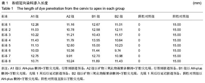
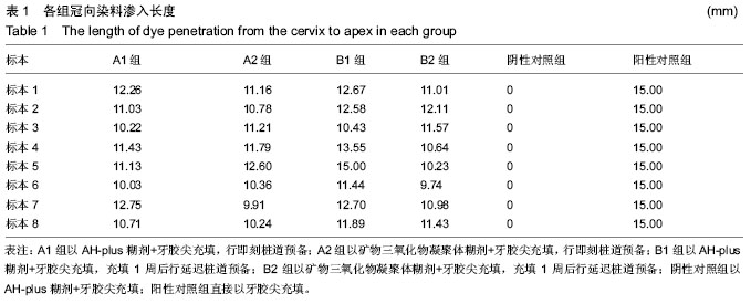
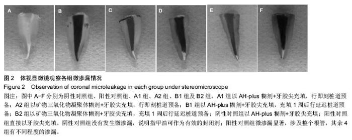
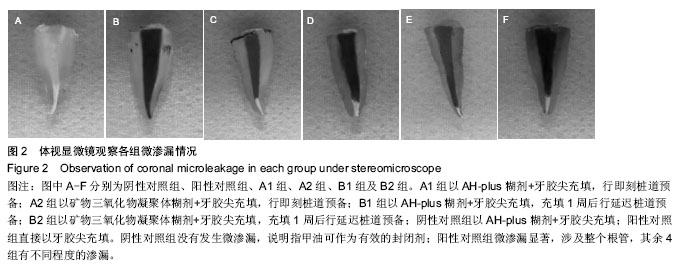
| [1] 章润贞,夏荣,冀章章.四种暂封材料对根管治疗后冠方微渗漏的影响[J].中国组织工程研究, 2015,19(3):405-409.[2] 张海龙.陈喜波.白红艳,等.3种糊剂在离体牙热牙胶填充中根尖封闭效果的观察[J].中国现代医学杂志, 2015, 25(28):86-89.[3] 仇珺,曹海炜,田宇,等.两种方法比较3种根管封闭剂体外抗菌效果的研究[J].牙体牙髓牙周病学杂志, 2015,25(4): 238-243.[4] 伍廷芸,黄扬睿,朱友家,等.不同根充材料对于根尖横剖面封闭性能的效果评价[J].武汉大学学报(医学版), 2015, 35(1):135-139.[5] Lodiene G,Kleivmyr M,Bruzell E.Sealing ability of mineral trioxide aggregate, glass ionomer cement and composite resin when repairing large furcal perforation. Br Dent J,2011;3(12):1-6.[6] Raji VS,Karunakar P,Madhavi N.Mineral trioxide aggregate in management of immature teeth with open apices a report of clinical cases.J Pierre Fauchard Acad. 2013;27(1):2-8.[7] 李淼,关朕,赵坤.矿物三氧化物凝聚体与玻璃离子修补髓室底穿孔的系统评价[J].中国现代医学杂志, 2015,25(11): 67-71.[8] Garip H,Garip Y,Oruço?lu H,et al.Effect of the angle of apical resection on apical leakage, measured with a computerized fluid filtration device.Oral Surg Oral Med Oral Pathol Oral RadiolEndod. 2011;111 (3): e50-54.[9] Lodiene G,Kleivmyr M,Bruzell E,et al.Sealing ability of mineral trioxide aggregate, glass ionomer cement and composite resin when repairing large furcal perforations. Br Dent J.2011;210(5):E7.[10] Vasconcelos BC,Bernardes RA,Duarte MA.Apical sealing of root canal fillings performed with five different endodontic sealers: analysis by fluid filtration. J Appl Oral Sci. 2011; 19(4):324-328.[11] Sarkar NK,Caicedo R,Ritwik P,et al.Physicochemical basis of the biologic properties of mineral trioxide aggregate.J Endod.2005;31(2):97-100.[12] Pappen AF,Bravo M,Gonzalez-Lopez S,et al.An in vitro study of coronal leakage after intraradicular preparation of cast-dowel space.J Prosthet Dent. 2005; 94(3):214-218.[13] Kazem M,Mahjour F,Dianat O,et al.Root-end filling with cement-based materials: An in vitro analysis of bacterial and dye microleakage.Dent Res J (Isfahan). 2013;10(1):46-51. [14] Goldman M,Simmonds S,Rush R.The usefulness of dye-penetration studies reexamined.Oral Surg Oral Med Oral Pathol.1989;67(3):327-332.[15] Madison S,Zakariasen KL.Linear and volumetric analysis of apical leakage in teeth prepared for posts.J Endod.1984;10(9):422-427.[16] Abramovitz I,Tagger M,Tamse A,et al.The effect of immediate vs. delayed post space preparation on the apical seal of a root canal filling: a study in an increased-sensitivity pressure-driven system.J Endod. 2000;26(8):435-439.[17] Rossomando KJ,Wendt SL Jr.Thermocycling and dwell times in microleakage evaluation for bonded restorations. Dent Mater.1995;11(1):47-51.[18] De Munck J,Van Landuyt K,Peumans M,et al.A critical review of the durability of adhesion to tooth tissue: methods and results.J Dent Res.2005;84(2):118-132.[19] Chen G,Chang YC.Effect of immediate and delayed post space preparation on apical leakage using three root canal obturation techniques after rotary instrumentation. J Formos Med Assoc. 2011;110(7): 454-549.[20] Bodrumlu E,Tunga U,Alaçam T.Influence of immediate and delayed post space preparation on sealing ability of resilon. Oral Surg Oral Med Oral Pathol Oral Radiol Endod.2007;103(6):e61-64.[21] 肖月,于佳,王健平,等.桩道及桩核不同时间差预备对根充材料封闭根方微渗漏的影响[J].中国组织工程研究, 2012, 16(29):5366-5370. [22] Jainaen A,Palamara JE,Messer HH.Push-out bond strengths of the dentine-sealer interface with and without a main cone.IntEndod J.2007;40(11):882-890.[23] 仇珺,曹海炜,田宇,等.两种方法比较3种根管封闭剂体外抗菌效果的研究[J].牙体牙髓牙周病学杂志, 2015,25(4): 238-243.[24] 王建平,Geogi George.纳米羟基磷灰石复合根充糊剂的抑菌性及微渗漏[J].中国组织工程研究, 2014,18(21): 3350-3354.[25] Prado CJ,Estrela C,Panzeri H,et al.Permeability of remaining endodontic obturation after post preparation. Gen Dent.2006;54(1):41-43.[26] Kwan EH,Harrington GW.The effect of immediate post preparation on apical seal. J Endod. 1981;7(7): 325-329.[27] Nawal RR,Parande M,Sehgal R,et al.A comparative evaluation of antimicrobial efficacy and flow properties for Epiphany, Guttaflow and AH-Plus sealer.Int Endod J.2011;44(4):307-313.[28] Rosen E,Goldberger T,Taschieri S,et al.The Prognosis of Altered Sensation after Extrusion of Root Canal Filling Materials: A Systematic Review of the Literature.J Endod.2016.pii: S0099-2399(16)30107-8. doi: 10.1016/j.joen.2016.03.018. [Epub ahead of print][29] Zarei M,Javidi M,Kazemi Z,et al.In Vitro Evaluation of Apical Sealing Ability of HEROfill® Obturator Versus Cold Lateral Condensation in Curved Root Canals.J Dent (Tehran). 2015;12(8):599-606.[30] Espir CG,Guerreiro-Tanomaru JM,Spin-Neto R,et al.Solubility and bacterial sealing ability of MTA and root-end filling materials.J Appl Oral Sci. 2016;24(2): 121-125. [31] Godlewski B,Klauz G,Czepko R.Thoracic Nerve Root Schwannoma Filling the Spinal Canal Almost Entirely Without any Neurological Deficits.Anesth Pain Med. 2016 ;6(1):e33886. [32] Christopher SR,Mathai V,Nair RS,et al.The effect of three different antioxidants on the dentinal tubular penetration of Resilon and Real Seal SE on sodium hypochlorite-treated root canal dentin: An in vitro study.J Conserv Dent.2016;19(2):161-165.[33] Segato RA,Pucinelli CM,Ferreira DC,et al. Physicochemical Properties of Root Canal Filling Materials for Primary Teeth.Braz Dent J. 2016;27(2): 196-201. [34] Can ED,Kele? A,Aslan B.Evaluation of the root filling quality of three root canal filling systems with micro-CT.Int Endod J.2016.doi: 10.1111/iej.12644. [Epub ahead of print][35] Lambor RT,de Ataide Ide N,Chalakkal P,et al.An in vitro comparison between the apical sealing abilities of resilon with Epiphany sealer and gutta-percha with AH plus sealer.Indian J Dent Res.2012;23(5):694.[36] Sarkar NK,Caicedo R,Ritwik P,et al.Physicochemical basis of the biologic properties of mineral trioxide aggregate. J Endod.2005;31(2):97-100. |