Chinese Journal of Tissue Engineering Research ›› 2016, Vol. 20 ›› Issue (25): 3738-3743.doi: 10.3969/j.issn.2095-4344.2016.25.014
Previous Articles Next Articles
Tissue-engineered acellular dermis repairs a skin ulcer: its preparation and healing mechanism
Du Peng, Guan Zhen, Wang Xiao-chuan
- Affiliated Hospital of Kunming University of Science and Technology (First People’s Hospital of Yunnan & Kunhua Hospital), Kunming 650001, Yunnan Province, China
-
Received:2016-03-29Online:2016-06-17Published:2016-06-17 -
About author:Du Peng, Master, Attending physician, Affiliated Hospital of Kunming University of Science and Technology (First People’s Hospital of Yunnan & Kunhua Hospital), Kunming 650001, Yunnan Province, China Guan Zhen, Studying for master’s degree, Affiliated Hospital of Kunming University of Science and Technology (First People’s Hospital of Yunnan & Kunhua Hospital), Kunming 650001, Yunnan Province, China Du Peng and Guan Zhen contributed equally to this work. -
Supported by:the Fund Project of Science Academy of Yuannan Provincial Education Commission, No. 20144C004Y
CLC Number:
Cite this article
Du Peng, Guan Zhen, Wang Xiao-chuan. Tissue-engineered acellular dermis repairs a skin ulcer: its preparation and healing mechanism[J]. Chinese Journal of Tissue Engineering Research, 2016, 20(25): 3738-3743.
share this article
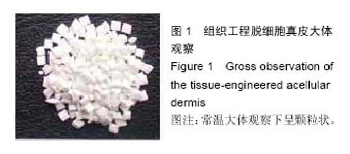
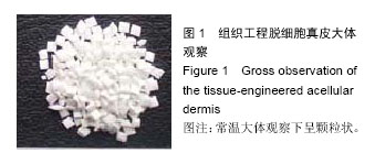
2.1 实验动物数量分析 实验选用SD大鼠16只,分为2组,实验过程无脱失,全部进入结果分析。 2.2 组织工程脱细胞真皮大体观察 获得的组织工程脱细胞真皮为白色状,常温大体观察呈颗粒状,见图1。"
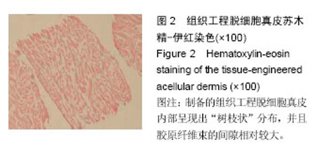
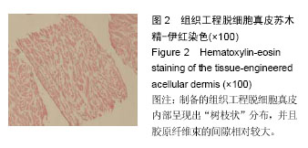
2.3 组织工程脱细胞真皮苏木精-伊红染色 结果显示:制备的组织工程脱细胞真皮内部呈现出“树枝状”分布,并且胶原纤维束的间隙相对较大,见图2。"
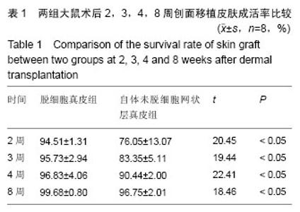
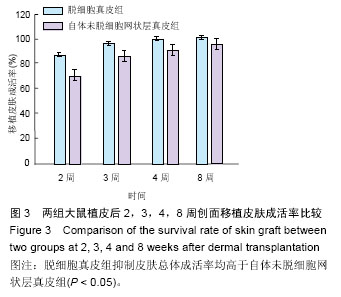
2.4 两组大鼠植皮后2,3,4,8周创面移植皮肤成活率比较 脱细胞真皮组术后2,3,4,8周创面移植皮肤成活率,显著高于自体未脱细胞网状层真皮组(P < 0.05),见表1,图3。"

"
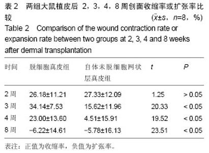
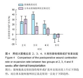
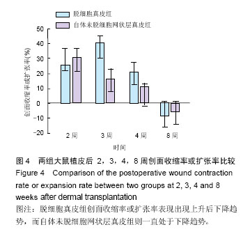
2.5 两组大鼠植皮后2,3,4,8周创面收缩率或扩张率比较 两组大鼠术后2周创面收缩率差异无显著性意义(P > 0.05);实验组3,4,8周创面收缩率或扩展率,显著高于自体未脱细胞网状层真皮组(P < 0.05),见表2,图4。"

"
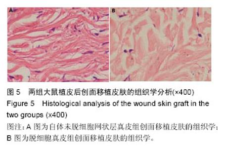
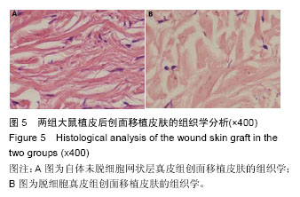
2.6 两组大鼠植皮后创面移植皮肤的组织学分析 脱细胞真皮组大鼠修复后组织呈现条索状,“树枝样”纵横交叉分布,而自体未脱细胞网状层真皮组则呈现均质 化改变,见图5。 2.7 两组大鼠创周正常皮肤胶原纤维束平均直径及间隙率比较 脱细胞真皮组创周正常皮肤胶原纤维束平均直径[(9.61±0.82) μm]及间隙率[(23.82±5.00)%],显著高于自体未脱细胞网状层真皮组胶原纤维束平均直径[(7.26±1.39) μm]和间隙率[(16.94±4.17)%],差异有显著性意义(t=22.43,15.25,均P < 0.05)。"
| [1] Jiang Y, Huang S, Fu X, et al. Epidemiology of chronic cutaneous wounds in China.Wound Repair Regen. 2011;19(2):181-188.[2] 王学锋,崔向琴,刘怀英.三种药物外敷治疗放疗所致皮肤损伤的疗效观察[J].实用心脑肺血管病杂志, 2011,19(7): 1172.[3] Dionyssiou D, Demiri E, Foroglou P, et al.The effectiveness of intralesional injection of platelet-rich plasma in accelerating the healing of chronic ulcers: an experimental and clinical study.Int Wound J.2013;10(4): 397-406.[4] 王若峥,吾甫尔,王多明,等. 比亚芬防治鼻咽癌同期调强放疗和化疗放射性皮肤损伤的疗效观察[J].中国肿瘤临床, 2007, 34(20):1181-1184.[5] Veves A, Falanga V, Armstrong DG, et al.Graftskin, a human skin equivalent, is effective in the management of noninfected neuropathic diabetic footulcers: a prospective randomized multicenter clinical trial. Diabetes Care.2001;24(2):290-295.[6] 俸瑞发,秦琴,黄明,等. 乳腺癌术后胸壁巨大放射性溃疡的多皮瓣联合修复[J].中华乳腺病杂志,2009, 3 (6): 679-683.[7] Azari O, Babaei H, Derakhshanfar A, et al. Effects of transplanted mesenchymal stem cells isolated from Wharton's jelly of caprine umbilical cord oncutaneous wound hea-ling; histopathological evaluation. Vet Res Commun.2011;35(4);211-222.[8] Skardal A, Mack D, Kapetanovic E, et al. Bioprinted amniotic fluid-derived stem cells accelerate healing of large skin wounds.Stem Cells Transl Med.2012;1 (11): 792-802.[9] Kim JW, Lee JH, Lyoo YS, et al. The effects of topical mesenchymal stem cell transplantation in canine experimental cutaneous wounds. Vet Dermatol. 2013;24(2):242-e53.[10] Maharlooei MK, Bagheri M, Solhjou Z, et al. Adipose tissue derived mesenchymal stem cell(AD-MSC) promotes skin wound healing in diabetic rats.Diabetes Res Clin Pract.2011;93(2):228-234.[11] 王明勇,黄华英,孙晓磊,等.乳猪提取物对糖尿病大鼠创面的影响[J].中国医药导报,2012,14(11):1966-1968.[12] 赛佳明,张慧琴.腺相关病毒分别介导转化生长因子β1和血管内皮生长因子联合转染促进糖尿病皮肤溃疡愈合的生物学效应[J].创伤外科杂志, 2007, 9(5):450-454.[13] Huang SP, Hsu CC, Chang SC, et al. Adipose-derived stem cells seeded on acellular dermal matrix grafts enhance wound healing in a murine model of a full-thickness defect.J Ann Plast Surg.2012;69(6): 656-662.[14] Kim SW, Zhang HZ, Guo L, et al.Amniotic mesenchymal stem cells enhance wound healing in diabetic NOD / SCID mice through high angiogenic and engraftment capabilities.PLoS One.2012;7(7): e41105.[15] 李琳,冉林晋.鞣酸软膏预防重症患者臀部腌红的效果观察[J].中国误诊学杂志, 2012,12(4):811.[16] Kong P, Xie X, Li F, et al. Placenta mesenchymal stem cell accelerates wound healing by enhancing angiogenesis in diabetic Goto- Kakizaki(GK) rats.Biochem BiophysRes Commun.2013;438(2): 410-419.[17] 曾龙英.3M无痛皮肤保护膜预防大便失禁患者肛周湿疹的效果[J].中华现代护理杂志,2011,17(28):3440-3441.[18] Kuo YR, Wang CT, Cheng JT, et al. Bone marrow-derived mesenchymal stem cells enhanced diabetic wound healing through recruitment of tissue regeneration in a rat model of streptozotocin-induced diabetes.Plast Reconstr Surg.2011;128( 4):872-880.[19] O'Loughlin A, Kulkarni M, Creane M, et al.Topical administration of allogeneic mesenchymal stromal cells seeded in a collagen scaffold augments wound healing and increases angiogenesis in the diabetic rabbit ulcer.Diabetes.2013;62(7):2588-2594.[20] 张娜,吴娟.失禁相关性皮炎的护理研究进展[J].中华护理杂志,2012,47(11):1066-1069.[21] Ngo MD, Aberman HM, Hawes ML, et al. Evaluation of human acellular dermis versus porcine acellular dermis in an in vivo model for incisional hernia repair. Cell Tissue Bank.2011;12 (2):135-145.[22] 肖强,张盈帆,潘银根,等.异种脱细胞真皮基质作为软组织填充物的生物相容性研究[J].中国美容整形外科杂志, 2009, 20(4):236-239.[23] Garcia O, Scott JR.Analysis of acellular dermal matrix integration and revascularization following tissue expander breast reconstruction in a clinically relevant large-animal model .Plast Reconstr Surg.2013;131(5): 741e-751e.[24] 孟祥海,王晓林,李学拥,等.复合皮移植修复烧伤功能部位创面疗效评价[J].中国修复重建外科杂志,2012, 26(2): 219-222.[25] 左海斌. 新型颗粒状脱细胞真皮的制备、表征及其在创面修复中的应用[D]. 第三军医大学,2011.[26] Shi LJ, Wang Y, Yang C, et al.Application of acellular dermal matrix in reconstruction of oral mucosal defects in 36 cases. J Oral Maxillofac Surg.2012;70 (11):e586-591.[27] Luo W, Zheng X, Chen L, et al.The use of human acellular dermal matrix in the prevention of infra -auricular depressed deformities and Frey's syn drome following total parotidectomy. Oral Surg Oral Med Oral Pathol Oral Radiol.2012;114 (2) : e9-13.[28] 张庆泉,孙岩,王强,等.异种(牛)脱细胞真皮基质修复咽喉肿瘤切除术后黏膜缺损的效果评估[J].中国组织工程研究与临床康复,2008,12(6):1081-1084.[29] 卫红梅, 朱国际.过敏性紫癜患儿血清补体水平变化的临床研究[J].中国血液流变学杂志,2009,19(2): 279-280.[30] 张洁, 周怡, 周晓荣, 等.PCPA对于外周血CD4+T细胞Thl/Th2分化失衡影响的初步研究[J]. 免疫学杂志,2011, 27(8):645-648, 661.[31] Ru L, Abudouhaer A, Guo YF, Clinical significance of serumlevels of IGF-1 and IGFBP-3 in children with Henoch-Schonlein purpura or Henoch- Schonlein purpura nephritis. JZhongguo Dang Dai Er Ke Za Zhi. 2013; 15(11): 1009-1013.[32] 常红, 张秋业, 程娜.过敏性紫癜患儿外周血单个核细胞TLR2、TLR4表达及其与Th1和Th2型免疫应答相关性观察[J]. 中华微生物学和免疫学杂志, 2013, 33(11): 839-844.[33] 高辉香, 田玲玲, 常红.过敏性紫癜患儿外周血单核细胞TLR2、TLR4、TLR6及与Treg相关细胞因子的表达[J].中国组织工程研究,2014,18(38):6222-6227.[34] 彭丽, 常桂芬, 宋丽君.过敏性紫癜患儿食物不耐受检测及其意义[J].中国实验诊断学, 2011, 15(2):285-287.[35] Govaere E, Van Gysel D, Verhamme KM, et al. The association of allergic symptoms with sensitization to inhalant allergens in childhood. J Pediatr Allergy Immunol.2009;20(5): 448-457.[36] Calvo V, Loricera J, Martin L, et al. Henoch - Schonlein pur-pura nephritis and IgA nephropathy: a comparative clinical study.J Clinical and Experimental Rheumatology. 2013;31(75): S45-51. |
| [1] | Zhang Tongtong, Wang Zhonghua, Wen Jie, Song Yuxin, Liu Lin. Application of three-dimensional printing model in surgical resection and reconstruction of cervical tumor [J]. Chinese Journal of Tissue Engineering Research, 2021, 25(9): 1335-1339. |
| [2] | Jiang Hongying, Zhu Liang, Yu Xi, Huang Jing, Xiang Xiaona, Lan Zhengyan, He Hongchen. Effect of platelet-rich plasma on pressure ulcers after spinal cord injury [J]. Chinese Journal of Tissue Engineering Research, 2021, 25(8): 1149-1153. |
| [3] | Li Jing, Xie Jianshan, Cui Huilin, Cao Ximei, Yang Yanping, Li Hairong. Expression and localization of diacylglycerol kinase zeta and protein kinase C beta II in mouse back skin with different coat colors [J]. Chinese Journal of Tissue Engineering Research, 2021, 25(8): 1196-1200. |
| [4] | Zeng Yanhua, Hao Yanlei. In vitro culture and purification of Schwann cells: a systematic review [J]. Chinese Journal of Tissue Engineering Research, 2021, 25(7): 1135-1141. |
| [5] | Ma Binxiang, He Wanqing, Zhou Guangchao, Guan Yonglin. Triptolide improves motor dysfunction in rats following spinal cord injury [J]. Chinese Journal of Tissue Engineering Research, 2021, 25(5): 701-706. |
| [6] | Xu Dongzi, Zhang Ting, Ouyang Zhaolian. The global competitive situation of cardiac tissue engineering based on patent analysis [J]. Chinese Journal of Tissue Engineering Research, 2021, 25(5): 807-812. |
| [7] | Wu Zijian, Hu Zhaoduan, Xie Youqiong, Wang Feng, Li Jia, Li Bocun, Cai Guowei, Peng Rui. Three-dimensional printing technology and bone tissue engineering research: literature metrology and visual analysis of research hotspots [J]. Chinese Journal of Tissue Engineering Research, 2021, 25(4): 564-569. |
| [8] | Chang Wenliao, Zhao Jie, Sun Xiaoliang, Wang Kun, Wu Guofeng, Zhou Jian, Li Shuxiang, Sun Han. Material selection, theoretical design and biomimetic function of artificial periosteum [J]. Chinese Journal of Tissue Engineering Research, 2021, 25(4): 600-606. |
| [9] | Liu Fei, Cui Yutao, Liu He. Advantages and problems of local antibiotic delivery system in the treatment of osteomyelitis [J]. Chinese Journal of Tissue Engineering Research, 2021, 25(4): 614-620. |
| [10] | Li Xiaozhuang, Duan Hao, Wang Weizhou, Tang Zhihong, Wang Yanghao, He Fei. Application of bone tissue engineering materials in the treatment of bone defect diseases in vivo [J]. Chinese Journal of Tissue Engineering Research, 2021, 25(4): 626-631. |
| [11] | Zhang Zhenkun, Li Zhe, Li Ya, Wang Yingying, Wang Yaping, Zhou Xinkui, Ma Shanshan, Guan Fangxia. Application of alginate based hydrogels/dressings in wound healing: sustained, dynamic and sequential release [J]. Chinese Journal of Tissue Engineering Research, 2021, 25(4): 638-643. |
| [12] | Chen Jiana, Qiu Yanling, Nie Minhai, Liu Xuqian. Tissue engineering scaffolds in repairing oral and maxillofacial soft tissue defects [J]. Chinese Journal of Tissue Engineering Research, 2021, 25(4): 644-650. |
| [13] | Xing Hao, Zhang Yonghong, Wang Dong. Advantages and disadvantages of repairing large-segment bone defect [J]. Chinese Journal of Tissue Engineering Research, 2021, 25(3): 426-430. |
| [14] | Zhang Lishu, Liu Anqi, He Xiaoning, Jin Yan, Li Bei, Jin Fang. Alpl gene affects the therapeutic effect of bone marrow mesenchymal stem cells on ulcerative colitis [J]. Chinese Journal of Tissue Engineering Research, 2021, 25(25): 3970-3975. |
| [15] | Xu Xiaoming, Chen Yan, Song Qian, Yuan Lu, Gu Jiaming, Zhang Lijuan, Geng Jie, Dong Jian. Human placenta derived mesenchymal stem cell gel promotes the healing of radiation skin damage in SD rats [J]. Chinese Journal of Tissue Engineering Research, 2021, 25(25): 3976-3980. |
| Viewed | ||||||
|
Full text |
|
|||||
|
Abstract |
|
|||||