Chinese Journal of Tissue Engineering Research ›› 2015, Vol. 19 ›› Issue (38): 6081-6086.doi: 10.3969/j.issn.2095-4344.2015.38.003

Previous Articles     Next Articles

Preparation and characterization of inducible bone repair composite scaffold with genipin-crosslinked bionic bone structure

Li Gen1, 2, Li Wen-jie2, Tang Jun-jie2, Wang Jiu-na2, Zhao Ling2, Qin Wen2, Zhao Xing-xu1, Zhao Hong-bin1, 2     

  1. 1College of Life Science and Technology, Gansu Agricultural University, Lanzhou 730070, Gansu Province, China;
    2General Hospital of Lanzhou Military Command, Lanzhou 730050, Gansu Province, China
  • Online:2015-09-17 Published:2015-09-17
  • Contact: Zhao Hong-bin, Associate chief technician, General Hospital of Lanzhou Military Command, Lanzhou 730050, Gansu Province, China
  • About author:Li Gen, Studying for master’s degree, College of Life Science and Technology, Gansu Agricultural University, Lanzhou 730070, Gansu Province, China; General Hospital of Lanzhou Military Command, Lanzhou 730050, Gansu Province, China
  • Supported by:

    the Science and Technology Project of Gansu Province, No. 1203FKDA036

Abstract:

BACKGROUND: Although there is a certain progress in the preparation of tissue-engineered bone tissue using a variety of materials, some deficiencies have appeared such as mismatching between scaffold degradation rate and new bone formation rate, slow tissue growth, toxic metabolites.

OBJECTIVE: To build a new type of inducible bone repair composite scaffold with bionic bone structurematerials and to evaluate its physicochemical and biological properties.

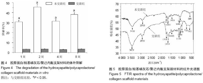
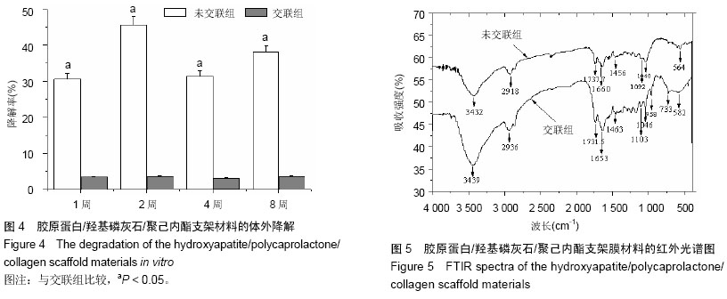
METHODS: Icariin encapsulated by chitosan was used to prepare drug-loaded microspheres, and the drug release rate of the microspheres was detected. Chitosan microspheres were mixed with collagen to build the core part of scaffold materials. Hydroxyapatite (HA), polycaprolactone (PCL) and collagen were mixed in hexafluoride isopropanol (HFIP) to prepare the HA/PCL/collagen outer part of composite scaffold material at the rate of 0:3:3, 1:3:3, 2:3:3, 3:3:3. Each proportional electrospinning was used for one layer, and finally the 4-layer outer tube of the scaffold was produced. The tube core and outer tube were crosslinked by 1% genipin. Universal material testing machine, surface contact angle meter, infrared spectroscopy, scanning electron microscope, water absorption, permeability, porosity, in vitro degradation tests for cross-linked and uncross-linked were used to 
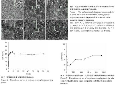
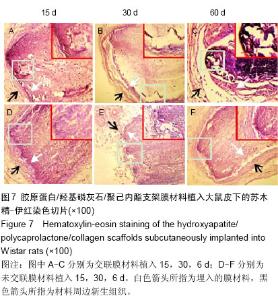
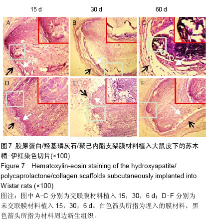
observe the structure and characteristics of tubular materials. Bone marrow mesenchymal stem cells were seeded on the surface of cross-linked and uncross-linked bone repair materials to evaluate the biocompatibility of the scaffolds. Cross-linked and uncross-linked bone repair materials were implanted subcutaneously into Wistar rats to evaluate the histocompatibility of the scaffolds.

RESULTS AND CONCLUSION: The drug in the scaffold had a suitable release; the bone scaffold material had good uniformity, and cross-linked scaffolds materials had better mechanical properties, water absorption and permeability than the uncross-linked (P < 0.05). The degradation rate of the cross-linked group was significantly lower than that of the uncross-linked group (P < 0.05). Hematoxylin-eosin staining showed that the bone marrow mesenchymal stem cells could adhere well to the cross-linked and uncross-linked materials. No inflammatory reactions occurred after subcutaneous implantation of cross-linked and uncross-linked materials. These findings indicate that the cross-linked scaffold for inducible bone tissue engineering has good biocompatibility and mechanical properties.

中国组织工程研究杂志出版内容重点:生物材料;骨生物材料; 口腔生物材料; 纳米材料; 缓释材料; 材料相容性;组织工程

Key words: Durapatite, Collagen, Cross-Linking Reagents

CLC Number: