Chinese Journal of Tissue Engineering Research ›› 2015, Vol. 19 ›› Issue (1): 18-23.doi: 10.3969/j.issn.2095-4344.2015.01.004
Previous Articles Next Articles
Age-associated proliferation and differentiation changes of rat bone marrow mesenchymal stem cells
Li Hai-rui, Zheng Dong, Jiang Can, Guo Jun, Zhang Ai-dong, Li Zi-cheng
- Department of Cardiology, the First Affiliated Hospital of Jinan University, Guangzhou 510630, Guangdong Province, China
-
Revised:2014-11-20Online:2015-01-01Published:2015-01-01 -
Contact:Guo Jun, M.D., Associate chief physician, Associate professor, Department of Cardiology, the First Affiliated Hospital of Jinan University, Guangzhou 510630, Guangdong Province, China -
About author:Li Hai-rui, Studying for master’s degree, Department of Cardiology, the First Affiliated Hospital of Jinan University, Guangzhou 510630, Guangdong Province, China -
Supported by:the National Natural Science Foundation of China for the Youth, No. 81100078; the Key Science Research Project of the Ministry of Education, No. 211207; the Zhujiang Scientific New Star Plan of Guangzhou, No. 2012J2200063; the Doctoral Startup Program of Guangdong Science and Technology Commission, No. S2011040001392
CLC Number:
Cite this article
Li Hai-rui, Zheng Dong, Jiang Can, Guo Jun, Zhang Ai-dong, Li Zi-cheng. Age-associated proliferation and differentiation changes of rat bone marrow mesenchymal stem cells[J]. Chinese Journal of Tissue Engineering Research, 2015, 19(1): 18-23.
share this article
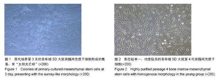
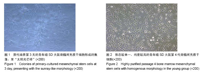
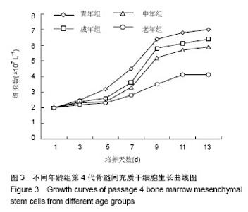
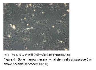
2.1 观察骨髓间充质干细胞生长情况 原代细胞接种后24 h换液换掉大部分非贴壁细胞,倒置相差显微镜下可见稀少的呈长梭形的贴壁细胞。3 d后梭形贴壁细胞开始增多,集落周围的贴壁细胞逐渐开始融合(图1),进入对数增长期,8-12 d达到80%的融合,10-12 d后基本进入细胞生长的平台期。 传代细胞增殖更加迅速,8-10 d即可长满整个皿底。随着换液与传代的次数增加,骨髓间充质干细胞逐渐得到纯化,表现为圆形和不规则形细胞明显减少,三四代后基本除尽,镜下可见以梭形为主,大小均一的贴壁骨髓间充质干细胞(图2)。 青年组大鼠的骨髓间充质干细胞增殖能力强于对照组,具体表现为无论是原代或是传代细胞的贴壁时间均缩短,细胞集落形成早且集落较多,细胞形态较规则,形成细胞单层的时间缩短,每六七天即可传代1次,从第4代骨髓间充质干细胞生长曲线来看,青年组较其他组骨髓间充质干细胞提前2 d左右进入对数生长期,老年组第4代骨髓间充质干细胞对数生长期表现相对不甚明显,进入平台期的细胞数量亦随年龄的增加而降低(图3),并且细胞体形较大,部分细胞呈不规则形,经过6代以后的长期传代容易老化(图4)。"
"


"
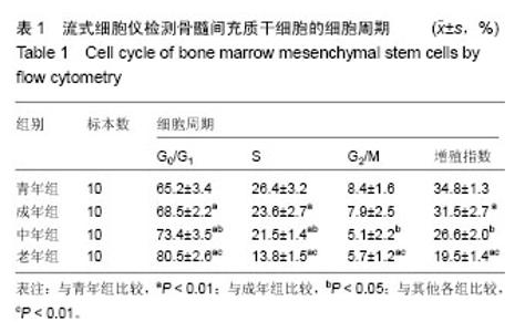
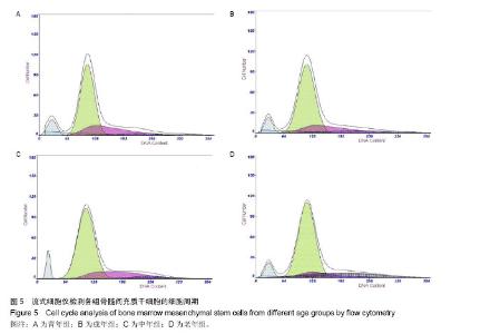
2.2 骨髓间充质干细胞的细胞周期检测结果 与其他各组比较,老年组骨髓间充质干细胞的G0/G1期细胞百分率显著升高(P < 0.01),S期、G2/M期细胞百分率及增殖指数较其他各组显著降低(P < 0.01);总的说来,骨髓间充质干细胞处于G0/G1期的细胞百分率随年龄的增加而增加,增殖指数随年龄的增加而减少(P < 0.01),见表1,图5。"

"
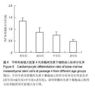
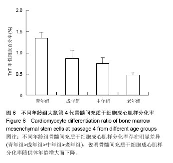
2.3 混合培养后骨髓间充质干细胞向心肌细胞分化的测定 DAPI的激发光波长为358 nm,发射光波长为461 nm,荧光激发下发蓝光;TRITC的激发光波长为550 nm,发射光波长为570 nm,荧光激发下发红光,两者的激发光谱并不重合。通过在同一视野下转换激发光,查找有TnT表达的DAPI标记的骨髓间充质干细胞,镜下可见部分蓝色细胞核周围的胞浆发出红色荧光。高倍镜下,部分发生向心肌细胞分化的骨髓间充质干细胞可见较清楚的心肌细胞的肌丝结构,有的可见较清楚的心肌细胞特征性横纹结构;这些心肌细胞特异性蛋白表达阳性的骨髓间充质干细胞均与相邻心肌细胞直接接触。 在混合培养的第6天青年组骨髓间充质干细胞的TnT阳性率为(1.35±0.14)%,显著高于其他组(P < 0.01),成年组与中年组TnT阳性率差异无显著性意义(P > 0.05),老年组骨髓间充质干细胞的TnT阳性率为(0.47±0.08)%,显著低于其他各组,差异有显著性意义(P < 0.01)。总的来看,各组骨髓间充质干细胞的成心肌样细胞的分化率随年龄的增长而减少(图6)。"
| [1] Ma Z, Liu Q, Liu H,et al.Cardiogenic Regulation of Stem-Cell Electrical Properties in a Laser-Patterned Biochip.Cell Mol Bioeng. 2012;5(3):327-336. [2] Ma Z, Liu Q, Liu H,et al.Laser-patterned stem-cell bridges in a cardiac muscle model for on-chip electrical conductivity analyses. Lab Chip. 2012;12(3):566-573. [3] Peters NS, Wit AL.Gap junction remodeling in infarction: does it play a role in arrhythmogenesis.J Cardiovasc Electrophysiol. 2000;11(4):488-490. [4] Qi BD, Meng BX, Yang Y,et al.Study of immuno-tolerance mechanism of the third-party bone marrow-derived mesenchymal stem cells in allogenic transplantation. Zhonghua Zheng Xing Wai Ke Za Zhi. 2011;27(3):207-212. [5] Wang Y, Liu J, Xu C,et al.Bone marrow transplantation combined with mesenchymal stem cells induces immune tolerance without cytotoxic conditioning.J Surg Res. 2011; 171(1):e123-131. [6] Pan H, Zhao K, Wang L,et al.Mesenchymal stem cells enhance the induction of mixed chimerism and tolerance to rat hind-limb allografts after bone marrow transplantation.J Surg Res. 2010;160(2):315-324. [7] Pittenger MF, Mackay AM, Beck SC,et al.Multilineage potential of adult human mesenchymal stem cells.Science. 1999;284(5411):143-147. [8] Qayyum AA, Mathiasen AB, Kastrup J.Stem cell therapy to treat heart ischaemia: implications for diabetes cardiovascular complications.Curr Diab Rep. 2014;14(12):554. [9] Yamawaki-Ogata A, Hashizume R, Fu XM, et al. Mesenchymal stem cells for treatment of aortic aneurysms. World J Stem Cells. 2014;6(3):278-287. [10] Liu B, Duan CY, Luo CF,et al.Effectiveness and safety of selected bone marrow stem cells on left ventricular function in patients with acute myocardial infarction: A meta-analysis of randomized controlled trials.Int J Cardiol. 2014;177(3): 764-770. [11] Soleimani M, Nadri S.A protocol for isolation and culture of mesenchymal stem cells from mouse bone marrow.Nat Protoc. 2009;4(1):102-106. [12] Peng L, Jia Z, Yin X, et al.Comparative analysis of mesenchymal stem cells from bone marrow, cartilage, and adipose tissue.Stem Cells Dev. 2008;17(4):761-773. [13] Wang C, Xu Y, Song WG, et al.Isolation and culturation, phenotype detection of rat bone marrow mesenchymal stem cells.Xi Bao Yu Fen Zi Mian Yi Xue Za Zhi. 2007;23(5): 466-468. [14] Ehler E, Moore-Morris T, Lange S.Isolation and culture of neonatal mouse cardiomyocytes.J Vis Exp. 2013;(79): e50154. [15] Song YH, Pinkernell K, Alt E.Stem cell induced cardiac regeneration: fusion/mitochondrial exchange and/or transdifferentiation?Cell Cycle. 2011;10(14):2281-2286. [16] Makkar RR, Price MJ, Lill M,et al. Intramyocardial injection of allogenic bone marrow-derived mesenchymal stem cells without immunosuppression preserves cardiac function in a porcine model of myocardial infarction.J Cardiovasc Pharmacol Ther. 2005;10(4):225-233. [17] Yin N, Lu R, Lin J,et al.Islet-1 promotes the cardiac-specific differentiation of mesenchymal stem cells through the regulation of histone acetylation.Int J Mol Med. 2014;33(5): 1075-1082. [18] Hou J, Lü AL, Liu BW,et al.Combination of BMP-2 and 5-AZA is advantageous in rat bone marrow-derived mesenchymal stem cells differentiation into cardiomyocytes.Cell Biol Int. 2013;37(12):1291-1299. [19] Cheng Z, Ou L, Zhou X,et al.Targeted migration of mesenchymal stem cells modified with CXCR4 gene to infarcted myocardium improves cardiac performance.Mol Ther. 2008;16(3):571-579. [20] Cheng Z, Liu X, Ou L,et al.Mobilization of mesenchymal stem cells by granulocyte colony-stimulating factor in rats with acute myocardial infarction.Cardiovasc Drugs Ther. 2008; 22(5):363-371. [21] Ripa RS, Haack-Sørensen M, Wang Y, et al. Bone marrow derived mesenchymal cell mobilization by granulocyte-colony stimulating factor after acute myocardial infarction: results from the Stem Cells in Myocardial Infarction (STEMMI) trial.Circulation. 2007;116(11 Suppl):I24-30. [22] Rangappa S, Entwistle JW, Wechsler AS,et al. Cardiomyocyte-mediated contact programs human mesenchymal stem cells to express cardiogenic phenotype.J Thorac Cardiovasc Surg. 2003;126(1):124-132. [23] Fukuhara S, Tomita S, Yamashiro S,et al.Direct cell-cell interaction of cardiomyocytes is key for bone marrow stromal cells to go into cardiac lineage in vitro.J Thorac Cardiovasc Surg. 2003;125(6):1470-1480. [24] Qu Z, Xu H, Tian Y,et al.Atorvastatin improves microenvironment to enhance the beneficial effects of BMSCs therapy in a rabbit model of acute myocardial infarction.Cell Physiol Biochem. 2013;32(2):380-389. [25] Li X, Yu X, Lin Q,et al.Bone marrow mesenchymal stem cells differentiate into functional cardiac phenotypes by cardiac microenvironment.J Mol Cell Cardiol. 2007;42(2):295-303. [26] 郭军,贾光宏,林国生,等.体外模拟心肌微环境下骨髓间充质干细胞向心肌样细胞分化的实验研究[J].心脏杂志,2004,16(6): 501-504. [27] Stenderup K, Rosada C, Justesen J,et al.Aged human bone marrow stromal cells maintaining bone forming capacity in vivo evaluated using an improved method of visualization. Biogerontology. 2004;5(2):107-118. [28] Mueller SM, Glowacki J.Age-related decline in the osteogenic potential of human bone marrow cells cultured in three-dimensional collagen sponges.J Cell Biochem. 2001; 82(4):583-590. [29] Zaim M, Karaman S, Cetin G, et al.Donor age and long-term culture affect differentiation and proliferation of human bone marrow mesenchymal stem cells.Ann Hematol. 2012;91(8): 1175-1186. [30] Wu J, Niu J, Li X,et al.TGF-β1 induces senescence of bone marrow mesenchymal stem cells via increase of mitochondrial ROS production.BMC Dev Biol. 2014;14:21. [31] Lu Y, Liu JJ, Bi XY,et al.Pyridostigmine ameliorates cardiac remodeling induced by myocardial infarction via inhibition of the transforming growth factor-β1/TGF-β1-activated kinase pathway.J Cardiovasc Pharmacol. 2014;63(5):412-420. [32] Bischoff DS, Makhijani NS, Yamaguchi DT.Constitutive expression of human telomerase enhances the proliferation potential of human mesenchymal stem cells.Biores Open Access. 2012;1(6):273-279. [33] Madonna R, Taylor DA, Geng YJ,et al.Transplantation of mesenchymal cells rejuvenated by the overexpression of telomerase and myocardin promotes revascularization and tissue repair in a murine model of hindlimb ischemia.Circ Res. 2013;113(7):902-914. [34] Madonna R, Willerson JT, Geng YJ.Myocardin a enhances telomerase activities in adipose tissue mesenchymal cells and embryonic stem cells undergoing cardiovascular myogenic differentiation.Stem Cells. 2008;26(1):202-211. [35] Terada N, Hamazaki T, Oka M,et al.Bone marrow cells adopt the phenotype of other cells by spontaneous cell fusion. Nature. 2002;416(6880):542-545. [36] Haneef K, Naeem N, Khan I,et al.Conditioned medium enhances the fusion capability of rat bone marrow mesenchymal stem cells and cardiomyocytes.Mol Biol Rep. 2014;41(5):3099-3112. [37] Ying QL, Nichols J, Evans EP,et al.Changing potency by spontaneous fusion.Nature. 2002;416(6880):545-548. |
| [1] | Pu Rui, Chen Ziyang, Yuan Lingyan. Characteristics and effects of exosomes from different cell sources in cardioprotection [J]. Chinese Journal of Tissue Engineering Research, 2021, 25(在线): 1-. |
| [2] | Lin Qingfan, Xie Yixin, Chen Wanqing, Ye Zhenzhong, Chen Youfang. Human placenta-derived mesenchymal stem cell conditioned medium can upregulate BeWo cell viability and zonula occludens expression under hypoxia [J]. Chinese Journal of Tissue Engineering Research, 2021, 25(在线): 4970-4975. |
| [3] | Hou Jingying, Yu Menglei, Guo Tianzhu, Long Huibao, Wu Hao. Hypoxia preconditioning promotes bone marrow mesenchymal stem cells survival and vascularization through the activation of HIF-1α/MALAT1/VEGFA pathway [J]. Chinese Journal of Tissue Engineering Research, 2021, 25(7): 985-990. |
| [4] | Shi Yangyang, Qin Yingfei, Wu Fuling, He Xiao, Zhang Xuejing. Pretreatment of placental mesenchymal stem cells to prevent bronchiolitis in mice [J]. Chinese Journal of Tissue Engineering Research, 2021, 25(7): 991-995. |
| [5] | Liang Xueqi, Guo Lijiao, Chen Hejie, Wu Jie, Sun Yaqi, Xing Zhikun, Zou Hailiang, Chen Xueling, Wu Xiangwei. Alveolar echinococcosis protoscolices inhibits the differentiation of bone marrow mesenchymal stem cells into fibroblasts [J]. Chinese Journal of Tissue Engineering Research, 2021, 25(7): 996-1001. |
| [6] | Fan Quanbao, Luo Huina, Wang Bingyun, Chen Shengfeng, Cui Lianxu, Jiang Wenkang, Zhao Mingming, Wang Jingjing, Luo Dongzhang, Chen Zhisheng, Bai Yinshan, Liu Canying, Zhang Hui. Biological characteristics of canine adipose-derived mesenchymal stem cells cultured in hypoxia [J]. Chinese Journal of Tissue Engineering Research, 2021, 25(7): 1002-1007. |
| [7] | Geng Yao, Yin Zhiliang, Li Xingping, Xiao Dongqin, Hou Weiguang. Role of hsa-miRNA-223-3p in regulating osteogenic differentiation of human bone marrow mesenchymal stem cells [J]. Chinese Journal of Tissue Engineering Research, 2021, 25(7): 1008-1013. |
| [8] | Lun Zhigang, Jin Jing, Wang Tianyan, Li Aimin. Effect of peroxiredoxin 6 on proliferation and differentiation of bone marrow mesenchymal stem cells into neural lineage in vitro [J]. Chinese Journal of Tissue Engineering Research, 2021, 25(7): 1014-1018. |
| [9] | Zhu Xuefen, Huang Cheng, Ding Jian, Dai Yongping, Liu Yuanbing, Le Lixiang, Wang Liangliang, Yang Jiandong. Mechanism of bone marrow mesenchymal stem cells differentiation into functional neurons induced by glial cell line derived neurotrophic factor [J]. Chinese Journal of Tissue Engineering Research, 2021, 25(7): 1019-1025. |
| [10] | Duan Liyun, Cao Xiaocang. Human placenta mesenchymal stem cells-derived extracellular vesicles regulate collagen deposition in intestinal mucosa of mice with colitis [J]. Chinese Journal of Tissue Engineering Research, 2021, 25(7): 1026-1031. |
| [11] | Pei Lili, Sun Guicai, Wang Di. Salvianolic acid B inhibits oxidative damage of bone marrow mesenchymal stem cells and promotes differentiation into cardiomyocytes [J]. Chinese Journal of Tissue Engineering Research, 2021, 25(7): 1032-1036. |
| [12] | Li Cai, Zhao Ting, Tan Ge, Zheng Yulin, Zhang Ruonan, Wu Yan, Tang Junming. Platelet-derived growth factor-BB promotes proliferation, differentiation and migration of skeletal muscle myoblast [J]. Chinese Journal of Tissue Engineering Research, 2021, 25(7): 1050-1055. |
| [13] | Liu Cong, Liu Su. Molecular mechanism of miR-17-5p regulation of hypoxia inducible factor-1α mediated adipocyte differentiation and angiogenesis [J]. Chinese Journal of Tissue Engineering Research, 2021, 25(7): 1069-1074. |
| [14] | Wang Xianyao, Guan Yalin, Liu Zhongshan. Strategies for improving the therapeutic efficacy of mesenchymal stem cells in the treatment of nonhealing wounds [J]. Chinese Journal of Tissue Engineering Research, 2021, 25(7): 1081-1087. |
| [15] | Wang Shiqi, Zhang Jinsheng. Effects of Chinese medicine on proliferation, differentiation and aging of bone marrow mesenchymal stem cells regulating ischemia-hypoxia microenvironment [J]. Chinese Journal of Tissue Engineering Research, 2021, 25(7): 1129-1134. |
| Viewed | ||||||
|
Full text |
|
|||||
|
Abstract |
|
|||||