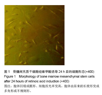
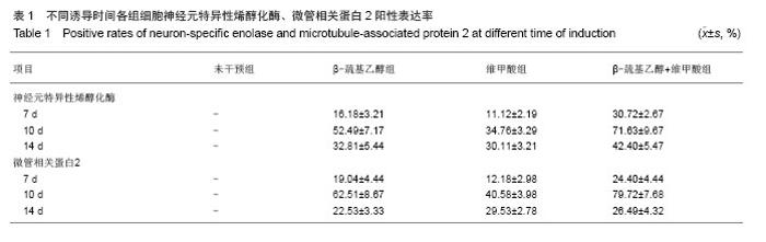
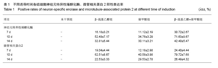
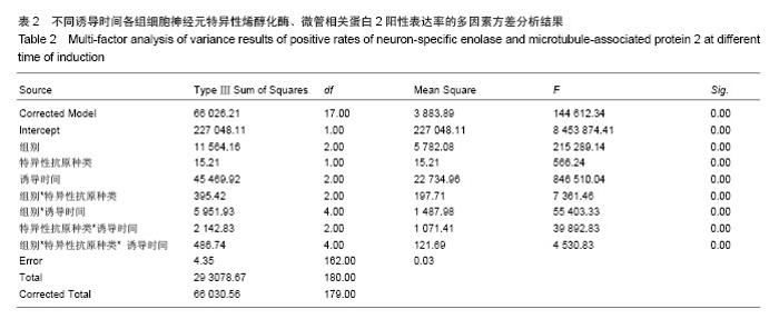
| [1]贾延劼,孙吉平,朱晓峰,等.Notch-1在骨髓间质干细胞分化为神经细胞中的作用[J].中华医学杂志,2006,86(41):2916-2921.
[2]李东正,王倩,李俊娜,等.骨髓间充质干细胞向神经细胞分化的研究[J].现代生物医学进展,2010,10(5):986-988.
[3]鞠文博,李淑波,周艳.β-巯基乙醇在大鼠骨髓基质细胞向神经干细胞诱导分化中的作用[J].中国组织工程研究与临床康复,2007, 11(20):3991-3994.
[4]程峰,郝怀勇,田和平.体外诱导骨髓间充质干细胞向神经细胞分化及凋亡的研究[J].南京医科大学学报:自然科学版,2010,30(1): 54-58.
[5]Wu J, Sun Z, Sun HS,et al. Intravenously administered bone marrow cells migrate to damaged brain tissue and improve neural function in ischemic rats.Cell Transplant. 2008;16(10): 993-1005.
[6]Wu QY, Li J, Feng ZT,et al. Bone marrow stromal cells of transgenic mice can improve the cognitive ability of an Alzheimer's disease rat model. Neurosci Lett. 2007;417(3): 281-285.
[7]Lu D, Mahmood A, Wang L,et al. Adult bone marrow stromal cells administered intravenously to rats after traumatic brain injury migrate into brain and improve neurological outcome. Neuroreport. 2001;12(3):559-563.
[8]Lu D, Li Y, Wang L,et al. Intraarterial administration of marrow stromal cells in a rat model of traumatic brain injury. J Neurotrauma. 2001;18(8):813-819.
[9]Kamada T, Koda M, Dezawa M,et al.Transplantation of bone marrow stromal cell-derived Schwann cells promotes axonal regeneration and functional recovery after complete transection of adult rat spinal cord. J Neuropathol Exp Neurol. 2005;64(1):37-45.
[10]Zhang P, He X, Liu K,et al. Bone marrow stromal cells differentiated into functional Schwann cells in injured rats sciatic nerve. Artif Cells Blood Substit Immobil Biotechnol. 2004;32(4):509-518.
[11]高平,孙占胜,王伯珉,等.骨髓间充质干细胞诱导成神经元样细胞移植治疗脊髓损伤[J].中国组织工程研究,2013,17(23): 4256- 4263.
[12]张瑞平,李健丁,刘强,等.蛛网膜下腔移植磁标骨髓间充质干细胞治疗脊髓损伤[J].中国组织工程研究,2013,17(1):74-78.
[13]Zhang C,He XJ,Li HP,et al.Chondroitinase ABC plus bone marrow mesenchymal stem cells for repair of spinal cord injury. Neural Regen Rese. 2013; 8(11): 965-974.
[14]Wu YX,Zhang JH,Ben XM.Neuronal-like cell differentiation of non-adherent bone marrow cell-derived mesenchymal stem cells. Neural Regen Rese. 2013, 8(22): 2078-2085. |