Chinese Journal of Tissue Engineering Research ›› 2014, Vol. 18 ›› Issue (33): 5334-5340.doi: 10.3969/j.issn.2095-4344.2014.33.015
Previous Articles Next Articles
Effects of proton pump inhibitor FR167356 on osseointegration of dental implant in osteoporosis rabbits
Zhang Peng1, Shi Wen-yi2, Guo Da-wei2, Yu Jiang-bo2, Song Ling2, Zhang Chun-yan2, Cao Yang2
- 1 College of Stomatology, Weifang Medical University, Weifang 261000, Shandong Province, China; 2 Department of Stomatology, Qingdao Municipal Hospital, Qingdao 266071, Shandong Province, China
-
Online:2014-08-13Published:2014-08-13 -
Contact:Guo Da-wei, Attending physician, Associate professor, Master’s supervisor, Department of Stomatology, Qingdao Municipal Hospital, Qingdao 266071, Shandong Province, China -
About author:Zhang Peng, Studying for master’s degree, College of Stomatology, Weifang Medical University, Weifang 261000, Shandong Province, China -
Supported by:Basic Research Project of Science and Technology of Qingdao, No. 12-1-4-16-(8)-jch; Health Science and Technology Project of Qingdao, No. 2012-WSZD005; Specialized President Funds by Qingdao Municipal Hospital, No. ZYZJJ-2013-W020-pei
CLC Number:
Cite this article
Zhang Peng, Shi Wen-yi, Guo Da-wei, Yu Jiang-bo, Song Ling, Zhang Chun-yan, Cao Yang. Effects of proton pump inhibitor FR167356 on osseointegration of dental implant in osteoporosis rabbits[J]. Chinese Journal of Tissue Engineering Research, 2014, 18(33): 5334-5340.
share this article
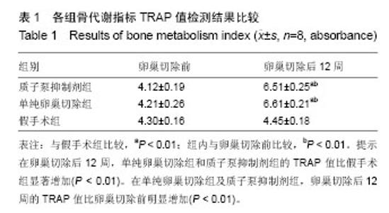
2.1 实验动物数量分析 实验选用新西兰大白兔24只,分为3组,实验过程无脱失,全部进入结果分析。 2.2 实验动物大体观察 所有动物经过2次手术都生存良好,第一次卵巢切除后第2天,家兔即可正常进食,术后3 d发现2只家兔腹部皮肤红肿,局部碘伏消毒并注射青霉素1周,炎症消退,其余22只家兔术区无感染。第2次种植体置入当天家兔即可正常进食,种植区局部碘伏消毒1周,伤口区未发现红肿,家兔可正常走动。 2.3 骨代谢指标检测结果 在卵巢切除后12周,单纯卵巢切除组和质子泵抑制剂组的TRAP值比假手术组显著增加(P < 0.01),质子泵抑制剂组与单纯卵巢切除组差异无显著性意义。在单纯卵巢切除组及质子泵抑制剂组,卵巢切除后12周的TRAP值比卵巢切除前明显增加(P < 0.01),假手术组卵巢切除前后差异无显著性意义(表1)。"
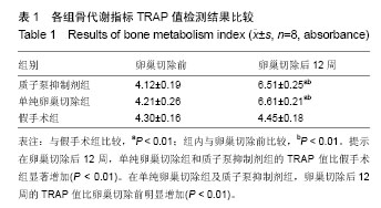

2.4 骨矿物质密度测量 在卵巢切除后12周,对家兔L5及股骨、胫骨进行骨矿物质密度测定。卵巢切除组3个不同部位的骨矿物质密度比假手术组降低了27.09%。说明骨质疏松模型的建立成功(表2)。"
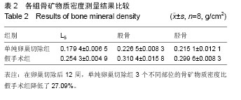
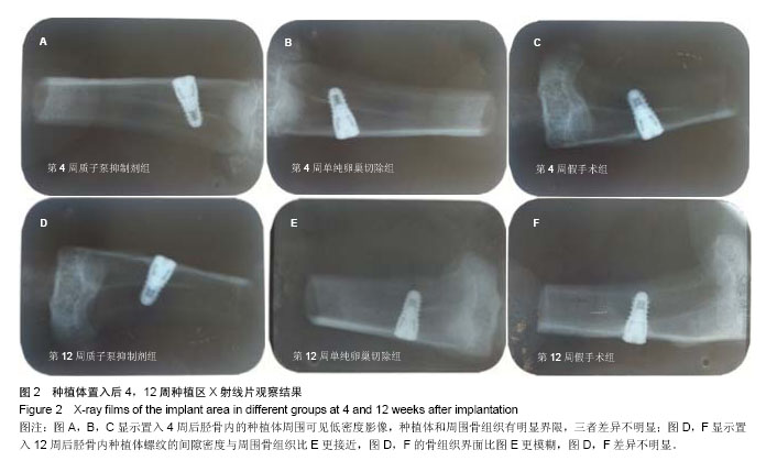
2.5 X射线片检查结果 种植体置入后第4周,两侧胫骨内的种植体周围可见低密度影像,种植体和周围骨组织有明显界限;第12周时,胫骨内种植体螺纹的间隙密度与周围骨组织接近,骨组织界面较模糊消失。同一时间点,单纯卵巢切除组种植体的高阻射影像、种植体螺纹间隙的低密度影像、骨组织的界限明显比其他2组清楚,假手术组和质子泵抑制剂组无明显差异(图2)。"
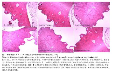
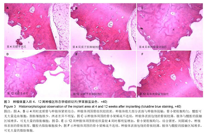
2.6 组织学观察结果 种植体置入后4周时,假手术组种植体与新生骨组织结合紧密,周围新生骨疏松多孔,松质骨区骨小梁较多且连接密集,包绕种植体表面的骨板较厚,少量脂肪细胞存在于髓腔。质子泵抑制剂组组织学改变与假手术组相似,皮质骨与种植体紧密结合,种植体周围骨组织较致密,种植体绝大部分表面与种植体接触,骨小梁粗细均匀,髓腔可见大量造血细胞,脂肪细胞极少。单纯卵巢切除组种植体周围皮质骨改变与前2组相似,但种植体与骨界面间有少量纤维组织,骨小梁稀疏且不均匀,种植体表面直接与骨髓腔接触,骨髓腔内含有大量的脂肪细胞。12周时,质子泵抑制剂组与假手术组组织学改变相似,种植体周围骨组织量较4周时都明显增加,骨小梁粗细均匀,结合紧密,间隙减小,但仍然有较多孔隙存在。种植体表面的骨板很厚,髓腔 内脂肪细胞极少。单纯卵巢切除组种植体周围的骨小梁稀疏不连续,种植体表面包绕的骨板较薄,植体与髓腔的接触区域增多,可见大量的脂肪细胞(图3)。 "

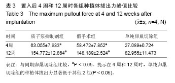
2.7 最大力值拔出实验结果 在拔出力学实验中,无论在4周和12周时,单纯卵巢切除组显著低于其他2组(P < 0.05),质子泵抑制剂组稍高于假手术组,但其差异无显著性意义(表3)。"
| [1]Hwang D, Wang HL. Medical contraindications to implant therapy: part II: relative contraindications. Implant Denti. 2007;16(1):13-23. [2]Bornstein MM, Cionca N, Mombelli A. Systemic conditions and treatments as risks for implant therapy. Int J Oral Maxillofac Implants. 2009;24 Suppl:12-27. [3]席光伟,王学玲,宫琳,等.辛伐他汀涂层内固定对大鼠骨质疏松骨折愈合晚期的影响[J].中国组织工程研究, 2013,17(51): 8827-8833. [4]Jiang GZ, Matsumoto H, Hori M, et al. Correlation among geometric, densitometric, and mechanical properties in mandible and femur of osteoporotic rats. J Bone Miner Metab. 2008;26(2):130-137. [5]Brennan-Calanan RM, Genco RJ, Wilding GE, et al. Osteoporosis and oral infection: independent risk factors for oral bone loss. J Dent Res. 2008;87(4):323-327. [6]Chang PC, Lang NP, Giannobile WV. Evaluation of functional dynamics during osseointegration and regeneration associated with oral implants. Clin Oral Implants Res. 2010; 21(1):1-12. [7]马俊岭,郭海英,阳晓东.骨质疏松症的流行病学概况[J].中国全科医学,2009,12(9):1744-1746. [8]Narai S, Nagahata S. Effects of alendronate on the removal torque of implants in rats with induced osteoporosis. Int J Oral Maxillofac Implants. 2003;18(2):218-223. [9]Raggatt LJ, Partridge NC. Cellular and molecular mechanisms of bone remodeling. J Biol Chem. 2010;285(33): 25103-25108. [10]Al-Bataineh MM, Gong F, Marciszyn A, et al. Regulation of the Proximal Tubule Vacuolar H+-ATPase by PKA and AMP-Activated Protein Kinase. Am J Physiol Renal Physiol. 2014;306(9):F981-995. [11]Laitala T, Väänänen HK. Inhibition of bone resorption in vitro by antisense RNA and DNA molecules targeted against carbonic anhydrase II or two subunits of vacuolar H (+)- ATPase. J Clin Invest. 1994;93(6):2311. [12]Frattini A, Orchard PJ, Sobacchi C, et al. Defects in TCIRG1 subunit of the vacuolar proton pump are responsible for a subset of human autosomal recessive osteopetrosis. Nature Genet. 2000;25(3):343-346. [13]Kartner N, Manolson MF. Novel techniques in the development of osteoporosis drug therapy: the osteoclast ruffled-border vacuolar H+-ATPase as an emerging target. Expert Opin Drug Discov. 2014;9(5):505-522. [14]Niikura K, Takano M, Sawada M. A novel inhibitor of vacuolar ATPase, FR167356, which can discriminate between osteoclast vacuolar ATPase and lysosomal vacuolar ATPase. Br J Pharmacol. 2004;142(3):558-566. [15]方忠,杨琴,李锋,等.去势法兔骨质疏松模型建立的探讨[J].中国骨质疏松杂志,2005,11(2):202-205. [16]吴松涛,孟维艳,蔡青,等.PTH (1-34)对兔骨质疏松种植体骨结合影响的研究[J].实用口腔医学杂志, 2011,27(2):181-184. [17]Huss M, Wieczorek H. Inhibitors of V-ATPases: old and new players. J Exp Biol. 2009;212(3):341-346. [18]Price PA, June HH, Buckley JR, et al. SB 242784, a selective inhibitor of the osteoclastic V-H+-ATPase, inhibits arterial calcification in the rat. Circ Res. 2002;91(6):547-552. [19]Nyman JKE, Väänänen HK. A rationale for osteoclast selectivity of inhibiting the lysosomal V-ATPase a3 isoform. Calcif Tissue Int. 2010;87(3):273-283. [20]Tarakida A, Higuchi T, Mizunuma H. [Evidence of hormone replacement therapy for osteoporosis]. Clin Calcium. 2008; 18(10):1434-1441. [21]Palacios S. Advances in hormone replacement therapy: making the menopause manageable. BMC Women's Health. 2008;8(1):22. [22]侯玉东,余萍萍,韩晓鹏,等.雌激素干预吸烟骨质疏松大鼠种植体周围的骨沉积[J].中国组织工程研究,2012,16(46):8556-8560. [23]Minsk L, Polson AM. Dental implant outcomes in postmenopausal women undergoing hormone replacement. Compend Contin Educ Dent. 1998;19(9):859-862. [24]Wang Y, Yang X, Li X, et al. Knowledge and personal use of menopausal hormone therapy among Chinese obstetrician- gynecologists: results of a survey. Menopause. 2014. [Epub ahead of print]. [25]Dikicier E, Karaçayl? Ü, Dikicier S, et al. Effect of systemic administered zoledronic acid on osseointegration of a titanium implant in ovariectomized rats. J Craniomaxillofac Surg. 2014. pii: S1010-5182(14)00041-9. [26]张玉芹,邓久鹏,戚孟春,等.局部应用唑来膦酸对骨质疏松大鼠种植体骨结合的影响[J].中国老年学杂志, 2012,32(15):3227- 3229. [27]李春艳,周延民,王林,等.阿仑膦酸钠对骨质疏松种植体骨结合影响的力学研究[J].华西口腔医学杂志,2011,29(3):328-329. [28]Brown JP, Morin S, Leslie W, et al. Bisphosphonates for treatment of osteoporosis Expected benefits, potential harms, and drug holidays. Can Fam Physician. 2014;60(4):324-333. [29]李国宾,贾素侠,陈旻生,等.辛伐他汀对骨质疏松患者种植体周围骨的影响[J].中国实用医药,2012,7(18):49-50. [30]Ahn SJ, Leesungbok R, Lee SW, et al. Differences in implant stability associated with various methods of preparation of the implant bed: An in vitro study. J Prosthet Dent. 2012;107(6): 366-372. [31]蒋柳宏,景向东,闫春歌,等.仙灵骨葆对骨质疏松状态下纯钛种植体骨结合的影响[J].新中医,2008,40(4):100-101. [32]Ustun Y, Erdogan O, Kurkcu M, et al. Effects of low-intensity pulsed ultrasound on dental implant osseointegration: a preliminary report. Eur J Dent. 2008;2:254. [33]Danoux CB, Barbieri D, Yuan H, et al. In vitro and in vivo bioactivity assessment of a polylactic acid/hydroxyapatite composite for bone regeneration. Biomatter. 2014;4(1): e27664. [34]张帆,陈晖,程志鹏.加速口腔种植体愈合的相关性研究现状[J]. 全科医学临床与教育,2009,7(5):503-305. [35]Sørensen MG, Henriksen K, Neutzsky‐Wulff AV, et al. Diphyllin, a Novel and Naturally Potent V‐ATPase Inhibitor, Abrogates Acidification of the Osteoclastic Resorption Lacunae and Bone Resorption. J Bone Mineral Res. 2007; 22(10):1640-1648. [36]Niikura K. Comparative analysis of the effects of a novel vacuolar adenosine 5'-triphosphatase inhibitor, FR202126, and doxycycline on bone loss caused by experimental periodontitis in rats. J Periodontol. 2006;77(7):1211-1216. [37]Kempen DHR, Lu L, Heijink A, et al. Effect of local sequential VEGF and BMP-2 delivery on ectopic and orthotopic bone regeneration. Biomaterials. 2009;30(14):2816-2825. [38]Niikura K, Takeshita N, Takano M. A vacuolar ATPase inhibitor, FR167356, prevents bone resorption in ovariectomized rats with high potency and specificity: potential for clinical application. J Bone Miner Res. 2005;20(9):1579-1588. [39]Nishisho T, Hata K, Nakanishi M, et al. The a3 isoform vacuolar type H+-ATPase promotes distant metastasis in the mouse B16 melanoma cells. Mol Cancer Res. 2011;9(7): 845-855. [40]Pérez-Sayáns M, Somoza-Martín JM, Barros-Angueira F, et al. V-ATPase inhibitors and implication in cancer treatment. Cancer Treat Rev. 2009;35(8):707-713. |
| [1] | Tang Hui, Yao Zhihao, Luo Daowen, Peng Shuanglin, Yang Shuanglin, Wang Lang, Xiao Jingang. High fat and high sugar diet combined with streptozotocin to establish a rat model of type 2 diabetic osteoporosis [J]. Chinese Journal of Tissue Engineering Research, 2021, 25(8): 1207-1211. |
| [2] | Li Zhongfeng, Chen Minghai, Fan Yinuo, Wei Qiushi, He Wei, Chen Zhenqiu. Mechanism of Yougui Yin for steroid-induced femoral head necrosis based on network pharmacology [J]. Chinese Journal of Tissue Engineering Research, 2021, 25(8): 1256-1263. |
| [3] | Hou Guangyuan, Zhang Jixue, Zhang Zhijun, Meng Xianghui, Duan Wen, Gao Weilu. Bone cement pedicle screw fixation and fusion in the treatment of degenerative spinal disease with osteoporosis: one-year follow-up [J]. Chinese Journal of Tissue Engineering Research, 2021, 25(6): 878-883. |
| [4] | Li Shibin, Lai Yu, Zhou Yi, Liao Jianzhao, Zhang Xiaoyun, Zhang Xuan. Pathogenesis of hormonal osteonecrosis of the femoral head and the target effect of related signaling pathways [J]. Chinese Journal of Tissue Engineering Research, 2021, 25(6): 935-941. |
| [5] | Xiao Fangjun, Chen Shudong, Luan Jiyao, Hou Yu, He Kun, Lin Dingkun. An insight into the mechanism of Salvia miltiorrhiza intervention on osteoporosis based on network pharmacology [J]. Chinese Journal of Tissue Engineering Research, 2021, 25(5): 772-778. |
| [6] | Liu Bo, Chen Xianghe, Yang Kang, Yu Huilin, Lu Pengcheng. Mechanism of DNA methylation in exercise intervention for osteoporosis [J]. Chinese Journal of Tissue Engineering Research, 2021, 25(5): 791-797. |
| [7] | Li Chenjie, Lü Linwei, Song Yang, Liu Jingna, Zhang Chunqiu. Measurement and statistical analysis of trabecular morphological parameters of titanium alloy peri-prosthesis under preload [J]. Chinese Journal of Tissue Engineering Research, 2021, 25(4): 516-520. |
| [8] | Shi Xiaoxiu, Mao Shilong, Liu Yang, Ma Xingshuang, Luo Yanfeng. Comparison of tantalum and titanium (alloy) as orthopedic materials: physical and chemical indexes, antibacterial and osteogenic ability [J]. Chinese Journal of Tissue Engineering Research, 2021, 25(4): 593-599. |
| [9] | Nie Shaobo, Li Jiantao, Sun Jien, Zhao Zhe, Zhao Yanpeng, Zhang Licheng, Tang Peifu. Mechanical stability of medial support nail in treatment of severe osteoporotic intertrochanteric fracture [J]. Chinese Journal of Tissue Engineering Research, 2021, 25(3): 329-333. |
| [10] | Zhong Yuanming, Wan Tong, Zhong Xifeng, Wu Zhuotan, He Bingkun, Wu Sixian. Meta-analysis of the efficacy and safety of percutaneous curved vertebroplasty and unilateral pedicle approach percutaneous vertebroplasty in the treatment of osteoporotic vertebral compression fracture [J]. Chinese Journal of Tissue Engineering Research, 2021, 25(3): 456-462. |
| [11] | Zhu Yun, Chen Yu, Qiu Hao, Liu Dun, Jin Guorong, Chen Shimou, Weng Zheng. Finite element analysis for treatment of osteoporotic femoral fracture with far cortical locking screw [J]. Chinese Journal of Tissue Engineering Research, 2021, 25(24): 3832-3837. |
| [12] | Feng Guancheng, Fang Jianming, Lü Haoran, Zhang Dongsheng, Wei Jiadong, Yu Bingbing. How does bone cement dispersion affect the early outcome of percutaneous vertebroplasty [J]. Chinese Journal of Tissue Engineering Research, 2021, 25(22): 3450-3457. |
| [13] | Liu Chang, Li Datong, Liu Yuan, Kong Lingbo, Guo Rui, Yang Lixue, Hao Dingjun, He Baorong. Poor efficacy after vertebral augmentation surgery of acute symptomatic thoracolumbar osteoporotic compression fracture: relationship with bone cement, bone mineral density, and adjacent fractures [J]. Chinese Journal of Tissue Engineering Research, 2021, 25(22): 3510-3516. |
| [14] | Huo Hua, Cheng Yuting, Zhou Qian, Qi Yuhan, Wu Chao, Shi Qianhui, Yang Tongjing, Liao Jian, Hong Wei. Effects of drug coating on implant surface on the osseointegration [J]. Chinese Journal of Tissue Engineering Research, 2021, 25(22): 3558-3564. |
| [15] | Cai Qunbin, Yang Lijuan, Li Qiumin, Chen Xinmin, Zheng Liqin, Huang Peizhen, Lin Ziling, Jiang Ziwei . Feasibility of internal fixation removal of intertrochanteric fractures in elderly patients based on fracture mechanics [J]. Chinese Journal of Tissue Engineering Research, 2021, 25(21): 3313-3318. |
| Viewed | ||||||
|
Full text |
|
|||||
|
Abstract |
|
|||||