Chinese Journal of Tissue Engineering Research ›› 2014, Vol. 18 ›› Issue (23): 3682-3690.doi: 10.3969/j.issn.2095-4344.2014.23.013
Previous Articles Next Articles
Biological characteristics and dopaminergic neural-like cell differentiation potential of human amniotic membrane-derived mesenchymal stem cells
Zhou Wen-ran1, Li Xin1, Wang Wen-bo1, Xie Yan-xia1, Tang Na1, Yan Ying1, 2
- 1Institute for Stem Cell and Regenerative Medicine, Zhongjian Medical Group, Beijing 100094, China; 2Saint Lukes Cancer Institute and School of Medicine, University Missouri-Kansas City, Kansas City, MO 64108, USA
-
Revised:2014-03-14Online:2014-06-04Published:2014-06-04 -
Contact:Yan Ying, M.D., Professor, Doctoral supervisor, Institute for Stem Cell and Regenerative Medicine, Zhongjian Medical Group, Beijing 100094, China; Saint Lukes Cancer Institute and School of Medicine, University Missouri-Kansas City, Kansas City, MO 64108, USA -
About author:Zhou Wen-ran, Master, Associate investigator, Institute for Stem Cell and Regenerative Medicine, Zhongjian Medical Group, Beijing 100094, China
CLC Number:
Cite this article
Zhou Wen-ran, Li Xin, Wang Wen-bo, Xie Yan-xia, Tang Na, Yan Ying. Biological characteristics and dopaminergic neural-like cell differentiation potential of human amniotic membrane-derived mesenchymal stem cells[J]. Chinese Journal of Tissue Engineering Research, 2014, 18(23): 3682-3690.
share this article

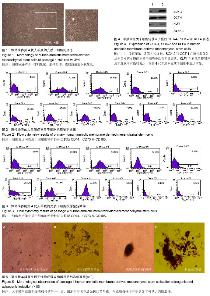
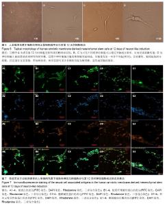
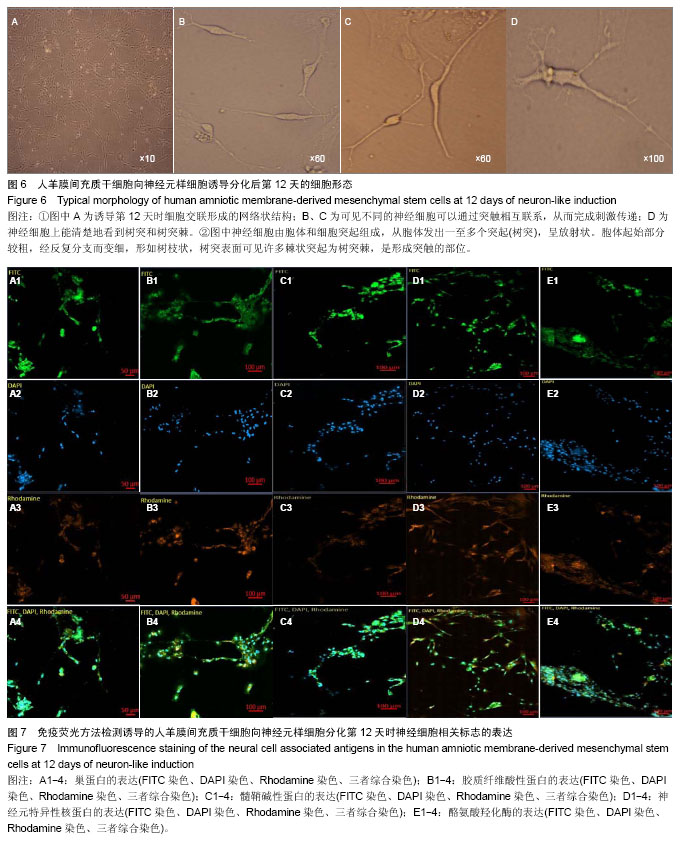
2.1 人羊膜间充质干细胞体外培养中的形态特点 原代培养:经Ⅱ型胶原酶消化和Percoll纯化后收集的有核细胞在培养24 h后,有少部分细胞贴壁。刚贴壁的细胞呈圆形,折光性强。随后细胞胞质逐渐展开,折光性减弱,逐渐形成扁平单层细胞,呈漩涡状生长或成簇生长, 7 d细胞融合度达到80%。 传代培养:传代细胞生长迅速,接种4 h后细胞贴壁,形态、大小均一,两三天可倍增,细胞可以稳定生长传代。用于诱导分化的第4代羊膜间充质干细胞形态(图1)。 2.2 人羊膜间充质干细胞相关抗原的表达 原代羊膜间充质干细胞(图2)和第4代羊膜间充质干细胞(图3)细胞表面抗原和神经细胞相关抗原表达的特点。 羊膜间充质干细胞不表达造血细胞特异性抗原CD45,CD14,CD11c和CD34(图中未展示)。原代和第4代羊膜间充质干细胞均表达间充质干细胞特异性标志抗原CD44,CD73和CD105,传代后的羊膜间充质干细胞上述抗原表达趋向均一,表达比例也有所增加。神经细胞相关抗原巢蛋白、CD133、β-微管蛋白Ⅲ和胶质纤维酸性蛋白在原代羊膜间充质干细胞中表达比例分别为2.44%、10.19%、47.06%和73.07%,而在第4代羊膜间充质干细胞中表达比例分别为3.66%、29.72%、71.08%、97.17%。髓鞘碱性蛋白、酪氨酸羟化酶和神经元特异性核蛋白等神经细胞标记物在原代羊膜间充质干细胞中表达比例为1.58%、2.22%、13.95%,在第4代羊膜间充质干细胞中表达比例分别为0.26%、0.14%和0.14%。 2.3 原代和体外培养第4代羊膜间充质干细胞表达与胚胎干细胞密切相关的转录因子蛋白 Western Blot方法检测结果显示,上述与胚胎干细胞生物学特征密切相的转录因子蛋白OCT-4和SOX-2在原代和体外培养第4代羊膜间充质干细胞中均有明显表达。KLF4在原代羊膜间充质干细胞中有微弱表达,在第4代羊膜间充质干细胞中表达明显(图4)。 2.4 人羊膜间充质干细胞向成骨细胞的诱导分化 人羊膜间充质干细胞在诱导为骨细胞的初始阶段,细胞形态保持长梭形。随着诱导时间的延长,细胞增殖速度减慢,细胞趋向聚集,逐渐汇合呈铺路石状,细胞由长梭形变为胞体较小的立方形,同时在高倍镜下可见细胞呈复层生长。随后出现多角形成骨样细胞,胞质颜色变深,含粗大颗粒,胞核明显,胞外基质分泌逐渐增多,胶原堆积、钙盐沉积。诱导培养至10-12 d,细胞中有不透光钙结节形成(图5A),钙结节茜素红染色阳性(图5B)。 2.5 人羊膜间充质干细胞向成脂细胞的诱导分化 第4代羊膜间充质干细胞在成脂条件培养基诱导下,形态逐渐趋向圆形,胞体逐渐变大。在诱导第12天的细胞中可见大的脂肪滴,细胞介于不成熟脂肪细胞和成熟脂肪细胞之间阶段的标志(图5C),用0.3%油红(Oil Red-O)染色,显微镜下可见阳性细胞(图5D)。"
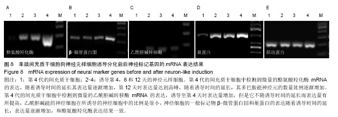
2.6 人羊膜间充质干细胞向多巴胺能神经元的诱导分化 诱导前的羊膜间充质干细胞呈成纤维细胞样的典型间充质细胞形态,诱导后第3天可见羊膜间充质干细胞细胞质向胞核收缩,呈典型核周体形态,6 d后可见多数细胞呈类神经元样细胞形态,胞体呈圆形,突起伸长,在突起末端出现分支,部分相邻细胞的突起连接成网状。随着诱导时间延长,细胞之间形成的网络状结构更加明显。诱导第12天时细胞交联形成的网络状结构。此时细胞形态改变更为明显,多数细胞转变为双极或多极神经元细胞样形态。细胞伸出突触,在高倍镜下可见较典型的神经细胞形态(图6)。 免疫荧光染色结果显示诱导后的细胞表达神经细胞特异性标志β-微管蛋白Ⅲ,神经元特异性核蛋白、酪氨酸羟化酶、胶质纤维酸性蛋白、髓鞘碱性蛋白(图7)。"

2.7 RT-PCR检测诱导后神经元细胞相关神经标记mRNA的表达分析 分别提取培养至第4代的羊膜间充质干细胞以及经过神经培养基诱导第4天、第8天和第12天的神经元样细胞cDNA进行微管蛋白基因、酪氨酸羟化酶基因、多巴胺转运体基因、乙酰胆碱转移酶基因、多巴胺受体D2型基因、巢蛋白基因的PCR扩增。PCR产物进行1%琼脂糖凝胶电泳,微管蛋白基因、酪氨酸羟化酶基因以及巢蛋白基因mRNA的表达量随着诱导时间的延长逐渐增高,但未检测到多巴胺转运体和多巴胺受体D2型基因的mRNA表达。在第4代间充质干细胞中未检测到乙酰胆碱转移酶基因mRNA的表达,诱导之后出现阳性表达(图8)。"
| [1] 李艳华,白慈贤,谢超,等.成人骨髓间充质干细胞体外定向诱导分化为胰岛样细胞团的研究[J].自然科学进展,2003,13(6):593-597.
[2] 艾国平,粟永萍,闫国和,等.骨髓间充质干细胞的分离与培养[J]. 第三军医大学学报,2001,23(5):553-555.
[3] 李秀森,郭子宽,杨靖清,等.骨髓间充质干细胞的生物学特性[J]. 解放军医学杂志,2000,25(5):346-348.
[4] 方利君,付小兵,孙同柱,等.骨髓间充质干细胞分化为血管内皮细胞的实验研究[J].中华烧伤杂志, 2003,19(1):22-24.
[5] 刘晓丹,郭子宽,李秀森,等.人骨髓间充质干细胞分离与培养方法的建立[J].军事医学科学院院刊,2000,24(4):282-284.
[6] 方利君,付小兵,孙同柱,等.在体诱导骨髓间充质干细胞分化为表皮细胞的初步观察[J].中华创伤杂志,2003,19(4):19-21.
[7] 郭礼和.人羊膜上皮细胞具有胚胎干细胞和移植免疫耐受等优良特性[J].中国细胞生物学学报,2011,33(4):451-454.
[8] 喻皇飞,陈代雄.人羊膜细胞神经生物学性状研究进展[J].重庆医学,2011,40(32):3315-3317.
[9] Sakuragawa N, Thangavel R,Mizuguchi M, et al. Expression of markers for both neuronal and glial cells in human amniotic epithelial cells. Neurosci Lett. 1996;209(1):9-12.
[10] Sakuragawa N, Kakinuma K, Kikuchi A, et al. Human amnion mesenchyme cells express phenotypes of neuroglial progenitor cells. J Neurosci Res. 2004;78(2):208-214.
[11] Niknejad H, Peirovi H, Ahmadiani A, et al. Differentiation factors that influence neuronal markers expression in vitro from human amniotic epithelial cells. Eur Cell Mater. 2010;19:22-29.
[12] Miki T,Lehmann T, Cai H, et al.Stem cell characteristics of amniotic epithelial cells.Stem Cells. 2005;23(10):1549-1559.
[13] Ishii T, Ohsugi K, Nakamura S, et al. Gene expression of oligodendrocyte markers in human amniotic epithelial cells using neural cell-type-specific expression system. Neurosci Lett. 1999;268(3):131-134.
[14] Kim J, Park S, Kang HM, et al. Human insulin secreted from insulinogenic xenograft restores normoglycemia in type I diabetic mice without immunosuppression. Cell Transplant. 2012;21(10):2131-2147
[15] Kang JW, Koo HC, Hwang SY,et al. unomodulatory effects of human amniotic membrane-derived mesenchymal stem cells. J Vet Sci. 2012;13(1):23-31.
[16] König J, Huppertz B, Desoye G, et al. Amnion-derived mesenchymal stromal cells show angiogenic properties but resist differentiation into mature endothelial cells. Stem Cells Dev. 2012;21(8):1309-1320.
[17] Lisi A, Briganti E, Ledda M, et al. A combined synthetic-fibrin scaffold supports growth and cardiomyogenic commitment of human placental derived stem cells. PLoS One. 2012;7(4): e34284.
[18] Paracchini V, Carbone A, Colombo F, et al.Amniotic mesenchymal stem cells: A new source for hepatocyte-like cells and induction of cftr expression by coculture with cystic fibrosis airway epithelial cells. J. Biomed. Biotechnol. 2012; 2012:575471.
[19] Díaz-Prado S, Muiños-López E, Hermida-Gómez T, et al. Human amniotic membrane as an alternative source of stem cells for regenerative medicine. Differentiation. 2011;81(3): 162-171.
[20] Sivasubramaniyan K, Lehnen D, Ghazanfari R, et al. Phenotypic and functional heterogeneity of human bone marrow- and amnion-derived MSC subsets. Ann N Y Acad Sci. 2012;1266:94-106.
[21] Kawanabe N, Murata S, Fukushima H,et al. Stage-specific embryonic antigen-4 identifies human dental pulp stem cells. Exp Cell Res. 2012;318(5):453-463.
[22] Kim SW, Zhang HZ, Kim CE, et al. Amniotic mesenchymal stem cells have robust angiogenic properties and are effective in treating hindlimb ischaemia. Cardiovasc Res. 2012;93(3): 525-534.
[23] Kronsteiner B, Wolbank S, Peterbauer A, et al. Human mesenchymal stem cells from adipose tissue and amnion influence T-cells depending on stimulation method and presence of other immune cells. Stem Cells Dev. 2011; 20(12):2115-2126.
[24] 冶娟,慕晓玲.人胎盘源性间充质干细胞体外分离培养及多向分化潜能的研究[J].石河子大学学报(自然科学版),2012, 30(3): 351-355.
[25] Cargnoni A, Ressel L, Rossi D, et al. Conditioned medium from amniotic mesenchymal tissue cells reduces progression of bleomycin-induced lung fibrosis. Cytotherapy. 2012;14(2): 153-161.
[26] Manuelpillai U, Moodley Y, Borlongan CV, et al. Amniotic membrane and amniotic cells: Potential therapeutic tools to combat tissue inflammation and fibrosis? Placenta. 2011; 32(Suppl.4):S320-S325.
[27] 彭琳,王建,卢光琇.人羊膜间充质细胞的分离培养及向胰岛样细胞诱导分化[J].南方医科大学学报, 2011,31(01):5-10.
[28] 李海建,张广静,张兰,等.人羊膜间充质细胞分离培养及其干细胞特性研究[J].现代生物医学进展,2011,11(02):227-229.
[29] Takashima S,Ise H,Zhao P, et al. Human amniotic epithelial cells possess hepatocyte-like characteristics and functions. Cell Struct Funct. 2004;29(3):73-84.
[30] 蔡哲,周忠蜀,向青,等.人羊膜间充质细胞的神经生物学特性及其治疗帕金森模型小鼠的实验研究[J].中国康复理论与实践, 2010, 16(4):318-321+402-403.
[31] 陈娟.干细胞移植在缺血性脑卒中治疗中的应用[J].中国组织工程研究,2012,16(19):3576-3583
[32] 胡炜,杨枫唐,尤佳,等.羊膜间充质干细胞向运动神经元前体细胞的分化[J].中国组织工程研究,2012,16(36):6767-6773.
[33] 刘娟.体外诱导人羊膜细胞向神经细胞分化[D]. 遵义医学院, 2012.
[34] Hu W, Guan FX, Li Y, et al. New methods for inducing the differentiation of amniotic-derived mesenchymal stem cells into motor neuron precursor cells. Tissue Cell. 2013; 45(5): 295-305.
[35] Sun C, Shao J, Su L, et al. Cholinergic Neuron-Like Cells Derived From Bone Marrow Stromal Cells Induced by Tricyclodecane-9-yl-xanthogenate Promote Functional Recovery and Neural Protection After Spinal Cord Injury. Cell Transplantation. 2013;22(6):961-975.
[36] 金钧,黄坚,王俊,等.体外羊膜间充质干细胞分离培养及向神经元样细胞的分化[J].中国组织工程研究与临床康复,2010,14(32): 5939-5943.
[37] Deng J, Petersen B E, Steindler D A, et al. Mesenchymal stem cells spontaneously express neural proteins in culture and are neurogenic after transplantation. Stem cells. 2006; 24(4):1054-1064.
[38] Pacary E, Legros H, Valable S, et al. Synergistic effects of CoCl2 and ROCK inhibition on mesenchymal stem cell differentiation into neuron-like cells. Journal of cell science. 2006;119(13):2667-2678.
[39] Lee FJ,Liu F. Genetic factors involved in the pathogenesis of Parkinson's disease. Brain Res Rev. 2008;58(2):354-364.
[40] Kakishita K, Nakao N, Sakuragawa N, et al. Implantation of human amniotic epithelial cells prevents the degeneration of nigral dopamine neurons in rats with 6-hydroxydopamine lesions. Brain Res. 2003;980(1):48-56.
[41] 郭礼和,赵刚,刘天津.人羊膜细胞临床前研究:治疗神经退行性疾病方面的研究进展[J].中国细胞生物学学报,2011,33(6):720- 723. |
| [1] | Pu Rui, Chen Ziyang, Yuan Lingyan. Characteristics and effects of exosomes from different cell sources in cardioprotection [J]. Chinese Journal of Tissue Engineering Research, 2021, 25(在线): 1-. |
| [2] | Lin Qingfan, Xie Yixin, Chen Wanqing, Ye Zhenzhong, Chen Youfang. Human placenta-derived mesenchymal stem cell conditioned medium can upregulate BeWo cell viability and zonula occludens expression under hypoxia [J]. Chinese Journal of Tissue Engineering Research, 2021, 25(在线): 4970-4975. |
| [3] | Zhang Xiumei, Zhai Yunkai, Zhao Jie, Zhao Meng. Research hotspots of organoid models in recent 10 years: a search in domestic and foreign databases [J]. Chinese Journal of Tissue Engineering Research, 2021, 25(8): 1249-1255. |
| [4] | Wang Zhengdong, Huang Na, Chen Jingxian, Zheng Zuobing, Hu Xinyu, Li Mei, Su Xiao, Su Xuesen, Yan Nan. Inhibitory effects of sodium butyrate on microglial activation and expression of inflammatory factors induced by fluorosis [J]. Chinese Journal of Tissue Engineering Research, 2021, 25(7): 1075-1080. |
| [5] | Wang Xianyao, Guan Yalin, Liu Zhongshan. Strategies for improving the therapeutic efficacy of mesenchymal stem cells in the treatment of nonhealing wounds [J]. Chinese Journal of Tissue Engineering Research, 2021, 25(7): 1081-1087. |
| [6] | Liao Chengcheng, An Jiaxing, Tan Zhangxue, Wang Qian, Liu Jianguo. Therapeutic target and application prospects of oral squamous cell carcinoma stem cells [J]. Chinese Journal of Tissue Engineering Research, 2021, 25(7): 1096-1103. |
| [7] | Xie Wenjia, Xia Tianjiao, Zhou Qingyun, Liu Yujia, Gu Xiaoping. Role of microglia-mediated neuronal injury in neurodegenerative diseases [J]. Chinese Journal of Tissue Engineering Research, 2021, 25(7): 1109-1115. |
| [8] | Li Shanshan, Guo Xiaoxiao, You Ran, Yang Xiufen, Zhao Lu, Chen Xi, Wang Yanling. Photoreceptor cell replacement therapy for retinal degeneration diseases [J]. Chinese Journal of Tissue Engineering Research, 2021, 25(7): 1116-1121. |
| [9] | Jiao Hui, Zhang Yining, Song Yuqing, Lin Yu, Wang Xiuli. Advances in research and application of breast cancer organoids [J]. Chinese Journal of Tissue Engineering Research, 2021, 25(7): 1122-1128. |
| [10] | Wang Shiqi, Zhang Jinsheng. Effects of Chinese medicine on proliferation, differentiation and aging of bone marrow mesenchymal stem cells regulating ischemia-hypoxia microenvironment [J]. Chinese Journal of Tissue Engineering Research, 2021, 25(7): 1129-1134. |
| [11] | Zeng Yanhua, Hao Yanlei. In vitro culture and purification of Schwann cells: a systematic review [J]. Chinese Journal of Tissue Engineering Research, 2021, 25(7): 1135-1141. |
| [12] | Kong Desheng, He Jingjing, Feng Baofeng, Guo Ruiyun, Asiamah Ernest Amponsah, Lü Fei, Zhang Shuhan, Zhang Xiaolin, Ma Jun, Cui Huixian. Efficacy of mesenchymal stem cells in the spinal cord injury of large animal models: a meta-analysis [J]. Chinese Journal of Tissue Engineering Research, 2021, 25(7): 1142-1148. |
| [13] | Hou Jingying, Yu Menglei, Guo Tianzhu, Long Huibao, Wu Hao. Hypoxia preconditioning promotes bone marrow mesenchymal stem cells survival and vascularization through the activation of HIF-1α/MALAT1/VEGFA pathway [J]. Chinese Journal of Tissue Engineering Research, 2021, 25(7): 985-990. |
| [14] | Shi Yangyang, Qin Yingfei, Wu Fuling, He Xiao, Zhang Xuejing. Pretreatment of placental mesenchymal stem cells to prevent bronchiolitis in mice [J]. Chinese Journal of Tissue Engineering Research, 2021, 25(7): 991-995. |
| [15] | Liang Xueqi, Guo Lijiao, Chen Hejie, Wu Jie, Sun Yaqi, Xing Zhikun, Zou Hailiang, Chen Xueling, Wu Xiangwei. Alveolar echinococcosis protoscolices inhibits the differentiation of bone marrow mesenchymal stem cells into fibroblasts [J]. Chinese Journal of Tissue Engineering Research, 2021, 25(7): 996-1001. |
| Viewed | ||||||
|
Full text |
|
|||||
|
Abstract |
|
|||||