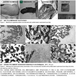
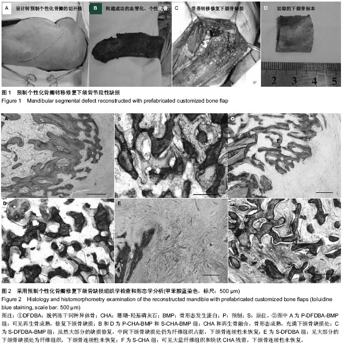
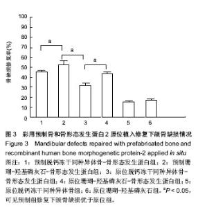
| [1] Lim YS, Kim JS, Kim NG, et al. Free Flap Reconstruction of Head and Neck Defects after Oncologic Ablation: One Surgeon's Outcomes in 42 Cases. Arch Plast Surg. 2014; 41(2):148-152.
[2] Li W, Xu Z, Liu F, et al. Vascularized free forearm flap versus free anterolateral thigh perforator flaps for reconstruction in patients with head and neck cancer: assessment of quality of life. Head Neck. 2013;35(12):1808-1813.
[3] Cai M, Lu X, Yang D, et al. Application of a Novel Intraorally Customized Transport Distraction Device in the Reconstruction of Segmental Mandibular Defect. J Craniofac Surg. 2014 Apr 2.
[4] Cai M, Lu X, Shen G, et al. Customized bifocal and trifocal transport distraction osteogenesis device for extensive mandibular reconstruction. J Craniofac Surg. 2011;22(2): 562-565.
[5] Ko EC, Chang CM, Chang P, et al. Tibial Cancellous Bone Grafting in Jaw Reconstruction: 10 Years of Experience in Taiwan. Clin Implant Dent Relat Res. 2013.
[6] Chao JW, Rohde CH, Chang MM, et al. Oral rehabilitation outcomes after free fibula reconstruction of the mandible without condylar restoration. J Craniofac Surg. 2014;25(2): 415-417.
[7] Zhu J, Xiao Y, Liu F, et al. Measures of health-related quality of life and socio-cultural aspects in young patients who after mandible primary reconstruction with free fibula flap. World J Surg Oncol. 2013;11:250.
[8] Ozmen S, Uygur S, Eryilmaz T, et al. Osseous regeneration of the free fibula flap resembling a firm mass after mandible reconstruction. J Craniofac Surg. 2012;23(3):949.
[9] Serra T, Ortiz-Hernandez M, Engel E, et al. Relevance of PEG in PLA-based blends for tissue engineering 3D-printed scaffolds. Mater Sci Eng C Mater Biol Appl. 2014;38:55-62.
[10] Warnke PH, Springer IN, Wiltfang J, et al. Growth and transplantation of a custom vascularised bone graft in a man. Lancet. 2004;364(9436):766-770.
[11] Heliotis M, Lavery KM, Ripamonti U, et al. Transformation of a prefabricated hydroxyapatite/osteogenic protein-1 implant into a vascularised pedicled bone flap in the human chest. Int J Oral Maxillofac Surg. 2006;35(3):265-269.
[12] Warnke PH, Wiltfang J, Springer I, et al. Man as living bioreactor: fate of an exogenously prepared customized tissue-engineered mandible. Biomaterials. 2006;27(17): 3163-3167.
[13] Terheyden H, Jepsen S, Rueger DR. Mandibular reconstruction in miniature pigs with prefabricated vascularized bone grafts using recombinant human osteogenic protein-1: a preliminary study. Int J Oral Maxillofac Surg. 1999;28(6):461-463.
[14] Terheyden H, Knak C, Jepsen S, et al. Mandibular reconstruction with a prefabricated vascularized bone graft using recombinant human osteogenic protein-1: an experimental study in miniature pigs. Part I: Prefabrication. Int J Oral Maxillofac Surg. 2001;30(5):373-379.
[15] Warnke PH, Springer IN, Acil Y, et al. The mechanical integrity of in vivo engineered heterotopic bone. Biomaterials. 2006; 27(7):1081-1087.
[16] Terheyden H, Menzel C, Wang H, et al. Prefabrication of vascularized bone grafts using recombinant human osteogenic protein-1--part 3: dosage of rhOP-1, the use of external and internal scaffolds. Int J Oral Maxillofac Surg. 2004;33(2):164-172.
[17] Zhou M, Peng X, Mao C, et al. Primate mandibular reconstruction with prefabricated, vascularized tissue-engineered bone flaps and recombinant human bone morphogenetic protein-2 implanted in situ. Biomaterials. 2010;31(18):4935-4943.
[18] Boyne PJ, Salina S, Nakamura A, et al. Bone regeneration using rhBMP-2 induction in hemimandibulectomy type defects of elderly sub-human primates. Cell Tissue Bank. 2006;7(1): 1-10.
[19] Takahashi Y, Yamamoto M, Yamada K, et al. Skull bone regeneration in nonhuman primates by controlled release of bone morphogenetic protein-2 from a biodegradable hydrogel. Tissue Eng. 2007;13(2):293-300.
[20] Seto I, Asahina I, Oda M, et al. Reconstruction of the primate mandible with a combination graft of recombinant human bone morphogenetic protein-2 and bone marrow. J Oral Maxillofac Surg. 2001;59(1):53-61.
[21] Amini AR, Laurencin CT, Nukavarapu SP. Bone tissue engineering: recent advances and challenges. Crit Rev Biomed Eng. 2012;40(5):363-408.
[22] Zhan K, Bai L, Xu J. Role of vascular endothelial progenitor cells in construction of new vascular loop. Microvasc Res. 2013;90:1-11.
[23] Liu X, Zhao K, Gong T, et al. Delivery of growth factors using a smart porous nanocomposite scaffold to repair a mandibular bone defect. Biomacromolecules. 2014;15(3):1019-1030.
[24] Romagnoli C, D'Asta F, Brandi ML. Drug delivery using composite scaffolds in the context of bone tissue engineering. Clin Cases Miner Bone Metab. 2013;10(3):155-161.
[25] Tannoury CA, An HS. Complications with the use of bone morphogenetic protein 2 (BMP-2) in spine surgery. Spine J. 2014;14(3):552-559.
[26] Herford AS, Boyne PJ, Williams RP. Clinical applications of rhBMP-2 in maxillofacial surgery. J Calif Dent Assoc. 2007; 35(5):335-341.
[27] Santos MI, Reis RL. Vascularization in bone tissue engineering: physiology, current strategies, major hurdles and future challenges. Macromol Biosci. 2010;10(1):12-27.
[28] Fernandes RP, Yetzer JG. Reconstruction of acquired oromandibular defects. Oral Maxillofac Surg Clin North Am. 2013;25(2):241-249. |