Chinese Journal of Tissue Engineering Research ›› 2014, Vol. 18 ›› Issue (10): 1591-1596.doi: 10.3969/j.issn.2095-4344.2014.10.018
Previous Articles Next Articles
Proliferative abilities of placenta-derived mesenchymal stem cells at different passages
Bai Jin-ping1, Li Xiu-ying2, Li Xue3, Yang Qi-wei2, Liu Ying-nan2, Wang Yi-min 2, 4
- 1 Department of Pathology, School of Basic Medical Sciences, Jilin University, Changchun 130021, Jilin Province, China; 2 Central Laboratory, China-Japan Union Hospital, Jilin University, Changchun 130033, Jilin Province, China; 3 Department of Pharmacology, Yanbian University, Yanbian 133002, Jilin Province, China; 4 Jilin Zhongke Bio-engineering Co., Ltd., Changchun 130012, Jilin Province, China
-
Online:2014-03-05Published:2014-03-05 -
Contact:Wang Yi-min, M.D., Doctoral supervisor, Central Laboratory, China-Japan Union Hospital, Jilin University, Changchun 130033, Jilin Province, China; Jilin Zhongke Bio-engineering Co., Ltd., Changchun 130012, Jilin Province, China -
About author:Bai Jin-ping, Studying for master’s degree, Department of Pathology, School of Basic Medical Sciences, Jilin University, Changchun 130021, Jilin Province, China
CLC Number:
Cite this article
Bai Jin-ping, Li Xiu-ying, Li Xue, Yang Qi-wei, Liu Ying-nan, Wang Yi-min. Proliferative abilities of placenta-derived mesenchymal stem cells at different passages[J]. Chinese Journal of Tissue Engineering Research, 2014, 18(10): 1591-1596.
share this article
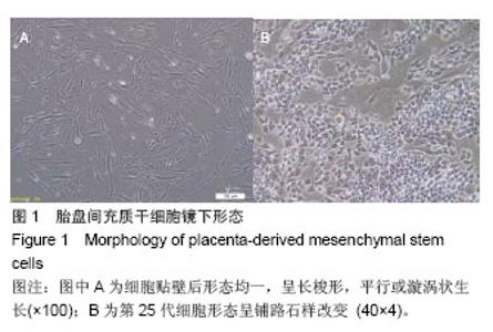
2.1 胎盘间充质干细胞形态观察结果 胎盘间充质干细胞为贴壁细胞,细胞贴壁后形态均一,呈长梭形,平行或漩涡状生长,培养3 d后细胞融合度达70%-80%,传代后细胞生长迅速(图1A),第25代胎盘间充质干细胞镜下形态见图1B。"
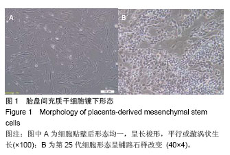
2.2 间充质干细胞表面标志物的表达 通过流式细胞仪检测,阳性表达CD44、CD105和CD90,不表达CD14、CD34和CD45。表明培养的细胞具备干细胞的表面标志物,分离得到的细胞为胎盘间充质干细胞(图2)。"


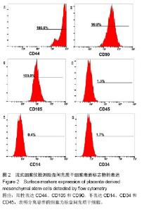
2.3 成脂成骨细胞分化 胎盘间充质干细胞在加入成脂诱导液后,第4天开始出现卵圆形透亮的小脂滴,随着时间的推移,脂滴逐渐变成大的脂肪滴,诱导21 d后固定,加入油红O染色,在显微镜下可观看到红色脂肪滴。在加入成骨诱导液后,诱导第8天,开始出现小的结节,钙结节逐渐增多,在第35天用茜素红染色,显微镜下可观察到钙盐沉积形成的结节(图3)。"
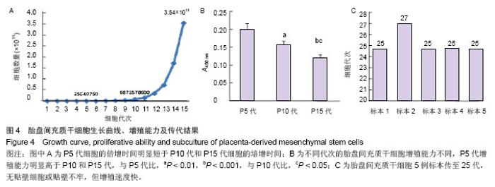
2.4 细胞计数 细胞从第1代开始计数,直到第15代共获得3.5×10¹¹个细胞,根据公式绘制成细胞的生长曲线(图4A),并通过计数结果计算出P5代细胞的倍增时间是 38.9 h,P10代细胞的倍增时间是48.9 h, P15代细胞的培增时间为69 h。不同代次胎盘间充质干细胞的增殖能力是不同的。 2.5 BrdU检测增殖 使用BrdU掺入到DNA合成期,检测P5、P10和P15代细胞的增殖能力,结果显示P5代细胞与P10和P15代的细胞相比较,其增殖能力较高(P < 0.01, P < 0.001),P10代细胞的增殖能力也高于P15代(P < 0.05,图4B)。 2.6 细胞传代能力 5例标本均进行长期培养传代(图4C),其中2例标本传代到P25代,贴壁细胞减少,继续培养72 h后几乎无贴壁细胞。其余3例标本,分别在P25、P25 和P27代出现细胞形态改变,呈铺路石样、贴壁不牢、易消化,但是增殖速度快,1∶3传代培养48 h后,融合度达80%-90%(图1B)。"

| [1]Maxson S, Lopez EA, Yoo D,et al. Role of Mesenchymal Stem Cells in Wound Repair. Stem Cells Trans Med. 2012; 1: 142-149. [2]Patel DM, Shah J,Srivastava AS. Therapeutic Potential of Mesenchymal Stem Cells in Regenerative Medicine. Stem Cells International. 2013;(2013):15. [3]Phinney DG, Prockop DJ.Concise review: mesenchymal stem/multipotent stromal cells: the state of transdifferentiation and modes of tissue repair-current views. Stem Cells.2007;25: 2896-2902. [4]Pittenger MF, Mackay AM, Beck SC, et al.Multilineage Potential of Adult Human Mesenchymal Stem Cells. Science. 1999;284(5411):143-147. [5]Bilkovski R, Schulte DM, Oberhauser F,et al. Role of WNT-5a in the determination of human mesenchymal stem cells into preadipocytes.J Biol Chem.2010; 285: 6170-6178. [6]Hematti P. Role of mesenchymal stromal cells in solid organ transplantation. Transplant Rev (Orlando).2008;22: 262-273. [7]Kim DW, Staples M, Shinozuka K, et al. Wharton’s jelly derived Mesenchymal stem cells: phenotypic characterization and optimizing their therapeutic potential for clinical applications. Int J Mol Sci. 2013; 14(6):11692-11712. [8]Kuo CK,Tuan RS.Tissue engineering with mensenchymal stem cells. IEEE Eng Med Bio Mag.2003;22(5):51. [9]Shin M,Yoshimoto H,Vacanti JP.In vivo bone tissue engineering using mesenchymal stem cells on a novel electrospun nano fibrous scaffold.Tissue Eng.2004; 10(1-2): 33. [10]Marie PJ.Cell and gene therapy for bone repair. Osteoporos Int. 2011; 22: 2023-2026. [11]Kon E, Filardo G, Roffi A, et al. Bone regeneration with mesenchymal stem cells. Clin Cases Miner Bone Metab. 2012; 9:24-27. [12]Baksh D,Song L,Tuan RS.Adult mesenchymal stem cells: characterization, differentiation, and application in cell and gene therapy.J Cell Mol Med.2004; 8(3):301-316. [13]Matikainen T, Laine J.Placenta: An alternative source of stem cells. Toxicol Appl Pharmacol. Toxicol Appl Pharmacol. 2005; 207(2 suppl):544-549. [14]Erices A, Conget P, Minguell JJ.Mesenchymal progenitor cells in human umbilical cord blood. Br J Haematol.2000; 109:235-242. [15]Ilancheran S, Moodley Y, Manuelpillai U.Human fetal membranes: a source of stem cells for tissue regeneration and repair? Placenta.2009; 30: 2-10. [16]Dai LJ, Moniri MR, Zeng ZR,et al. Potential implications of mesenchymal stem cells in cancer therapy. Cancer Lett.2011; 305: 8-20. [17]Toubai T, Paczesny S, Shono Y, et al. Mesenchymal stem cells for treatment and prevention of graft-versus-host disease after allogeneic hematopoietic cell transplantation. Current stem cell research & therapy.2009; 4: 252-259. [18]Parekkadan B, Milwid JM.Mesenchymal stem cells as therapeutics. Annu Rev Biomed Eng. 2010;12:87-117. [19]D'Agostino B, Sullo N, Siniscalco D, et al.Mesenchymal stem cell therapy for the treatment of chronic obstructive pulmonary disease. Expert Opin Biol Ther. 2010; 10:681. [20]Liu YH, Vaghjiani V, Tee JY, et al.Amniotic epithelial cells from the human placenta potently suppress a mouse model of multiple sclerosis.PLoS One. 2012;7(4):1-8. [21]Hayashi N,Takahashi K,Abe Y, et al. Placental/umbilical cord blood- derived mesenchymal stem cell-like stromal cells support hematopoietic recovery of X-irradiated human CD34~+ cells. Life Sciences. 2009; 84(17-18):598-605. [22]Pittenger MF, Mackay AM, Beck SC, et al. Multilineage po tential of adu lt hum an mesenchyma l stem cell. Science. 1999;284(2): 143-147. [23]Bianco P, Gehron RP. Marrow stromal stem cells. J Clin Invest. 2000; 105:1663-1668. [24]Vaanaenaen HK. Mesenchymal stem cells. Ann Med.2005; 37: 469-479. [25]Kolf CM, Cho E, Tuan RS. Mesenchymal stromal cells. Biology of adult mesenchymal stem cells: regulation of niche, self-renewal and differentiation. Arthritis Res Ther.2007;9: 204. [26]He Q, Wan C, Li G. Concise review: multipotent mesenchymal stromal cells in blood. Stem Cells.2007;25: 69-77. [27]Van Den Berk LC, Figdor CG, et al. Mesenchymal stromal cells:tissue engineers and immune response modulators. Arch Immunol Ther Exp(Warsz).2008;56: 325-329. [28]Zhu SF, Zhong ZN, Fu XF, et al.Comparison of cell proliferation, apoptosis, cellular morphology and ultrastructure between human umbilical cord and placenta-derived mesenchymal stem cells. Neuroscience letters.2013; 541: 77-82. [29]Kadam S, Muthyala S, Nair P, et al.Human placenta-derived mesenchymal stem cells and islet-like cell clusters generated from these cells as a novel source for stem cell therapy in diabetes.Rev Diabet Stud. 2010;7(2):168-182. [30]Parolini O, Alviano F, Bergwerf I,et al.Toward cell therapy using placenta-derived cells: disease mechanisms, cell biology, preclinical studies, and regulatory aspects at the round table. Stem Cells and Development.2010; 19(2): 143-154. [31]Tuan RS, Boland G, Tuli R. Adult mesenchymal stem cells and cell-based tissue engineering. Arthritis Res Ther.2003; 5:32-45. [32]Fukuchi Y, Nakajima H, Sugiyama D, et al. Human placenta-derived cells have mesenchymal stem/progenitor cell potential.Stem Cells.2004;22: 649-658. [33]Yen BL, Huang HI, Chien CC, et al.Isolation of multipotent cells from human term placenta. Stem Cells.2005;23(1):3-9. [34]Miao Z, Jin J, Chen L, et al. Isolation of mesenchymal stem cells from human placenta: Comparison with human bone marrow mesenchymal stem cells. Cell Biol Int. 2006;30(9): 681-687. [35]Vikram Sabapathy, Saranya Ravi, Vivi Srivastava,et al . Long-term cultured human term placenta-derived mesenchymal stem cells of maternal origin displays plasticity.Stem Cells International.2012;(2012):11. [36]Nazarov I, Lee JW, Soupene E, et al. Multipotent Stromal Stem Cells from Human Placenta Demonstrate High Therapeutic Potential. Stem Cells Translational Medicine. 2012;1(5):359-372. [37]Groza I, Pall E, Groza D,et al. Evaluation of multipotent mesenchymal stem cells from human placenta. Lucr?ri Tiin?ifice Medicin? Veterinar Vol. 2010; Xliii (2):9-14. [38]Parolini O, Alviano F, Bagnara G, et al.Isolation and characterization of cells from human term placenta: Outcome of the First International workshop on placenta derived stem cells. Stem Cells. 2008;26:300-311. [39]Prockop DJ, Sekiya I, Colter DC. Isolation and characterization of rapidly self-renewing stem cells from cultures of human marrow stromal cells. Cytotherapy.2001;3 (5):393-396. [40]Majumdar MK,Keane-Moore M,Buyaner D,et al. Characterization and functionality of cell surface molecules on human mesenchymal stem cells. J Biomed Sci.2003;10: 228-241. [41]Gekas C, Rhodes KE, Mikkola HK,et al. Isolation and analysis of hematopoietic stem cells from the placenta. J Vis Exp. 2008; 6:742. [42]李丽春,何治,李剑平.胎盘间充质干细胞的体外诱导分化[J].中国组织工程研究, 2013,17(6):1116-1119. [43]Chamberlain G, Fox J, Ashton B, et al. Concise review: mesenchymal stem cells: their phenotype, diferentiation capacity, immunological features, and potential for homing. Stem Cells. 2007; 25(11): 2739-2749. [44]Oswald J,Boxberger S,Jorgensen B,et al.Mesenchymal stem cells can be differentiated into endothelial cells in vitro. Stem Cells.2004;22(3):377-384. [45]Chien CC, Yen BL, Lee FK, et al.In vitro differentiation of human placenta-derived multipotent cells into hepatocyte-like cells. Stem Cells. 2006;24(7):1759-1760. [46]Pravin D. Potdar,Preeti Prasannan. Differentiation of Human Dermal Mesenchymal Stem Cells into Cardiomyocytes by Treatment with 5-Azacytidine: Concept for Regenerative Therapy in Myocardial Infarction. ISRN Stem Cells, 2013; (2013):9. [47]万振洲,冯亚松,彭海林,等.体外诱导胎盘间充质干细胞向表皮细胞的分化[J].中国组织工程研究与临床康复,2011, 15(49):9223- 9226. [48]Parolini O,Alviano F,Bagnara GP,et al.Isolation and char-acterization of cells from human term placenta:outcome of thefirst international workshop on placenta derived stem cells. Stem Cells. 2008;26(2):300-311. [49]张红艳,杨乃龙.胎盘来源间充质干细胞的生物学特征[J].中国组织工程究,2010,14(40):7535-7538. [50]Pipino C, Shangaris P, Resca E,et al.Placenta as a reservoir of stem cells: an underutilized resource? Br Med Bull. 2013; 105:43-68. |
| [1] | Lin Qingfan, Xie Yixin, Chen Wanqing, Ye Zhenzhong, Chen Youfang. Human placenta-derived mesenchymal stem cell conditioned medium can upregulate BeWo cell viability and zonula occludens expression under hypoxia [J]. Chinese Journal of Tissue Engineering Research, 2021, 25(在线): 4970-4975. |
| [2] | Pu Rui, Chen Ziyang, Yuan Lingyan. Characteristics and effects of exosomes from different cell sources in cardioprotection [J]. Chinese Journal of Tissue Engineering Research, 2021, 25(在线): 1-. |
| [3] | Zhang Xiumei, Zhai Yunkai, Zhao Jie, Zhao Meng. Research hotspots of organoid models in recent 10 years: a search in domestic and foreign databases [J]. Chinese Journal of Tissue Engineering Research, 2021, 25(8): 1249-1255. |
| [4] | Hou Jingying, Yu Menglei, Guo Tianzhu, Long Huibao, Wu Hao. Hypoxia preconditioning promotes bone marrow mesenchymal stem cells survival and vascularization through the activation of HIF-1α/MALAT1/VEGFA pathway [J]. Chinese Journal of Tissue Engineering Research, 2021, 25(7): 985-990. |
| [5] | Shi Yangyang, Qin Yingfei, Wu Fuling, He Xiao, Zhang Xuejing. Pretreatment of placental mesenchymal stem cells to prevent bronchiolitis in mice [J]. Chinese Journal of Tissue Engineering Research, 2021, 25(7): 991-995. |
| [6] | Liang Xueqi, Guo Lijiao, Chen Hejie, Wu Jie, Sun Yaqi, Xing Zhikun, Zou Hailiang, Chen Xueling, Wu Xiangwei. Alveolar echinococcosis protoscolices inhibits the differentiation of bone marrow mesenchymal stem cells into fibroblasts [J]. Chinese Journal of Tissue Engineering Research, 2021, 25(7): 996-1001. |
| [7] | Fan Quanbao, Luo Huina, Wang Bingyun, Chen Shengfeng, Cui Lianxu, Jiang Wenkang, Zhao Mingming, Wang Jingjing, Luo Dongzhang, Chen Zhisheng, Bai Yinshan, Liu Canying, Zhang Hui. Biological characteristics of canine adipose-derived mesenchymal stem cells cultured in hypoxia [J]. Chinese Journal of Tissue Engineering Research, 2021, 25(7): 1002-1007. |
| [8] | Geng Yao, Yin Zhiliang, Li Xingping, Xiao Dongqin, Hou Weiguang. Role of hsa-miRNA-223-3p in regulating osteogenic differentiation of human bone marrow mesenchymal stem cells [J]. Chinese Journal of Tissue Engineering Research, 2021, 25(7): 1008-1013. |
| [9] | Lun Zhigang, Jin Jing, Wang Tianyan, Li Aimin. Effect of peroxiredoxin 6 on proliferation and differentiation of bone marrow mesenchymal stem cells into neural lineage in vitro [J]. Chinese Journal of Tissue Engineering Research, 2021, 25(7): 1014-1018. |
| [10] | Zhu Xuefen, Huang Cheng, Ding Jian, Dai Yongping, Liu Yuanbing, Le Lixiang, Wang Liangliang, Yang Jiandong. Mechanism of bone marrow mesenchymal stem cells differentiation into functional neurons induced by glial cell line derived neurotrophic factor [J]. Chinese Journal of Tissue Engineering Research, 2021, 25(7): 1019-1025. |
| [11] | Duan Liyun, Cao Xiaocang. Human placenta mesenchymal stem cells-derived extracellular vesicles regulate collagen deposition in intestinal mucosa of mice with colitis [J]. Chinese Journal of Tissue Engineering Research, 2021, 25(7): 1026-1031. |
| [12] | Pei Lili, Sun Guicai, Wang Di. Salvianolic acid B inhibits oxidative damage of bone marrow mesenchymal stem cells and promotes differentiation into cardiomyocytes [J]. Chinese Journal of Tissue Engineering Research, 2021, 25(7): 1032-1036. |
| [13] | Guan Qian, Luan Zuo, Ye Dou, Yang Yinxiang, Wang Zhaoyan, Wang Qian, Yao Ruiqin. Morphological changes in human oligodendrocyte progenitor cells during passage [J]. Chinese Journal of Tissue Engineering Research, 2021, 25(7): 1045-1049. |
| [14] | Li Cai, Zhao Ting, Tan Ge, Zheng Yulin, Zhang Ruonan, Wu Yan, Tang Junming. Platelet-derived growth factor-BB promotes proliferation, differentiation and migration of skeletal muscle myoblast [J]. Chinese Journal of Tissue Engineering Research, 2021, 25(7): 1050-1055. |
| [15] | Liu Cong, Liu Su. Molecular mechanism of miR-17-5p regulation of hypoxia inducible factor-1α mediated adipocyte differentiation and angiogenesis [J]. Chinese Journal of Tissue Engineering Research, 2021, 25(7): 1069-1074. |
| Viewed | ||||||
|
Full text |
|
|||||
|
Abstract |
|
|||||