Chinese Journal of Tissue Engineering Research
Previous Articles Next Articles
Finite element model of distal tibial articular surface defect: Biomechanical analysis
Yu Hua1, Li Shao-xing2, Zhao Chang-yi3, Yan Jin-cheng1
- 1Department of Orthopedics, the Third Hospital of Hebei Medical University, Hebei Orthopaedic Biomechanics Laboratory, Shijiangzhuang 050051, Hebei Province, China; 2The Second Hospital of Hebei Medical University, Shijiangzhuang 050000, Hebei Province, China; 3Department of Anatomy, Hebei Medical University, Shijiazhuang 050017, Hebei Province, China
-
Received:2013-03-05Revised:2013-05-27Online:2013-10-22Published:2013-11-02 -
Contact:Yan Jin-cheng, M.D., Chief physician, Department of Orthopedics, the Third Hospital of Hebei Medical University, Hebei Orthopaedic Biomechanics Laboratory, Shijiangzhuang 050051, Heibei Province, China yanjincheng163@163.com -
About author:Yu Hua★, Master, Department of Orthopedics, the Third Hospital of Hebei Medical University, Hebei Orthopaedic Biomechanics Laboratory, Shijiangzhuang 050051, Heibei Province, China 1092230714@qq.com
CLC Number:
Cite this article
Yu Hua, Li Shao-xing, Zhao Chang-yi, Yan Jin-cheng . Finite element model of distal tibial articular surface defect: Biomechanical analysis[J]. Chinese Journal of Tissue Engineering Research, doi: 10.3969/j.issn.2095-4344.2013.43.012.
share this article
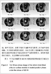
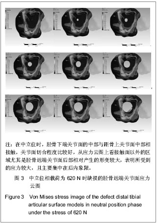
2.1 模型中立位时Von Mises应力云图、位移图 见图3,4。"
"
"

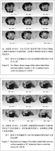
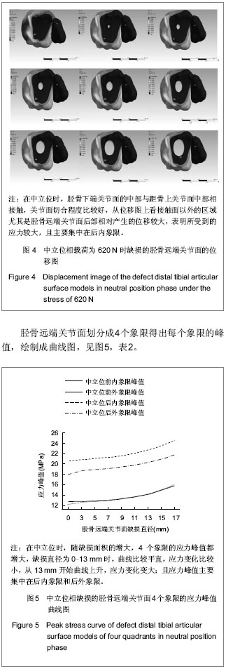
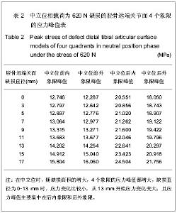
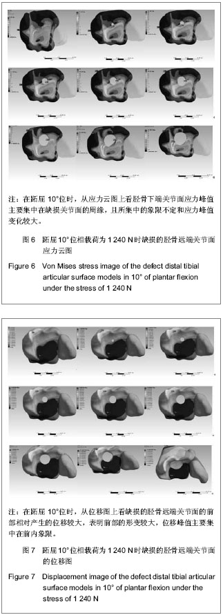
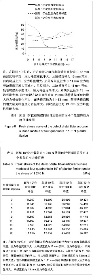
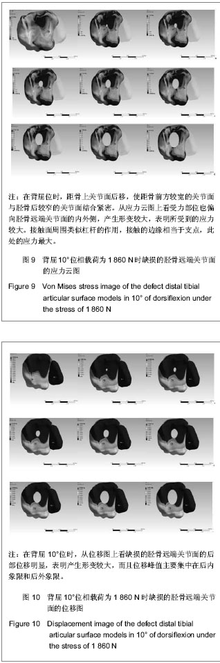
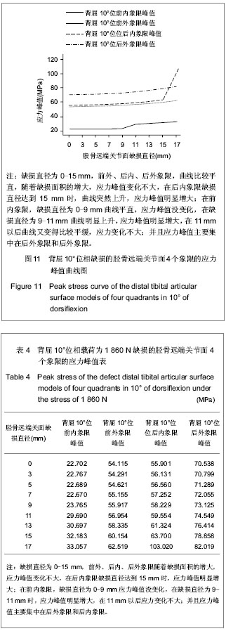
图5是表2的曲线图,图5和表2都表明:在中立位时,随缺损面积的增大,4个象限的应力峰值都增大,缺损直径为0-13 mm时,曲线比较平直,应力变化比较小,从13 mm开始曲线上升,应力变化变大;且应力峰值主要集中在后内象限和后外象限。 2.2 模型跖屈10°位时Von Mises应力 在跖屈10°位时,后内象限及前内象限缺损直径为0-13 mm曲线比较平直,应力峰值变化不大,从缺损直径为13 mm开始,曲线明显上升,应力峰值增大;后外象限直径为0-11 mm应力随着缺损面积增大而减小,呈反相关,从缺损直径为11 mm开始,随着缺损面积的增大,应力峰值逐渐增大,到缺损直径为13 mm达到最大值;前外象限在缺损直径为0-11 mm随着缺损面积的增大应力峰值变化不大,在缺损直径为11-13 mm, 随着缺损面积的增大应力峰值变化明显增大,到缺损直径为13 mm应力峰值达到最大值。应力云图、位移图,见6,7。胫骨远端关节面划分成4个象限得出每个象限的峰值,绘制成曲线图,见图8,表3。 2.3 模型背屈10°位时Von Mises应力 在背屈10°位时,缺损直径为0-15 mm,前外、后内、后外象限,曲线比较平直,随着缺损面积的增大,应力峰值变化不大,在后内象限缺损直径达到15 mm时,曲线突然上升,应力峰值明显增大;在前内象限,缺损直径为0-9 mm曲线平直,应力峰值没变化,在缺损直径为9-11 mm曲线明显上升,应力峰值明显增大,在11 mm以后曲线又变得比较平缓,应力变化不大;并且应力峰值主要集中在后外象限和后内象限。应力云图、位移图,见图9,10。"
胫骨远端关节面划分成4个象限得出每个象限的峰值,绘制成曲线图,见图11,表4。"
"
"
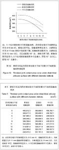
2.4 在胫骨远端关节面不同直径的缺损下胫距关节接触面积 3个位相的胫距关节的接触面积,在胫骨远端关节面缺损直径为0-5 mm时,曲线比较平直,接触面积变化很小;从缺损直径为5-11 mm曲线开始逐渐下降,接触面积逐渐变小,从缺损直径为13 mm以后,尤其是跖屈10°位,曲线突然下降,接触面积明显变小;3个位相的接触面积中立位最大,背屈10°位次之,跖屈10°位最小,见图12,表5。"

| [1] 胥少汀,葛宝丰,徐印坎.实用骨科学[M].3版.北京:人民军医出版社,2011:792. [2] 赵俊奇,邱莉.踝关节骨折伴下胫腓关节分离的治疗[J].中国实用医学, 2009,4(10):70-71. [3] 徐菲.踝足数值模型的建立及踝关节外侧失稳的距骨有限元分析[D].天津:天津医科大学,2009. [4] Greenwald AS, Matejczyk MB, Keppler L, et al. Preliminary observations on the weight-bearing surfaces of the human ankle joint. Surg Forum. 1976;27(62):505-506. [5] Matricali GA, Bartels W, Labey L, et al. High inter-specimen variability of baseline data for the tibio-talar contact area. Clin Biomech (Bristol, Avon). 2009;24(1):117-120. [6] Suckel A, Muller O, Wachter N, et al. In vitro measurement of intraarticular pressure in the ankle joint. Knee Surg Sports Traumatol Arthrosc. 2010;18(5):664-668. [7] 吴水培,俞立新,等.外踝缺损修复与重建前后力学变化的对比研究[J].2009,11(6):481-484. [8] 邹宇炜,杨新明,苏峰,等.外踝短缩对踝关节应力分布的影响[J].河北北方学院学报,2006,23(6):1-3. [9] 裴国献.数字骨科学的概念于临床初步应用[J].中华创伤骨科杂志,2008,10(2):101-102. [10] Bono CM, Khandha A. Residual sagittal motion after lumbar fusion:a finite element analysis with implications on radiographic flexion-extension criteria. J Spine. 2007;32(2): 417-422. [11] Ozan F, Yildiz H, Bora OA,et al.The effect of head trauma on fracture healing: biomechanical testing and finite element analysis. J Acta Orthop Traumatol Turc.2010;44(4):313-321. [12] Arabmotlagh M, Pilz M, Warzecha J,et al. Changes of femoral periprosthetic bone mineral density 6 years after treatment with alendronate following total hip arthroplasty. J Orthop Res,2009,27(2):183-188. [13] Dudda M, Gueleryuez A, Gautier E,et al. Risk factors for early dislocation after total hip arthroplasty:a matched case-control study.J Orthop Surg (Hong Kong).2010;18(2):179-183. [14] Wheeldon JA, Pintar FA, Knowles S, et al. Experimental flexion/extension data corridors for validation of finite element models of the young, normal cervical spine. J Biomech.2006; 39(2):375-380. [15] 马信龙,付鑫,马剑雄,等.人股骨近端空间结构重建新方法及有限元模型的建立[J].生物医学工程学杂志,2011,28(1):71-75. [16] 牛文鑫,杨云峰,俞光荣,等.人体足部三维有限元模型的有效构建方法及其合理性的实验分析研究[J].生物医学工程学杂志, 2009,26(1):80-84. [17] Cheung J T ,Zhang M , An KN. Effects of plantar fascia stiffness on the biomechanical responses of the ankle-foot complex. J Clin Biomech.2004;19(8):839. [18] Chen WP, Ju CW, Tang FT. Effects of total contact insoles on the plantar stress redistribution: a finite element analysis. Clin Biomech (Bristol, Avon). 2003;18(6):S17-24. [19] 王旭,马昕,陶凯,等.足踝有限元模型的建立与初步临床应用[J].中国生物医学工程学报,2008,27(2):287-292. [20] 中国解剖学会体质调查委员会.中国人解剖学数值[M].北京:人民卫生出版社,2002:27. [21] Burkwalter JA.骨科基础科学[M].北京:人民卫生出版社,2001: 231. [22] Turner CH, Cowin SC, Rho JY, et al. The fabric dependence of the orthotropic elastic constants of cancellous bone. J Biomech. 1990;23(6):549-561. [23] Duchemin L, Bousson V, Raossanaly C, et al. Prediction of mechanical properties of cortical bone by quantitative computed tomography. Med Eng Phys. 2008;30(3):321-328. [24] Anderson DD, Goldsworthy JK, Shivanna K, et al. Intra-articular contact stress distributions at the ankle throughout stance phase-patient-specific finite element analysis as a metric of degeneration propensity. Biomech Model Mechanobiol. 2006;5(2-3):82-89. [25] Corazza F,O’connor JJ,Leardini A,et al.Ligament fibre recruitment and forces for the anterior drawer test at the human ankle joint. J Biomech. 2003;36(3):363-372. [26] 唐芳根,袁芬连,钟敬亮.通络生骨胶囊促进兔膝关节全层软骨缺损修复的研究[J].中国实用方剂学,2010,16(12):153-156. [27] 王亦聪.骨与关节损伤.人民卫生出版社[M].第3版.北京:2001: 296. [28] Seireg A, Arvikar. The prediction of muscular lad sharing and joint forces in the lower extremities during walking. J Biomech. 1975;8(2):89-102. [29] Stauffer RN, Chao EY, Brewster RC. Force and motion analysis of the normal, diseased, and prosthetic ankle joint. Clin Orthop Relat Res. 1977;127:189-196. [30] Brekelmans WA, Poort HW, Slooff TJ. A new method to analyse the mechanical behaviour of skeletal parts. Acta Orthop Scand. 1972;43(5):301-317. [31] 李孝林.基于CT精细扫描构建人体胸腰段脊柱三维有限元模型的方法及意义[J].山东医药,2009,14(49):8-10. [32] 张功恒.寰枢椎三维有限元模型的建立及其应用[D].广西:广西医科大学,2009. [33] 林冬.一个退变颈椎三维有限元模型的建立和应用[D].四川:四川大学,2007. [34] 刘士明,周恩昌,张铮,等.肘关节三维有限元模型的建立及意义[J].山东医药,2009,27(49):22-23. [35] Amit G.Stress analysis of the standing foot following surgical plantar fascia release.J Biomech.2002;35(5):629-637. [36] 曹志林.肱二头肌长头腱有限元力学分析[D].吉林:吉林大学, 2009. [37] 史振满,史疆,王鑫,等.骨折髋支撑关节治疗股骨颈骨折的有限元力学分析[J].中国组织工程研究与临床康复,2010,14(13): 2462-2466. [38] Beaupre GS. Effect of fracture gap on stability of compression plate fixation: a finite element study.J Orthop Res. 2011; 29(1): 152. [39] Amin S, Kopperdhal DL, Melton LJ 3rd, et al. Association of hip strength estimates by finite-element analysis with fractures in women and men. J Bone Miner Res. 2011; 26(7):1593-1600. [40] Huang CH, Liau JJ, Huang CH, et al.Stress analysis of the anterior tibial post in posterior stabilized knee prostheses. J Orthop Res.2007;25(4):442-449. [41] 刘文芳,王菲,宋哲,等.以螺旋CT数据建立的颅底三维有限元模型[J].中国组织工程研究与临床康复,2010,14(52):9726-9729. [42] 陈伯华,孙鹏.颈椎三维有限元模型的建立及意义[J].中国脊柱脊髓杂志,2002,12(2):105-107. [43] 郭国新,郭继涛,李伟,等.基于有限元模型的踝关节生物力学分析[J].中国组织工程研究, 2012,16(17):3056-3060. [44] 刘峻滔,刘云鹏.跟骨 Bohler’s角的改变对踝关节接触面积及压影响的实验研究[J].滨州医学院学报,2011,34(3):198-200. [45] 郑文奎,刘勇,焦建宝,等. 胫骨骨折侧方成角畸形对胫股关节生物力学的影响[J]. 中国组织工程研究与临床康复,2011,14(28): 8988-8992. [46] 刘勇,彭阿钦,等.腓骨短缩对胫距关节生物力学接触特性的影响[J].中国矫形外科杂志,2007,15(2):134-136. [47] 适存,郭霞.肌肉骨骼系统基础生物力学[M].北京:人民卫生出版社,2008:150-170. [48] Ramse PL,Hamlton W.Changes in tibiotalar area of contact caused by lateral talar shift. J Bone Joint surg AM.1976; 58(3):356-357. [49] Greenwald AS, Matejczyk MB, Keppler L, et al. Preliminary observations on the weight-bearing surfaces of the human ankle joint. Surg Forum. 1976;27(62):505-506. [50] Egol KA ,Wolinsky P, Koval KJ.Open reduction and internal fixation of tibial pilon fractures.J Foot Ankle Clin.2000;5(4): 873-885. [51] McCann PA ,Jackson M ,Mitchell ST,et al .Complications of definitive open reduction and internal fixation of pilon fractures of the distal tibia. J Int Orthop. 2011;35 (3):413-418. [52] 陈志龙,蔡林.Pilon骨折骨折的治疗进展[J].中国骨与关节损伤杂志,2011,26((4):383-384. [53] 朱玮,窦帮,秦涛,等.关节镜辅助下微创经皮钢板内固定治疗Pilon骨折[J].实用骨科杂志,2011,17(3):266-269. [54] 王景超,朱勇,欧兆强. 闭合性Ⅲ型Pilon骨折不同时机手术的疗效分析[J].中国中医骨伤科杂志,2011,19(2):28-32. [55] 姜丹生,汪志明,施铁军.锁定加压钢板治疗 Pilon骨折[J].临床骨科杂志,2011,14(3):357. |
| [1] | Wang Jianping, Zhang Xiaohui, Yu Jinwei, Wei Shaoliang, Zhang Xinmin, Xu Xingxin, Qu Haijun. Application of knee joint motion analysis in machanism based on three-dimensional image registration and coordinate transformation [J]. Chinese Journal of Tissue Engineering Research, 2022, 26(在线): 1-5. |
| [2] | Wei Guoqiang, Li Yunfeng, Wang Yi, Niu Xiaofen, Che Lifang, Wang Haiyan, Li Zhijun, Shi Guopeng, Bai Ling, Mo Kai, Zhang Chenchen, Xu Yangyang, Li Xiaohe. Biomechanical analysis of non-uniform material femur under different loads [J]. Chinese Journal of Tissue Engineering Research, 2022, 26(9): 1318-1322. |
| [3] | Zhang Yufang, Lü Meng, Mei Zhao. Construction and verification of a full spine biomechanical model of adolescent scoliosis [J]. Chinese Journal of Tissue Engineering Research, 2022, 26(9): 1351-1356. |
| [4] | Zhang Jichao, Dong Yuefu, Mou Zhifang, Zhang Zhen, Li Bingyan, Xu Xiangjun, Li Jiayi, Ren Meng, Dong Wanpeng. Finite element analysis of biomechanical changes in the osteoarthritis knee joint in different gait flexion angles [J]. Chinese Journal of Tissue Engineering Research, 2022, 26(9): 1357-1361. |
| [5] | Bai Zixing, Cao Xuhan, Sun Chengyi, Yang Yanjun, Chen Si, Wen Jianmin, Lin Xinxiao, Sun Weidong. Construction and biomechanical analysis of ankle joint finite element model in gait cycle [J]. Chinese Journal of Tissue Engineering Research, 2022, 26(9): 1362-1366. |
| [6] | Liu Feng, Feng Yi. Finite element analysis of different Kirschner wire tension bands on transverse patella fractures during gait cycle [J]. Chinese Journal of Tissue Engineering Research, 2022, 26(9): 1367-1371. |
| [7] | Duan Chao, Shang Xiaoqiang, Duan Xianglin, Yang Ping, Tao Shengxiang. Stability of patellar claw versus loop plate combined with patellar claw for the treatment of comminuted fractures of the lower pole of the patella [J]. Chinese Journal of Tissue Engineering Research, 2022, 26(6): 934-937. |
| [8] | Li Guijun, Fang Xiaohui, Kong Weifeng, Yuan Xiaoqing, Jin Rongzhong, Yang Jun. Finite element analysis of the treatment of hallux valgus deformity by microplate combined with super strong suture elastic fixation [J]. Chinese Journal of Tissue Engineering Research, 2022, 26(6): 938-942. |
| [9] | Shao Yangyang, Zhang Junxia, Jiang Meijiao, Liu Zelong, Gao Kun, Yu Shuhan. Kinematics characteristics of lower limb joints of young men running wearing knee pads [J]. Chinese Journal of Tissue Engineering Research, 2022, 26(6): 832-837. |
| [10] | Wen Mingtao, Liang Xuezhen, Li Jiacheng, Xu Bo, Li Gang. Mechanical stability of Sanders II type calcaneal fractures fixed by two internal fixation methods [J]. Chinese Journal of Tissue Engineering Research, 2022, 26(6): 838-842. |
| [11] | Wang Hailong, Li Long, Maihemuti·Yakufu, Chen Hongtao, Liu Xu, Yilihamu·Tuoheti. Finite element analysis of stress distribution of acetabular prosthesis in the Lewinnek safety zone [J]. Chinese Journal of Tissue Engineering Research, 2022, 26(6): 843-847. |
| [12] | Huang Hao, Hong Song, Wa Qingde. Finite element analysis of the effect of femoral component rotation on patellofemoral joint contact pressure in total knee arthroplasty [J]. Chinese Journal of Tissue Engineering Research, 2022, 26(6): 848-852. |
| [13] | Zheng Yongze, Zheng Liqin, He Xingpeng, Chen Xinmin, Li Musheng, Li Pengfei, Lin Ziling. Extended finite element modeling analysis of femoral neck fracture based on ABAQUS software [J]. Chinese Journal of Tissue Engineering Research, 2022, 26(6): 853-857. |
| [14] | Wei Bing, Chang Shan. Finite element analysis of different angles of nail placement in sagittal plane of spinal fracture [J]. Chinese Journal of Tissue Engineering Research, 2022, 26(6): 864-869. |
| [15] | Liu Yuhang, Zhou Jianqiang, Xu Xuebin, Qu Xingyue, Li Ziyu, Li Kun, Wang Xing, Li Zhijun, Li Xiaohe, Zhang Shaojie. Establishment and validation of finite element model of lower cervical spine in 6-year-old children [J]. Chinese Journal of Tissue Engineering Research, 2022, 26(6): 870-874. |
| Viewed | ||||||
|
Full text |
|
|||||
|
Abstract |
|
|||||