Chinese Journal of Tissue Engineering Research ›› 2013, Vol. 17 ›› Issue (42): 7442-7448.doi: 10.3969/j.issn.2095-4344.2013.42.016
Previous Articles Next Articles
Interaction among a three-dimensional scaffold, vessels and cells in the culture of tumor cells
Hu Xue-feng, Zheng Li, Zhao Jin-min
- Guangxi Medical University, Nanning 530021, Guangxi Zhuang Autonomous Region, China
-
Received:2013-05-02Revised:2013-05-09Online:2013-10-15Published:2013-10-31 -
Contact:Zhao Jin-min, Professor, Chief physician, Master’s and doctoral supervisor, Guangxi Medical University, Nanning 530021, Guangxi Zhuang Autonomous Region, China zjm@gxmulh.com.cn -
About author:Hu Xue-feng★, Studying for master’s degree, Guangxi Medical University, Nanning 530021, Guangxi Zhuang Autonomous Region, China xiaohu529@163.com
CLC Number:
Cite this article
Hu Xue-feng, Zheng Li, Zhao Jin-min. Interaction among a three-dimensional scaffold, vessels and cells in the culture of tumor cells[J]. Chinese Journal of Tissue Engineering Research, 2013, 17(42): 7442-7448.
share this article
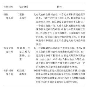
2.1 种子细胞 三维培养种子细胞是肿瘤工程构建的核心,是肿瘤工程研究领域中的基础环节。首先用于三维培养的细胞需满足以下3个基本前提:①具有特定的分化表型或定向分化潜能。②可靠的细胞来源。③不引发移植排斥反应。基于这些原因,人类肿瘤细胞是首选对象。首先,肿瘤细胞在体内具有不受控增殖性,在体外培养中仍如此。肿瘤细胞形成集落(克隆)的能力比正常细胞强,另外增殖数量增多扩展时接触抑制消除,细胞能相互重叠向三维空间发展,形成堆积物。其次,永生性也称不死性,在体外培养中表现为细胞可无限传代而不凋亡,这有利于细胞的培养及收集。再次,肿瘤细胞在缺氧状态下高表达血管生成因子,提示肿瘤细胞在肿瘤发生和演进阶段有间接促血管生成作用[6]。有研究发现两种G蛋白联受体,甲酰化肽受体和趋化因子受体4是血管生成的重要介导分子[7],这有利于肿瘤成活尤其利于应用组织工程构建体内肿瘤模型。第四,肿瘤细胞可合成一些多肽类生长因子,作用于自身相应的功能性受体,通过自分泌环路维持其增殖[8]。一些正性调控因子,如胰岛素样生长因子、白细胞介素6[9]、表皮生长因子等促进肿瘤细胞生长[10-11]。 2.2 土壤-生物支架材料 生物材料就是种子细胞生长的土壤,在肿瘤工程中起着替代细胞外基质或组织、器官基质的作用。 理想的肿瘤工程的支架材料具有以下特点:①无毒性,具有良好的生物组织相容性[12-13],不引起机体的免疫排斥反应。②有一定孔隙率,良好的表面活 性[14],维持生长其上的细胞形态和表型,为三维生长的细胞提供支架,使细胞间形成适宜的空间分布。③具有生物可降解性,可塑性和一定的机械强度[14]。④能增进细胞的黏附和增殖[15-16],诱导细胞定向分化。不同工程组织、器官的体外构建对生物材料生物活性的要求各不相同。目前,肿瘤细胞三维培养所用的生物材料众多,有细胞外基质、高分子聚合材料及有机材料复合物等。 肿瘤细胞三维培养所用的生物材料:"

| [1] Smalley KS,Lioni M,Herlyn M. Life ins't flat: Taking cancer biology to the next dimension.In Vitro Cell Dev Biol Anim. 2006;42(8-9): 242-247. [2] Petersen OW,Rnnov-Jessen L,Howlett AR,et al.Interaction with basement membrane serves to rapidly distinguish growth and differentiation pattern of normal and malignant human breast epithelial cells.Proc Natl Acad Sci U S A.1992;89 (19): 9064-9068. [3] Sun H,Liu W,Zhou G,et al.Tissue engineering of cartilage, tendon and bone. Front Med.2011;5(1):61-69. [4] Ghajar CM,Bissell MJ.Tumor engineering: the other face of tissue engineering.Tissue Eng Part A.2010;16(7):2153-2156. [5] Fischbach C,Chen R,Matsumoto T,et al.Engineering tumors with 3D scaffolds. Nature Methods.2007;4(10):855-860. [6] Bao S,Wu Q,Sathomsumetee S,et al. Stem cell-like glioma cells promote tumor angiogenesis through vascular endothelial growth factor.Cancer Res.2006;66(16):7843-7848. [7] Hu J,Deng X,Bian X,et al.The expression of functional chemokine receptor CXCR4 is associated with the metastatic potential of human nasopharyngeal carcinoma. Clin Cancer Res.2005;11(13):4658-4665. [8] Rosfiord EC,Dickson RB.Growth factors,apoptosis,and survival of mammary epithelial cells.J Mammary Gland Biol Neoplasia.1999;4(2):229-237. [9] Okamoto M,Hattori K,Oyasu R,et a1.Interleukin-6 functions as an autocrine growth factor in human bladder carcinoma cell lines in vitro.Int J Cancer. 1997;72(1):149-154. [10] Ruck A,Paulie S.The epidermal growth factor receptor is involved in autocrine growth of human bladder carcinoma cell lines.Anticancer Res.1997;17(3C): 1925-1931. [11] Negus RP,Balkwill FR.Cytokines in tumor growth,migration and metastasis. World J Urol.1996;14(3):157-165. [12] Stegemann JP,Kaszuba SN,Rowe SL.Review:advances in vascular tissue engineering using protein-based biomaterials. Tissue Eng.2007;13(11):2601-2613. [13] Meredith JE Jr,Fazeli B,Schwartz MA.The extracellular matrix as a cell survival factor.Mol Biol Cell.1993;4(9):953-961. [14] Edwards SL,Mitchell W,Matthews JB,et al.Design of nonwoven scaffold structures for tissue engineering of the anterior cruciate ligament.AUTEX Res J.2004;4(2):86-94. [15] Anselme K.Osteoblast adhesion on biomaterials. Biomaterials.2000;21(7): 667-681. [16] Gallagher WM,Lynch I,Allen LT,et al.Molecular basis of cell-biomaterial interaction: Insights gained from transcriptomic and proteomic studies. Biomaterials.2006; 27(35):5871-5882. [17] Revell CM,Athanasiou KA.Success rates and immunologic responses of autogenic, allogenic, and xenogenic treatments to repair articular cartilage defects.Tissue Eng Part B Rev. 2009;15(1):1-15. [18] Noth U,Rackwitz L,Heymer A,et al.Chondrogenic differentiation of human mesenchymal stem cells in collagen type I hydrogels.J Biomed Mater Res A. 2007;83(3):626-635. [19] Glowacki J,Mizuno S.Collagen scaffolds for tissue engineering. Biopolymers.2008;89(5):338-344. [20] Gotterbarm T,Richter W,Jung M,et al. An in vivo study of a growth-factor enhanced, cell free, two-layered collagen-tricalcium phosphate in deep osteochondral defects.Biomaterials.2006;27(18):3387-3395. [21] Cen L,Liu W,Cui L,et al.Collagen tissue engineering: Development of novel biomaterials and applications.Pediatr Res.2008;63(5):492-496. [22] Choe MM,Sporn PH,Swartz MA.An in vitro airway wall model of remodeling.Am J Physiol Lung Cell Mol Physiol.2003; 285(2): L427-L433. [23] Choe MM,Tomei AA,Swartz MA. Physiological 3D tissue model of the airway wall and mucosa.Nat Protoc.2006; 1(1):357-362. [24] Chen L,Xiao Z,Meng Y,et al.The enhancement of cancer stem cell properties of MCF-7 cells in 3D collagen scaffolds for modeling of cancer and anti-cancer drugs. Biomaterials. 2012; 33(5):1437-1444. [25] Tang J,Cui J,Chen R,et al.A three-dimensional cell biology model of human hepatocellular carcinoma in vitro.Tumor Biol.2011;32:469-479. [26] Loessner D,Stok KS,Lutolf MP,et al.Bioengineered 3D platform to explore cell-ECM interactions and drug resistance of epithelial ovarian cancer cells. Biomaterials.2010;31(32): 8494-8506. [27] Sahoo SK,Panda AK,Labhasetwar V.Characterization of porous PLGA/PLA microparticles as a scaffold for three dimensional growth of breast cancer cells. Biomacromolecules. 2005;6(2):1132-1139. [28] Zhou CZ,Confalonieri F,Jacquet M,et al.Silk fibroin: structural implications of a remarkable amino acid sequence. Proteins 2001;44(2):119-122. [29] Ki CS,Park SY,Kim HJ,et al.Development of 3-D nanofibrous fibroin scaffold with high porosity by electrospinning: implications for bone regeneration. Biotechnol Lett.2008; 30(3):405-410. [30] Altman GH,Diaz F,Jakuba C,et al.Silk-based biomaterials. Biomaterials. 2003;24(3):401-416. [31] Liu H,Fan H,Wang Y,et al. The interaction between a combined knitted silk scaffold and microporous silk sponge with human mesenchymal stem cells for ligament tissue engineering.Biomaterials.2008;29(6):662-674. [32] Wang Y,Kim HJ,Vunjak-Novakovic G,et al. Stem cell-based tissue engineering with silk biomaterials.Biomaterials. 2006; 27(36):6064-6082. [33] She Z,Jin C,Huang Z,et al. Silk fibroin/chitosan scaffold: preparation, characterization, and culture with HepG2 cell.J Mater Sci Mater Med.2008;19(12):3545-3553. [34] Fan H,Liu H,Wong EJW,et al.In vivo study of anterior cruciate ligament regeneration using mesenchymal stem cells and silk scaffold. Biomaterials.2008;29(23):3324-3337. [35] Tan PH,Aung KZ,Toh SL,et al.Three-dimensional porous silk tumor constructs in the approximation of in vivo osteosarcoma physiology. Biomaterials.2011;32: 6131-6137. [36] Willerth SM,Sakayama-Elbert SE.Approaches to neural tissue engineering using scaffolds for drug delivery.Adv Drug Deliv Rev.2007;59(4-5):325-338. [37] Santini MT,Rainaldi G,Romano R,et al.MG-63 human osteosarcoma cells grown in monolayer and as three-dimensional tumor spheroids present a different metabolic profile: a (1)H NMR study.FEBS Lett.2004; 557 (1-3):148-154. [38] Akeda K,Nishimura A,Satonaka H,et al.Three-dimensional alginate spheroid culture system of murine osteosarcoma. Oncol Rep.2009;22(5): 997-1003. [39] Plieva FM,Galaev IY,Mattiasson B.Macroporous gels prepared at subzero temperatures as novel materials for chromatography of particulate-containing fluids and cell culture applications.J Sep Sci.2007;30(11):1657-1671. [40] Tsang VL,Bhatia SN.Fabrication of three-dimensional tissues.Adv Biochem Eng Biotechnol.2007;103:189-205. [41] Brown RA.Phillips JB.Cell responses to biomimetic protein scaffolds used in tissue repair and engineering.Int Rev Cytol. 2007;262:75-150. [42] Schwarz RP,Goodw in TJ,Wolf DA.Cell culture for three-dimensional moldeling in rotating-wall vessels: an application of simulated microgravity.Tissue Cult Method. 1992;14(2):51-57. [43] Jessup JM,Goodwin TJ,Spaulding G.Prospects for use of microgravity-based bioreactors to study three-dimensional host-tumor interactions in human neoplasia. J Cell Biochem. 1993;51(3):290-300. [44] Radtke AL,Herbst-Kralovetz MM.Culturing and Applications of Rotating Wall Vessel Bioreactor Derived 3D Epithelial Cell Models.J Vis Exp.2012;(62): 3859-3868. [45] Freed LE,Vunjak-Novakovic G,Biron RJ,et al.Biodegradable polymer scaffolds for tissue engineering.Biotechnology(NY).1994;12(7):689-693. [46] Liu L,Guo Y,Chen XZ,et al.Three-dimensional dynamic culture of pre-osteoblasts seeded in HA-CS/Col/nHAP composite scaffolds and treated with a-ZAL.Acta Biochim Biophys Sin. 2012;44(8): 669-677. [47] Goral VN, Hsieh YC, Petzold ON, et al. Perfusion-based microfluidic device for three-dimensional dynamic primary human hepatocyte cell culture in the absence of biological or synthetic matrices or coagulants.Lab Chip.2010;10 (24): 3309-3432. [48] Yeatts AB, Geibel EM, Fears FF, et al. Humanmesenchymal stem cell position within scaffolds influences cell fate during dynamic culture. Biotechnol Bioeng.2012;109(9): 2381-2391. [49] Richardson TP,Peters MC,Ennett AB,et al. Polymeric system for dual growth factor delivery.Nat Biotechnol.2001;19(11): 1029-1034. [50] Ronnov-Jessen L,Petersen OW,Koteliansky VE,et al.The origin of the myofibroblasts in breast cancer. Recapitulation of tumor environment in culture unravels diversity and implicates converted fibroblasts and recruited smooth muscle cells.J Clin Invest.1995;95(2): 859-873. [51] Ghajar CM,SureshV,Peyton SR,et al.A novel threedimensional model to quantify metastatic melanoma invasion. Mol Cancer Ther.2007;6(2): 552-561. [52] Kenny PA,Lee GY,Myers CA,et al.The morphologies of breast cancer cell lines in three-dimensional assays correlate with their profiles of gene expression. Mol Oncol.2007;1(1): 84-96. [53] Miller BE,Miller FR,Heppner GH.Factors affecting growth and drug sensitivity of mouse mammary tumor lines in collagen gel cultures. Cancer Res.1985;45(5):4200-4205 [54] Petersen OW,Ronnov-Jessen L,Howlett AR,et al.Interaction with basement membrane serves to rapidly distinguish growth and differentiation pattern of normal and malignant human breast epithelial cells.Proc Natl Acad Sci USA. 1992;89(19): 9064-9068. [55] Weaver VM,Lelievre S,Lakins JN,et al.Beta4 integrin-dependent formation of polarized threedimensional architecture confers resistance to apoptosis in normal and malignant mammary epithelium.Cancer Cell.2002;2(3): 205-216. [56] Derda R,Laromaine A,Mammoto A,et al.Paper-supported 3D cell culture for tissue-based bioassays. Proc Natl Acad Sci USA.2009;106(44):18457-18462. [57] Petersen OW,Nielsen HL,Gudjonsson T,et al.Epithelial to mesenchymal transition in human breast cancer can provide a nonmalignant stroma.Am J Pathol.2003;162(2): 391-402. [58] Ebos JM,Lee CR,Cruz-Munoz W,et al.Accelerated metastasis after short-term treatment with a potent inhibitor of tumor angiogenesis.Cancer Cell.2009;15(3):232-239. [59] Ellis LM,Reardon DA.Cancer: the nuances of therapy. Nature. 2009;458(7236): 290-292. [60] Paez-Ribes M,Allen E,Hudock J,et al.Antiangiogenic therapy elicits malignant progression of tumors to increased local invasion and distant metastasis.Cancer Cell.2009;15(3): 220-231. [61] Fischbach-Teschl C,Stroock A.Microfluidic culture models of tumor angiogenesis.Tissue Eng Part A.2010;16(7):2143-2146 [62] Choi NW,Cabodi M,Held B,et al.Microfluidic scaffolds for tissue engineering. Nat Mater.2007;6(11):908-915. [63] Chrobak KM,Potter DR,Tien J.Formation of perfused, functional microvascular tubes in vitro.Microvasc Res.2006; 71(3): 185-196. [64] Verbridge S,Choi N,Zheng Y,et al. Oxygen-controlled three-dimensional cultures to analyze tumor angiogenesis.Tissue Eng Part A.2010;16(7):2133-2141. [65] Kikkawa Y,Hozumi K,Katagiri F,et al.Laminin-111-derived peptides and cancer.Cell Adh Migr, 2013;7(1):150-256. [66] Chen A,Cuevas I,Kenny PA,et al.Endothelial cell migration and vascular endothelial growth factor expression are the result of loss of breast tissue polarity.Cancer Res.2009;69(16): 6721-6729. |
| [1] | Yang Feng, Zhao Qian, Zhang Shixuan, Zhao Tienan, Feng Bo. Effectiveness and safety of rapamycin combined with CD133 antibody stent in preventing vascular restenosis [J]. Chinese Journal of Tissue Engineering Research, 2022, 26(4): 579-584. |
| [2] | Li Fang, Wu Ketong, Zhao Jun, Li Gang. Advances of endovascular stent and its treatment for aneurysms [J]. Chinese Journal of Tissue Engineering Research, 2021, 25(34): 5561-5569. |
| [3] | Xie Xiufeng, Zhang Yue, Qu Ze. Clinical outcomes of drug-eluting balloons and drug-eluting stents for the treatment of in-stent restenosis [J]. Chinese Journal of Tissue Engineering Research, 2020, 24(4): 555-560. |
| [4] | Gao Jian’an, Chen Xi, Zhang Longsheng, Liao Wenbo. Types and advantages of spinal implants in percutaneous kyphoplasty [J]. Chinese Journal of Tissue Engineering Research, 2020, 24(12): 1935-1940. |
| [5] | Wang Canli, Jiao Zhili, Sun Yong, Sun Song. Comparison of the effects of two kinds of platelet-rich fibrins on the proliferation activity of human gingival fibroblasts [J]. Chinese Journal of Tissue Engineering Research, 2019, 23(7): 1007-1012. |
| [6] | Yuan Bo, Wang Zhiwei, Tang Yifan, Zhou Shengyuan, Chen Xiongsheng, Jia Lianshun. Construction of polycaprolactone-tricalcium phosphate with different mixture ratios using three-dimensional printing technology and its osteoinductivity in vitro [J]. Chinese Journal of Tissue Engineering Research, 2019, 23(6): 821-826. |
| [7] | Zhao Liang, Li Xiafei, Zhou Kun, Yan Huanhuan, Zhang Qiqing. Preparation of acellular vascular scaffold using Triton-x100 and salvianolic acid B and its biomechanical performance [J]. Chinese Journal of Tissue Engineering Research, 2019, 23(6): 951-956. |
| [8] | Ling Hao, Wang Bao, Wu Lixia, Yan Hui, Song Chunli. Development of everolimus-eluting stents [J]. Chinese Journal of Tissue Engineering Research, 2019, 23(6): 978-984. |
| [9] | Liu Meilin, Fu Songtao, Wang Peisen, Hao Ran, Chang Bingmei, Hou Xiuying. Liposome prepared from human umbilical cord mesenchymal stem cell conditioned medium for repair of skin wound in rats [J]. Chinese Journal of Tissue Engineering Research, 2019, 23(5): 734-740. |
| [10] | Wei Wei, Liu Yanfei, Zhang Ling, Xiong Na . Self-assembling peptide hydrogel: hemostatic effect and mechanism [J]. Chinese Journal of Tissue Engineering Research, 2019, 23(2): 310-316. |
| [11] | Zhang Minbo, Peng Qifeng, Ma Yaping, Kong Weijun, Liao Wenbo. Physical properties and biocompatibility of 3D printed bone microparticle/poly(lactic-co-glycolic acid) scaffold [J]. Chinese Journal of Tissue Engineering Research, 2019, 23(14): 2215-2222. |
| [12] | Li Zhi, Tan Chunhua, Cai Xianhua, Wang Huasong, Ding Xiaoming, Zhao Yanhong. Fabrication and biocompatibility assessment of the scaffold with biomimetic interconnected macropore structure [J]. Chinese Journal of Tissue Engineering Research, 2019, 23(14): 2223-2227. |
| [13] | Luo Kai, Yang Yafeng, Ma Teng, Xia Bing, Huang Liangliang, Huang Jinghui, Luo Zhuojing. Effects of perfluorotributylamine/alginate/bioglass biomaterials on viability and osteogenic differentiation of adipose-derived stem cells [J]. Chinese Journal of Tissue Engineering Research, 2019, 23(13): 1995-2001. |
| [14] | Wang Yuanyuan, Song Wenshan, Yu Dejun, Dai Yuankun, Li Bafang. Preparation and evaluation of fish skin acellular dermal matrix for oral guided tissue regeneration [J]. Chinese Journal of Tissue Engineering Research, 2019, 23(10): 1526-1532. |
| [15] | Liu Shaoyang, Zou Hanlin . Biocompatibility of anodic titanium oxide: an experimental research [J]. Chinese Journal of Tissue Engineering Research, 2019, 23(10): 1575-1580. |
| Viewed | ||||||
|
Full text |
|
|||||
|
Abstract |
|
|||||